. 2001 Nov 15;29(22):E109.
doi: 10.1093/nar/29.22.e109.

Behavior of DNA fibers stretched by precise meniscus motion control

Affiliations

Behavior of DNA fibers stretched by precise meniscus motion control

K Otobe et al. Nucleic Acids Res. .

Abstract

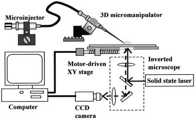
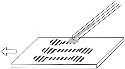
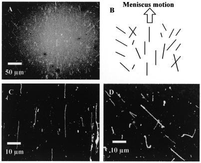
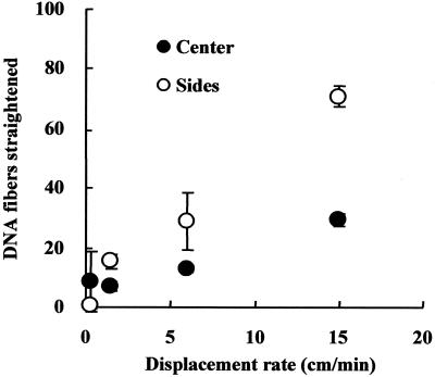
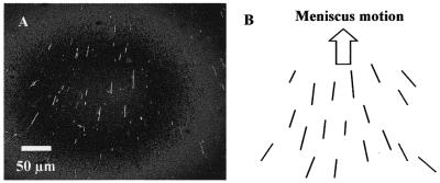
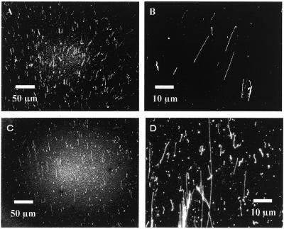
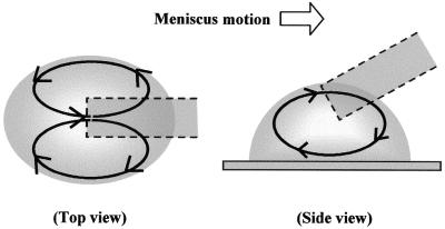
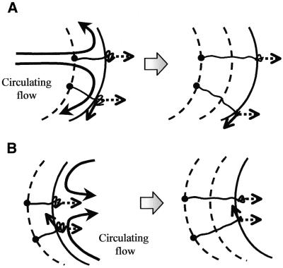
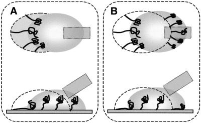
A modified DNA combing method, which can precisely locate straightened DNA fibers on a substrate, has been developed. Precise motion control of a DNA solution droplet on hydrophobic surfaces has allowed detailed analyses of DNA straightening behavior. Our method provides a technique for consistently straightening lambda phage DNA on a trace of droplet motion, though the straightened DNAs had several variations in their alignments. The dependence of the straightened DNA frequency upon motion rate, fluidity in the droplet and environmental humidity was investigated. Visualization of the solution flow in the moving droplet indicated that flows circulating parallel to the contour of the droplet markedly bias the direction of straightening in relation to the site in the droplet. As a result, the alignment variations caused by the site specificity of the bias direction revealed that environmental humidity significantly alters the straightening behavior.


Figures

Figure 1
Figure 1
Schematic set-up of the meniscus motion control system employing a micromanipulated capillary for straightening DNA fibers.
Figure 2
Figure 2
Illustration of the procedure for straightening DNAs using meniscus motion control. A droplet, whose size is adjusted by the microinjector, is dispensed and sandwiched between the capillary tip and the substrate. The droplet follows the tip because of its surface tension while the substrate is moved in a straight line in the direction of the arrow. Striped areas on the substrate represent traces of the droplet where DNAs have been straightened because of meniscus motion.
Figure 3
Figure 3
Straightened λ phage DNAs, combed by meniscus motion control in a <40% humidity environment. These images were captured by a fluorescence microscope utilizing a high sensitivity cooled CCD camera. (A) Overview of a trace of a droplet moved on a coverslip, observed through a 20× objective lens. (B) Schematic of the trend of straightened DNAs in the trace, illustrated on the basis of the direction of meniscus motion and the center line of the trace. (C) Detail of straightened DNAs, observed in the center area of the trace and magnified with a 100× objective lens. (D) Slanted, straightened DNAs seen in the right of the trace.
Figure 4
Figure 4
Influence of the displacement rate of meniscus motion on the number of straightened DNAs. Blank circles represent the numbers of straightened DNAs found in both sides of the traces and filled circles are those found in the center area. The numbers are averages counted in three traces on a coverslip. Bars at the circles indicate standard deviation.
Figure 5
Figure 5
Straightened λ phage DNAs combed by meniscus motion control in a >60% humidity environment. (A) Overview of a trace of a droplet moved on a coverslip, observed via a 20× objective lens. (B) Schematic of the tendency of straightened DNAs in the trace, illustrated on the basis of the direction of meniscus motion and the center line of the trace.
Figure 6
Figure 6
Behavior of straightened DNA with respect to differences in surface modification of substrates and in the origin of DNA. (A) Straightened λ phage DNAs; overview of a trace of a droplet moved on a coverslip coated with DDS. (B) Detail of the straightened phage DNAs. (C) Straightened genomic DNA of T.thermophilus HB8; overview of a trace. (D) Detail of the straightened genomic DNAs.
Figure 7
Figure 7
Illustration of solution flows in a droplet moved with a capillary tip. The top view shows two circulating flows, from the center to both sides of the droplet and then returning to the center. The side view illustrates a one-directional circulating flow from the bottom to the top of the droplet.
Figure 8
Figure 8
Influence of the circulation flows on the behavior of DNAs stretched by meniscus motion. Bold arrows represent circulating flows induced in the droplet, dotted arrows the flow directions corresponding to droplet motion and solid arrows the directions biased by the circulation flows on the surface. Large dots are adsorption points of the DNAs, broken lines indicate traces of the preceding interfaces and round solid lines show the interfaces between the meniscus and substrate. The meniscus moves from left to right in this illustration and the results are shown in the right figures. (A) Behavior of the DNAs adsorbed at the advancing edge of the droplet and then stretched by meniscus motion. The stretching direction slants towards the side of the droplet due to the circulation flow. (B) In contrast to the stretching at the advancing edge, at the receding edge the stretching direction slants towards the center of the droplet due to the circulation flow.
Figure 9
Figure 9
Behavior of DNAs straightened by droplet motion dependent on surface characteristics. Dotted lines represent the location of the preceding interface in which the extremities of the DNAs have been adsorbed on the surface and solid entangled lines represent stretching DNAs. Gray areas are those wetted by the droplet or soaked in DNA solution. The upper figures are top views of droplet movement and the lower ones side views. (A) Straightening DNAs on a poorly hydrophobic surface. Fixation of the contours of the DNAs, in accordance with their stretching, occurs solely at the receding edge since no fixation is allowed to occur in the wetted areas. (B) Straightening on a highly hydrophobic surface. Fixation occurs at both the advancing and receding edges of the droplet since water repulsion promotes fixation at the advancing edge.

References

    1. Wiegant J., Wiesmeijer,C.C., Hoovers,J.M.N., Schuuring,E., d’Azzo,A., Vrolijk,J., Tanke,H.J. and Raap,A.K. (1993) Multiple and sensitive fluorescence in situ hybridization with rhodamine-, fluorescein- and comarin-labeled DNAs. Cytogenet. Cell Genet., 63, 73–76. - PubMed
    1. Herrick J. and Bensimon,A. (1999) Imaging of single DNA molecule, application to high-resolution genomic studies. Chromosome Res., 7, 409–423. - PubMed
    1. Bustamante C., Vesenka,J., Tang,C.L., Rees,W., Guthold,M. and Keller,R. (1992) Circular DNA molecules imaged in air by scanning force microscopy. Biochemistry, 31, 22–26. - PubMed
    1. Hansma H.G., Bezanilla,M., Zenhausern,F., Adarian,M. and Sinsheimer,R.L. (1993) Atomic force microscopy of DNA in aqueous solutions. Nucleic Acids Res., 21, 505–512. - PMC - PubMed
    1. Wang W., Lin,J. and Schwartz,D.C. (1998) Scanning force microscopy of DNA molecules elongated by convective fluid flow in a evaporated droplet. Biophys. J., 75, 513–520. - PMC - PubMed

Publication types