. 2001 Nov;108(10):1495-504.
doi: 10.1172/JCI12190.

p38 MAP kinase modulates liver cell volume through inhibition of membrane Na+ permeability

Affiliations

p38 MAP kinase modulates liver cell volume through inhibition of membrane Na+ permeability

A P Feranchak et al. J Clin Invest. 2001 Nov.

Abstract

In hepatocytes, Na+ influx through nonselective cation (NSC) channels represents a key point for regulation of cell volume. Under basal conditions, channels are closed, but both physiologic and pathologic stimuli lead to a large increase in Na+ and water influx. Since osmotic stimuli also activate mitogen-activated protein (MAP) kinase pathways, we have examined regulation of Na+ permeability and cell volume by MAP kinases in an HTC liver cell model. Under isotonic conditions, there was constitutive activity of p38 MAP kinase that was selectively inhibited by SB203580. Decreases in cell volume caused by hypertonic exposure had no effect on p38, but increases in cell volume caused by hypotonic exposure increased p38 activity tenfold. Na+ currents were small when cells were in isotonic media but could be increased by inhibiting constitutive p38 MAP kinase, thereby increasing cell volume. To evaluate the potential inhibitory role of p38 more directly, cells were dialyzed with recombinant p38alpha and its upstream activator, MEK-6, which substantially inhibited volume-sensitive currents. These findings indicate that constitutive p38 activity contributes to the low Na+ permeability necessary for maintenance of cell volume, and that recombinant p38 negatively modulates the set point for volume-sensitive channel opening. Thus, functional interactions between p38 MAP kinase and ion channels may represent an important target for modifying volume-sensitive liver functions.


Figures

Figure 1
Figure 1
Cell volume recovery is Na+ dependent. HTC cell suspensions were exposed to hypertonic buffer (40% increase in NaCl, ∼395 mOsm) at time 0, and cell volume (20,000 cells for each timepoint) was measured with a Coulter Multisizer. Hypertonic exposure resulted in a rapid initial decrease in cell volume, followed by gradual recovery toward basal values. When Na+ in the buffer solution was replaced with the impermeant cation Tris+, volume recovery was inhibited. Values represent mean ± SEM for four trials, with 20,000 cells for each timepoint.
Figure 2
Figure 2
Hypertonic exposure activates NSC currents. (a) Representative whole-cell recording. Currents at –80 mV (downward deflection of the current tracing) correspond to INa+ (see Methods). Hypertonic exposure (sucrose 20 mM, ∼320 mOsm) resulted in activation of inward currents (top tracing). A voltage-step protocol (test potentials between –100 mV and +100 mV in 20-mV increments) was used to measure basal and hypertonic-induced currents (bottom tracings). (b) I-V relationship of whole-cell currents. Currents induced by hypertonic exposure were measured utilizing a voltage-ramp protocol (–100 mV to +100 mV over 200 ms). Hypertonicity increased current amplitude with either Na+ or K+ as the primary extracellular cation, characterized by reversal near 0 mV. When Na+ in the extracellular solution was partially replaced with the impermeant cation Tris+ (final [Na+], 40 mM), there was a significant decrease in inward current amplitude, and a shift in reversal potential to –26.6 ± 2.3 mV (n = 4), as expected for a cation-selective channel. (c) Decreasing [Na+] in the extracellular solution by partial replacement with the impermeant cation Tris+ resulted in a significant decrease in the magnitude of the inward currents measured at –80 mV. (d) Decreasing [Na+ + K+], by replacement with Tris+, resulted in a shift in reversal potential consistent with the predicted reversal potential for a cation-selective conductance (shown by dotted line). The expected reversal potential for a primary Cl conductance is shown by the dotted line at top.
Figure 3
Figure 3
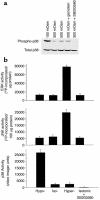
Osmolarity-sensitive changes in MAP kinase activity. HTC cells were exposed to isotonic (∼300 mOsm), hypotonic (∼100 mOsm), or hypertonic (∼500 mOsm) buffer solutions for 5 minutes, then immediately homogenized in lysis buffer. (a) Cell lysates were subjected to SDS-PAGE and Western blot analysis with either an antibody specific for the activated (phosphorylated at Thr-180 and Tyr-182) form of p38 (phospho-p38) or a control antibody that does not distinguish between phospho- and dephosphorylated p38 MAP kinase (total p38). Constitutive phospho-p38 activity was observed under isotonic conditions; values increased with hypotonic exposure. Activity was inhibited by the p38 inhibitor SB203580 (1 μM), but not by the tyrosine kinase inhibitor genistein (10 μM). None of the various exposures resulted in changes in total p38. (b) Average MAP kinase activity in response to osmotic changes performed as described in Methods. Under isotonic conditions, there was constitutive activity of p38, JNK, and ERK kinases. Hypertonic exposure stimulated a large increase in both ERK (fivefold) and JNK (fourfold) activity, but had little effect on p38 MAP kinase activity. In contrast, hypotonic exposure resulted in a large increase in p38 MAP kinase activity (tenfold), but had little effect on ERK or JNK activity compared with isotonic conditions. The putative p38 MAP kinase inhibitor SB203580 did not affect ERK or JNK activity, but completely inhibited constitutive p38 MAP kinase activity.
Figure 4
Figure 4
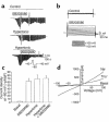
Inhibition of p38 MAP kinase increases membrane Na+ permeability. Whole-cell currents were measured under basal conditions and during exposure to the p38 inhibitor SB203580 (500 nM). (a) Representative whole-cell recordings. Currents at –80 mV (downward deflection of the current trace) correspond to INa+. Under basal conditions, currents were small (first tracing). Exposure to SB203580 stimulated an increase in currents (second tracing) with properties similar to those activated by hypertonic exposure (20 mM sucrose, third tracing). Exposure to SB203580 did not further increase current magnitude after cells were first exposed to hypertonicity (50 mM sucrose, ∼350 mOsm) to maximally activate currents (fourth tracing). (b) Voltage-step protocol (as described in Figure 2a) demonstrating currents under control conditions and following exposure to SB203580 (500 nM). (c) Cumulative data recorded as average current density at –80 mV. Both SB203580 (500 nM) and hypertonicity (50 mM sucrose) resulted in large increases in inward currents. The responses were not additive. (d) I-V relationship of whole-cell currents measured under basal conditions and during exposure to SB203580. With Na+ as the primary extracellular cation, currents were characterized by a nearly linear I-V relationship and reversal near 0 mV. Partial replacement of Na+ with Tris+ (final [Na+] = 20 mM) decreased inward currents and shifted the reversal potential to –40.5 ± 4.2 mV, as expected for a cation-selective conductance.
Figure 5
Figure 5
Comparison of hypertonic- and SB203580-induced currents. Average current density for currents activated by hypertonic exposure (20 mM sucrose) or SB203580 (500 nM) was measured at –80 mV. Both hypertonic- and SB203580-induced currents were inhibited by inclusion of EGTA (5 mM, no added Ca2+) in the patch pipette solution, and by amiloride (100 μM) in the extracellular solution (P < 0.01).
Figure 6
Figure 6
Inhibition of p38 increases cell volume. Cell volume was measured in the presence or absence of p38 inhibitor using a Coulter Multisizer. Compared with control, acute inhibition of p38 with SB203580 (500 nM) resulted in a rapid initial increase in cell size (5.8 ± 0.1%, n = 5, P < 0.01), reaching a maximum at 5 minutes. This was followed by a gradual return to basal values by 30 minutes.
Figure 7
Figure 7
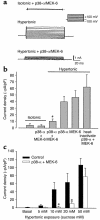
Intracellular dialysis with recombinant p38 MAP kinase protein inhibits volume-sensitive current activation. Under whole-cell patch-clamp conditions, the recombinant kinases p38α (5 μg/ml) and MEK-6 (5 μg/ml) were delivered, individually or together, to the cell interior by inclusion in the patch pipette, and cells were then exposed to hypertonic buffer (20 mM sucrose, ∼320 mOsm). (a) Whole-cell currents measured using the voltage-step protocol (as described in Figure 2a). Currents were small with intracellular dialysis of p38α and MEK-6 under isotonic conditions (top tracing). However, p38α and MEK-6 significantly inhibit the amplitude of hypertonic-induced currents (bottom tracing) as compared with control (middle tracing). (b) Cumulative data expressed as average current density at –80 mV. Control cells, dialyzed with heat-inactivated p38α and MEK-6, demonstrated characteristic current activation. Dialysis with either p38α or MEK-6 individually resulted in partial inhibition, which was not statistically significant compared with control. However, dialysis with p38α and MEK-6 together resulted in significant current inhibition. There was no effect of p38α and MEK-6 on basal (isotonic) currents. (c) Under control conditions, current density increased with increasing transmembrane osmolar gradients. In the presence of intracellular p38α + MEK-6, there was significant inhibition of current amplitude at lower transmembrane osmolar gradients (5–20 mM sucrose). However, this inhibitory effect was overcome at higher transmembrane osmolar gradients (50 mM sucrose). *P < 0.01.

Similar articles

Cited by

References

    1. Paul A, et al. Stress-activated protein kinases: activation, regulation and function. Cell Signal. 1997;9:403–410. - PubMed
    1. Bode JG, et al. The mitogen-activated protein (MAP) kinase p38 and its upstream activator MAP kinase kinase 6 are involved in the activation of signal transducer and activator of transcription by hyperosmolarity. J Biol Chem. 1999;274:30222–30227. - PubMed
    1. Han J, Richter B, Li Z, Kravchenko V, Ulevitch RJ. Molecular cloning of human p38 MAP kinase. . Biochim Biophys Acta. 1995; 1265:224–227. - PubMed
    1. Han J, Lee JD, Bibbs L, Ulevitch RJ. A MAP kinase targeted by endotoxin and hyperosmolarity in mammalian cells. Science. 1994;265:808–811. - PubMed
    1. Derijard B, et al. Independent human MAP-kinase signal transduction pathways defined by MEK and MKK isoforms [erratum 1995, 269(5220):17] Science. 1995;267:682–685. - PubMed

Publication types