. 2001 Nov 19;194(10):1407-19.
doi: 10.1084/jem.194.10.1407.

HIV-1 Vpr enhances viral burden by facilitating infection of tissue macrophages but not nondividing CD4+ T cells

Affiliations

HIV-1 Vpr enhances viral burden by facilitating infection of tissue macrophages but not nondividing CD4+ T cells

D A Eckstein et al. J Exp Med. .

Abstract

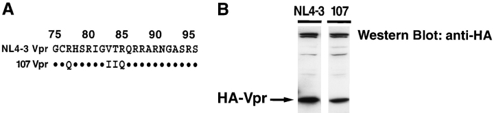
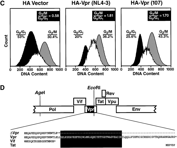
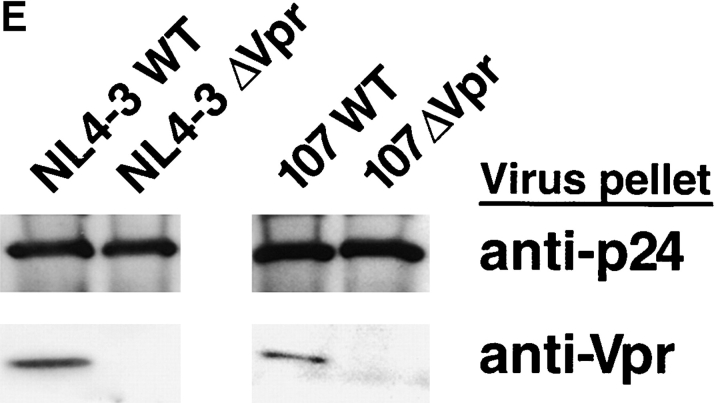
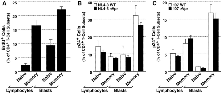
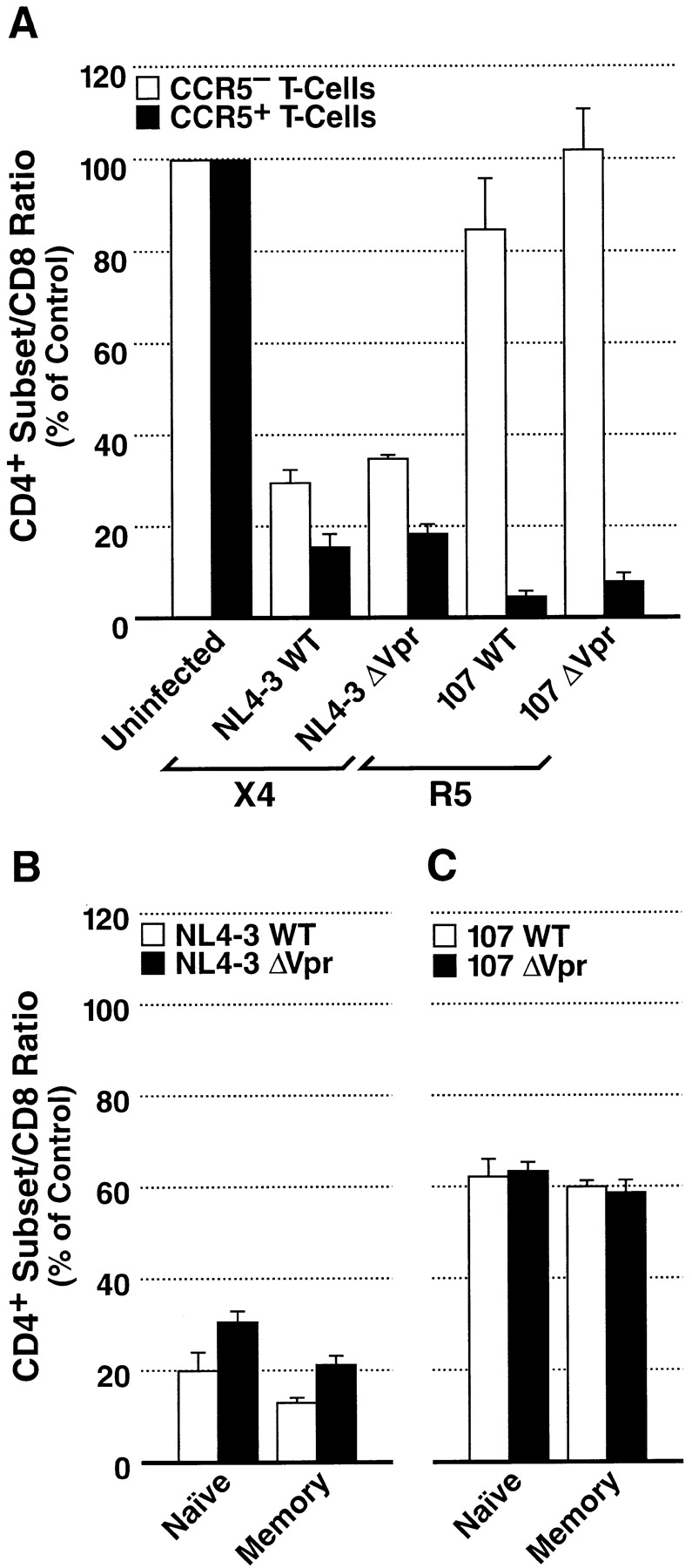
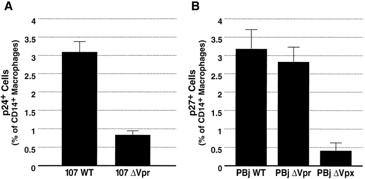
Prior experiments in explants of human lymphoid tissue have demonstrated that human immunodeficiency virus type 1 (HIV-1) productively infects diverse cellular targets including T cells and tissue macrophages. We sought to determine the specific contribution of macrophages and T cells to the overall viral burden within lymphoid tissue. To block infection of macrophages selectively while preserving infection of T cells, we used viruses deficient for viral protein R (Vpr) that exhibit profound replication defects in nondividing cells in vitro. We inoculated tonsil histocultures with matched pairs of congenic viruses that differed only by the presence of a wild-type or truncated vpr gene. Although these viruses exhibited no reduction in the infection or depletion of T cells, the ability of the Vpr-deficient R5 virus to infect tissue macrophages was severely impaired compared with matched wild-type R5 virus. Interestingly, the Vpr-deficient R5 virus also exhibited a 50% reduction in overall virus replication compared with its wild-type counterpart despite the fact that macrophages represent a small fraction of the potential targets of HIV-1 infection in these tissues. Collectively, these data highlight the importance of tissue macrophages in local viral burden and further implicate roles for CC chemokine receptor 5, macrophages, and Vpr in the life cycle and pathogenesis of HIV-1.


Figures

Figure 1.
Figure 1.
NL4–3- and 107-derived Vpr are functional and incorporated into mature virions. (A) HIV-1 Vpr from NL4–3 is identical to the BaL-derived, macrophage-tropic 107 variant except at residues 78 and 83–85. (B and C) DNA encoding HA-tagged NL4–3 Vpr or 107 Vpr and GFP was transfected into 293T cells. (B) Cells were lysed 36 h later, and expression of Vpr was determined by Western blot with anti-HA mAb. (C) Cells were harvested 36 h later, and labeled with propidium iodide. DNA content was measured in GFP+ cells by flow cytometry. (D) Cloning strategy for truncation of vpr within both NL4–3 and 107 proviral constructs. Note that the sequences of vif, pol, and tat are unaffected by these mutations within vpr. The shaded box identifies the deleted region within vpr. (E) DNA encoding each of the HIV-1 recombinants was transfected into 293T cells and supernatants were collected 48 h later. Virus production was normalized for p24 concentration, and equal amounts of virus were concentrated for each strain by ultra-centrifugation. The viral pellet was then subjected to Western blot analysis. Shown are representative experiments.
Figure 1.
Figure 1.
NL4–3- and 107-derived Vpr are functional and incorporated into mature virions. (A) HIV-1 Vpr from NL4–3 is identical to the BaL-derived, macrophage-tropic 107 variant except at residues 78 and 83–85. (B and C) DNA encoding HA-tagged NL4–3 Vpr or 107 Vpr and GFP was transfected into 293T cells. (B) Cells were lysed 36 h later, and expression of Vpr was determined by Western blot with anti-HA mAb. (C) Cells were harvested 36 h later, and labeled with propidium iodide. DNA content was measured in GFP+ cells by flow cytometry. (D) Cloning strategy for truncation of vpr within both NL4–3 and 107 proviral constructs. Note that the sequences of vif, pol, and tat are unaffected by these mutations within vpr. The shaded box identifies the deleted region within vpr. (E) DNA encoding each of the HIV-1 recombinants was transfected into 293T cells and supernatants were collected 48 h later. Virus production was normalized for p24 concentration, and equal amounts of virus were concentrated for each strain by ultra-centrifugation. The viral pellet was then subjected to Western blot analysis. Shown are representative experiments.
Figure 2.
Figure 2.
HIV-1 infection of CD4+ T lymphocytes and blasts. (A) Productively infected cell subsets in tonsil tissue following a 7-d infection with NL4–3WT (white bars) or NL4–3ΔVpr (black bars) as measured by intracellular p24 expression detected by FACS®. (B) Productively infected cell subsets in tonsil tissue after a 7-d infection with 107WT (white bars) or 107ΔVpr (black bars). Presented are mean values with SEM (n = 3) from a representative experiment.
Figure 3.
Figure 3.
BrdU incorporation and HIV-1 infection of naive and memory CD4+ T cell subsets. (A) Tissue samples were cultured continuously in medium containing BrdU for 7 d. Cells were isolated, immunostained for CD4, CD62L, CD45RA, and BrdU, and analyzed by FACS®. Shown are mean values with SEM (n = 3) from a representative experiment. (B and C) Tonsil histocultures were inoculated with equivalent titers of HIV-1, incubated for 7 d, and harvested. Cells were dispersed, immunostained for p24, CD62L, CD45RA, and CD4, and analyzed by FACS®. T cells were also stratified based on forward and side scatter properties defining lymphocyte and blast morphologies. (B) Productively infected subsets in tissue inoculated with NL4–3WT or NL4–3ΔVpr as measured by intracellular p24 expression. (C) Productively infected subsets in tissue inoculated with 107WT or 107ΔVpr. Shown are mean values with SEM (n = 3) from a representative experiment.
Figure 4.
Figure 4.
Cell-cycle analysis of p24+ or p24 CD4+ T cells. (A) Tonsil specimens were inoculated with equivalent titers of HIV-1 NL4–3WT (A and B) or NL4–3ΔVpr (C and D). After a 7-d incubation, samples were dispersed, stained for intracellular p24, CD4, and To-Pro-3 iodide, and analyzed by FACS®. DNA content is presented for cells stratified into p24+ (A and C) and p24 (B and D) fractions. Shown is a representative experiment.
Figure 5.
Figure 5.
Depletion of CD4+ T cells by HIV-1 in lymphoid histoculture. (A) After a 13-d infection, cells were isolated from histocultures and immunostained for surface expression of CD4, CD3, CD8, and CCR5. The mean CD4+ subset/CD8+ ratio relative to uninfected control tissue for CCR5 T cells (white bars) and CCR5+ T cells (black bars) was then calculated. Shown are mean values with SEM (n = 3) from a representative experiment. (B and C) Depletion of naive and memory CD4+ T cell subsets by HIV-1. After a 12-d infection with NL4–3WT and NL4–3ΔVpr (B) or 107WT and 107ΔVpr (C) cells were dispersed from histocultures and immunostained for CD4, CD62L, CD45RA, and CD8. The mean CD4+ subset/CD8+ ratio relative to uninfected control tissue for memory T cells (white bars) and naive T cells (black bars) was then calculated. Shown are mean values with SEM (n = 3) from a representative experiment.
Figure 6.
Figure 6.
Flow cytometric analysis of HIV-1 and SIV infection of macrophages in lymphoid tissue. After a 9-d incubation with 107WT or 107ΔVpr (A) or PBj6.6WT, PBj6.6ΔVpr or PBj6.6Δvpx (B), cells were dispersed and immunostained for surface expression of CD3 and CD14 as well as intracellular CD68 and p24 or p27. Shown are mean values with SEM (n = 3) from a representative experiment.
Figure 7.
Figure 7.
Kinetic analysis of HIV-1 replication in lymphoid histoculture. Tissue was inoculated with NL4–3WT or NL4–3ΔVpr (A) or 107WT or 107ΔVpr (B). Culture medium was sampled at the indicated time points and HIV-1 p24 concentration was measured by ELISA. Shown are mean values with SEM (n = 3) from a representative experiment.

References

    1. Bukrinsky, M.I., N. Sharova, M.P. Dempsey, T.L. Stanwick, A.G. Bukrinskaya, S. Haggerty, and M. Stevenson. 1992. Active nuclear import of human immunodeficiency virus type 1 preintegration complexes. Proc. Natl. Acad. Sci. USA. 89:6580–6584. - PMC - PubMed
    1. Balliet, J.W., D.L. Kolson, G. Eiger, F.M. Kim, K.A. McGann, A. Srinivasan, and R. Collman. 1994. Distinct effects in primary macrophages and lymphocytes of the human immunodeficiency virus type 1 accessory genes vpr, vpu, and nef: mutational analysis of a primary HIV-1 isolate. Virology. 200:623–631. - PubMed
    1. Heinzinger, N.K., M.I. Bukinsky, S.A. Haggerty, A.M. Ragland, V. Kewalramani, M.A. Lee, H.E. Gendelman, L. Ratner, M. Stevenson, and M. Emerman. 1994. The Vpr protein of human immunodeficiency virus type 1 influences nuclear localization of viral nucleic acids in nondividing host cells. Proc. Natl. Acad. Sci. USA. 91:7311–7315. - PMC - PubMed
    1. Connor, R.I., B.K. Chen, S. Choe, and N.R. Landau. 1995. Vpr is required for efficient replication of human immunodeficiency virus type-1 in mononuclear phagocytes. Virology. 206:935–944. - PubMed
    1. Popov, S., M. Rexach, G. Zybarth, N. Reiling, M.A. Lee, L. Ratner, C.M. Lane, M.S. Moore, G. Blobel, and M. Bukrinsky. 1998. Viral protein R regulates nuclear import of the HIV-1 pre-integration complex. EMBO J. 17:909–917. - PMC - PubMed

Publication types

Substances