. 2001 Dec;10(12):2419-25.
doi: 10.1110/ps.36701.

Crystal structure of calcium-free human sorcin: a member of the penta-EF-hand protein family

Affiliations

Crystal structure of calcium-free human sorcin: a member of the penta-EF-hand protein family

X Xie et al. Protein Sci. 2001 Dec.

Abstract

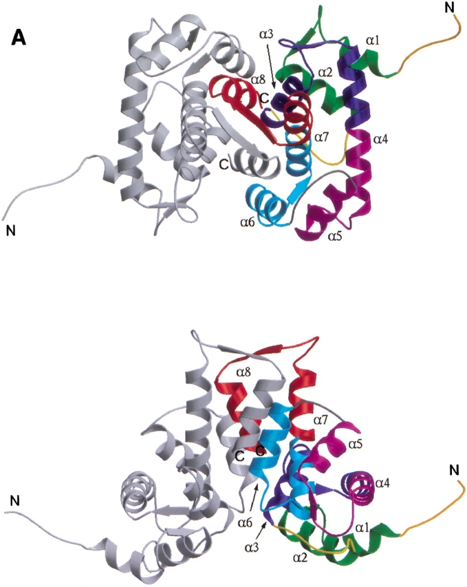
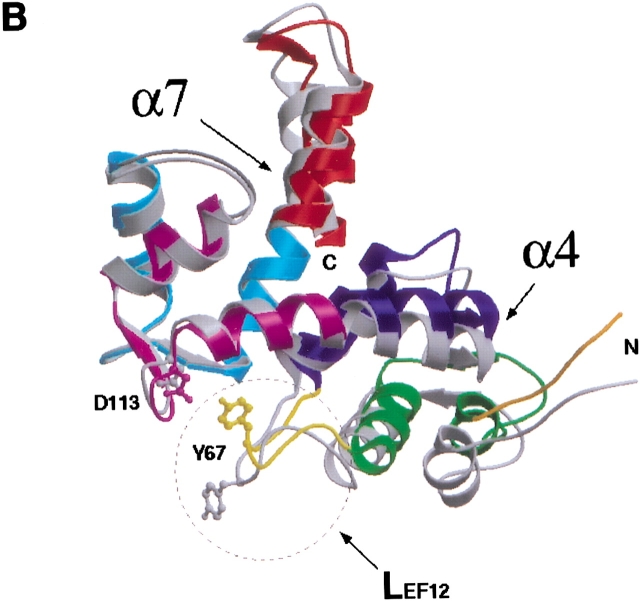
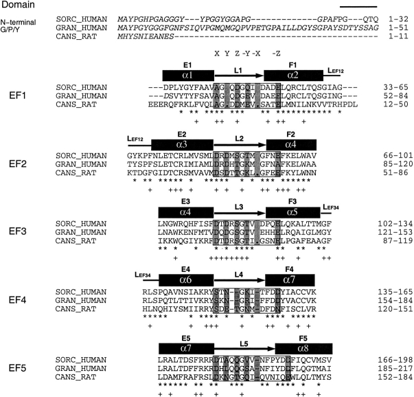
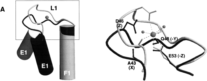
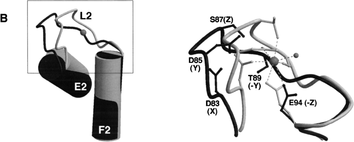
Sorcin is a 22 kD calcium-binding protein that is found in a wide variety of cell types, such as heart, muscle, brain and adrenal medulla. It belongs to the penta-EF-hand (PEF) protein family, which contains five EF-hand motifs that associate with membranes in a calcium-dependent manner. Prototypic members of this family are the calcium-binding domains of calpain, such as calpain dVI. Full-length human sorcin has been crystallized in the absence of calcium and the structure determined at 2.2 A resolution. Apart from an extended N-terminal portion, the sorcin molecule has a globular shape. The C-terminal domain is predominantly alpha-helical, containing eight alpha-helices and connecting loops incorporating five EF hands. Sorcin forms dimers through the association of the unpaired EF5, confirming this as the mode of association in the dimerization of PEF proteins. Comparison with calpain dVI reveals that the general folds of the individual EF-hand motifs are conserved, especially that of EF1, the novel EF-hand motif characteristic of the family. Detailed structural comparisons of sorcin with other members of PEF indicate that the EF-hand pair EF1-EF2 is likely to correspond to the two physiologically relevant calcium-binding sites and that the calcium-induced conformational change may be modest and localized within this pair of EF-hands. Overall, the results derived from the structural observations support the view that, in sorcin, calcium signaling takes place through the first pair of EF-hands.


Figures

Fig. 1.
Fig. 1.
(A) Ribbon representation of dimeric sorcin shown in two views. (Top) along the symmetry axis of the dimer with the EF5 near the viewer; (bottom) viewing perpendicular to the symmetry axis. Each monomer contains helices and loops that form five EF-hands. These EF-hands associate into pairs; EF1 pairs with EF2 whereas EF3 pairs with EF4, and EF5 pairs with its counterpart from the other monomer to form part of the dimer interface. One of the monomers is shown in monchrome (gray) whereas the other is color-coded to show the sub-domain structures: EF1 (green); EF2 (blue); EF3 (magenta); EF4 (cyan); EF5 (red); LEF12, the linker connecting the adjacent EF-hands within the first pair EF1–EF2 (yellow); LEF34, the linker connecting the adjacent EF-hands within the second pair EF3–EF4 (gray); and the short N-terminal fragment (dark orange). For each EF hand, the individual helices are denoted as E (N-terminal helix) and F (C-terminal helix) with an appropriate number indicating to which EF hand it belongs, whereas the loops flanked by the E and F helices are denoted as L with the same corresponding number. A sorcin monomer contains eight helices that are numbered from α1 to α8. Correspondence between this numbering scheme and the E/F notation: α1–E1, α2–F1, α3–E2, α4–fused F2 and E3, α5–F3, α6–E4, α7–fused F4 and E5, and α8–F5. (B) Superposition of the individual monomers of sorcin after the alignment of their EF3–EF4 sub-domains. The view is related by ∼90° rotation about the vertical axis to the bottom panel of Figure 1A ▶. The molecules are color-coded as in A. A circle is shown around LEF12 to highlight the structural difference between two individual monomers in this region. The side-chains of residues Tyr 67 and Asp 113 are shown in ball-and-stick representations. This figure was produced using the programs MOLSCRIPT (Kraulis 1991) and Raster3D (Merrit and Bacon 1997).
Fig. 1.
Fig. 1.
(A) Ribbon representation of dimeric sorcin shown in two views. (Top) along the symmetry axis of the dimer with the EF5 near the viewer; (bottom) viewing perpendicular to the symmetry axis. Each monomer contains helices and loops that form five EF-hands. These EF-hands associate into pairs; EF1 pairs with EF2 whereas EF3 pairs with EF4, and EF5 pairs with its counterpart from the other monomer to form part of the dimer interface. One of the monomers is shown in monchrome (gray) whereas the other is color-coded to show the sub-domain structures: EF1 (green); EF2 (blue); EF3 (magenta); EF4 (cyan); EF5 (red); LEF12, the linker connecting the adjacent EF-hands within the first pair EF1–EF2 (yellow); LEF34, the linker connecting the adjacent EF-hands within the second pair EF3–EF4 (gray); and the short N-terminal fragment (dark orange). For each EF hand, the individual helices are denoted as E (N-terminal helix) and F (C-terminal helix) with an appropriate number indicating to which EF hand it belongs, whereas the loops flanked by the E and F helices are denoted as L with the same corresponding number. A sorcin monomer contains eight helices that are numbered from α1 to α8. Correspondence between this numbering scheme and the E/F notation: α1–E1, α2–F1, α3–E2, α4–fused F2 and E3, α5–F3, α6–E4, α7–fused F4 and E5, and α8–F5. (B) Superposition of the individual monomers of sorcin after the alignment of their EF3–EF4 sub-domains. The view is related by ∼90° rotation about the vertical axis to the bottom panel of Figure 1A ▶. The molecules are color-coded as in A. A circle is shown around LEF12 to highlight the structural difference between two individual monomers in this region. The side-chains of residues Tyr 67 and Asp 113 are shown in ball-and-stick representations. This figure was produced using the programs MOLSCRIPT (Kraulis 1991) and Raster3D (Merrit and Bacon 1997).
Fig. 2.
Fig. 2.
Structure-based sequence alignment of human sorcin (SORC_HUMAN, SWISS-PROT accession code P30626; Wang et al. 1995), human grancalcin (HUMAN_GRAN, SWISS-PROT accession code P28676; Boyhan et al. 1992), and domain VI of rat calpain (CANS_RAT, SWISS-PROT accession code Q64537; Graham-Siegenthaler et al. 1994). Noted are the sorcin residues that are identical to grancalcin (asterisk), those identical to calpain dVI (plus), and those omitted from the final model in the crystal structures (italics). The alignment was obtained initially using ClustalW (Thompson et al. 1994) and was revised based on the superposition of the crystal structures of sorcin and calpain dVI; the gaps are noted (dash). Helices (dark rectangles) are labeled α1–α8; loops are also represented (dark arrows). The EF-hand motifs are supersecondary helix–loop–helix structural elements characterized by a canonical 12-residue loop that exhibits pentagonal bipyramidal coordination of the calcium ion (Strynadka and James 1989). The five equatorial calcium ion ligands are contributed by the side chains of two acidic residues (positions Y and −Z, the latter providing two ligands), one polar group (Ser or Thr, position Z), and the main chain carbonyl oxygen (position −Y). The two apical ligands are contributed by the side-chain of an acidic group (position X) and a water molecule (position −X). In many instances the EF-hands differ from the canonical sequence resulting in variants of calcium coordination or loss of calcium binding capability. In the novel EF-hand motif discovered in calpain dVI, EF1, the loop contains only 11 amino acids and lacks the canonical carboxylate side chain at positions X and Y. The ligand in position X is a carbonyl oxygen from Ala 25, whereas a water molecule occupies position Y. Sequences corresponding to the 12-residue consensus sequence of the canonical EF-hand are boxed. The residues that form the calcium coordination sphere are labeled as X, Y, Z, −Y, −X, and −Z based on the structure of calcium-bound calpain dVI and comparison with the canonical EF-hand motif is shown (gray shading). The solvent molecules that coordinate the calcium ion are shown (dots). All three proteins contain G/P/Y-rich N-terminal extensions of various lengths shown in the first line of the alignment. The C-terminal calcium-binding domains are grouped by their EF-hand motifs, in which the nomenclatures for all secondary structure elements are the same as indicated in Figure 1A ▶.
Fig. 3.
Fig. 3.
Schematic representation of EF-hands of sorcin compared with that of calpain dVI. The F-helix was used for alignment. Shown are sorcin (dark gray) and calpain dVI (light gray; RCSB Protein Data Bank entry code 1DVI; Blanchard et al. 1997). (Left) Helices E and F (cylinders), loop L (coil), and the calcium ion (gray sphere). (Right) The loop region (boxed in the left panel) is enlarged to show the detailed conformation and some of the residues (backbone carbonyl or side-chain) that are responsible for calcium coordination, in which the calcium ion (gray sphere) is shown connected (dashed lines) to its ligands and the water (small gray sphere). This figure was produced using the programs MOLSCRIPT (Kraulis 1991) and Raster3D (Merrit and Bacon 1997). (A) Superposition of EF1 of sorcin and calcium-bound EF1 of calpain dVI. The helices E1 and F1 in calpain are nearly perpendicular to each other, with F1 coplanar with the page and E1 pointing toward the viewer (left). In contrast, helices E1 and F1 in sorcin are closer to an anti-parallel orientation with respect to each other. (B) Superposition of EF2 of sorcin and calcium-bound EF2 of calpain dVI. The helices E2 and F2 in both calpain and sorcin are nearly perpendicular to each other, with F2 coplanar with the page and E2 pointing toward the viewer (left).
Fig. 3.
Fig. 3.
Schematic representation of EF-hands of sorcin compared with that of calpain dVI. The F-helix was used for alignment. Shown are sorcin (dark gray) and calpain dVI (light gray; RCSB Protein Data Bank entry code 1DVI; Blanchard et al. 1997). (Left) Helices E and F (cylinders), loop L (coil), and the calcium ion (gray sphere). (Right) The loop region (boxed in the left panel) is enlarged to show the detailed conformation and some of the residues (backbone carbonyl or side-chain) that are responsible for calcium coordination, in which the calcium ion (gray sphere) is shown connected (dashed lines) to its ligands and the water (small gray sphere). This figure was produced using the programs MOLSCRIPT (Kraulis 1991) and Raster3D (Merrit and Bacon 1997). (A) Superposition of EF1 of sorcin and calcium-bound EF1 of calpain dVI. The helices E1 and F1 in calpain are nearly perpendicular to each other, with F1 coplanar with the page and E1 pointing toward the viewer (left). In contrast, helices E1 and F1 in sorcin are closer to an anti-parallel orientation with respect to each other. (B) Superposition of EF2 of sorcin and calcium-bound EF2 of calpain dVI. The helices E2 and F2 in both calpain and sorcin are nearly perpendicular to each other, with F2 coplanar with the page and E2 pointing toward the viewer (left).

Similar articles

Cited by

References

    1. Blanchard, H., P. Grochulski, Y. Li, J.S. Arthur, P.L. Davies, J.S. Elce, and M. Cygler. 1997. Structure of a calpain Ca(2+)-binding domain reveals a novel EF-hand and Ca(2+)-induced conformational changes. Nat. Struct. Biol. 4 532–538. - PubMed
    1. Boyhan, A., C.M. Casimir, J.K. French, C.G. Teahan, and A.W. Segal. 1992. Molecular cloning and characterization of grancalcin, a novel EF-hand calcium-binding protein abundant in neutrophils and monocytes. J. Biol. Chem.. 267 2928–2933. - PubMed
    1. Brownawell, A.M., and C.E. Creutz. 1997. Calcium-dependent binding of sorcin to the N-terminal domain of synexin (annexin VII). J. Biol. Chem.. 272 22182–22190. - PubMed
    1. Brunger, A.T., P.D. Adams, G.M. Clore, W.L. DeLano, P. Gros, R.W. Grosse-Kunstleve, J.S. Jiang, J. Kuszewski, M. Nilges, N.S. Pannu, R.J. Read, L.M. Rice, T. Simonson, and G.L. Warren. 1998. Crystallography & NMR system: A new software suite for macromolecular structure determination. Acta Crystallogr D Biol Crystallogr. 54 905–921. - PubMed
    1. Graham-Siegenthaler, K., S. Gauthier, P.L. Davies, and J.S. Elce. 1994. Active recombinant rat calpain II. Bacterially produced large and small subunits associate both in vivo and in vitro. J Biol Chem. 269 30457–30460. - PubMed