Characterization of the structure and dynamics of amyloidogenic variants of human lysozyme by NMR spectroscopy
- PMID: 11714920
- PMCID: PMC2374041
- DOI: 10.1110/ps.28101
Characterization of the structure and dynamics of amyloidogenic variants of human lysozyme by NMR spectroscopy
Abstract
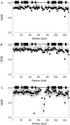
The structures and dynamics of the native states of two mutational variants of human lysozyme, I56T and D67H, both associated with non-neuropathic systemic amyloidosis, have been investigated by NMR spectroscopy. The (1)H and (15)N main-chain amide chemical shifts of the I56T variant are very similar to those of the wild-type protein, but those of the D67H variant are greatly altered for 28 residues in the beta-domain. This finding is consistent with the X-ray crystallographic analysis, which shows that the structure of this variant is significantly altered from that of the wild-type protein in this region. The (1)H-(15)N heteronuclear NOE values show that, with the exception of V121, every residue in the wild-type and I56T proteins is located in tightly packed structures characteristic of the native states of most proteins. In contrast, D67H has a region of substantially increased mobility as shown by a dramatic decrease in heteronuclear NOE values of residues near the site of mutation. Despite this unusual flexibility, the D67H variant has no greater propensity to form amyloid fibrils in vivo or in vitro than has I56T. This finding indicates that it is the increased ability of the variants to access partially folded conformations, rather than intrinsic changes in their native state properties, that is the origin of their amyloidogenicity.
Figures


Similar articles
-
Rationalising lysozyme amyloidosis: insights from the structure and solution dynamics of T70N lysozyme.J Mol Biol. 2005 Sep 30;352(4):823-36. doi: 10.1016/j.jmb.2005.07.040. J Mol Biol. 2005. PMID: 16126226
-
Characterisation of the structural, dynamic and aggregation properties of the W64R amyloidogenic variant of human lysozyme.Biophys Chem. 2021 Apr;271:106563. doi: 10.1016/j.bpc.2021.106563. Epub 2021 Feb 13. Biophys Chem. 2021. PMID: 33640796
-
In silico prediction of novel residues involved in amyloid primary nucleation of human I56T and D67H lysozyme.BMC Struct Biol. 2018 Jul 20;18(1):9. doi: 10.1186/s12900-018-0088-1. BMC Struct Biol. 2018. PMID: 30029603 Free PMC article.
-
Lysozyme: a paradigmatic molecule for the investigation of protein structure, function and misfolding.Clin Chim Acta. 2005 Jul 24;357(2):168-72. doi: 10.1016/j.cccn.2005.03.022. Clin Chim Acta. 2005. PMID: 15913589 Review.
-
The alternative conformations of amyloidogenic proteins and their multi-step assembly pathways.Curr Opin Struct Biol. 1998 Feb;8(1):101-6. doi: 10.1016/s0959-440x(98)80016-x. Curr Opin Struct Biol. 1998. PMID: 9519302 Review.
Cited by
-
Proteins with H-bond packing defects are highly interactive with lipid bilayers: Implications for amyloidogenesis.Proc Natl Acad Sci U S A. 2003 Mar 4;100(5):2391-6. doi: 10.1073/pnas.0335642100. Epub 2003 Feb 18. Proc Natl Acad Sci U S A. 2003. PMID: 12591960 Free PMC article.
-
Dynamics of apomyoglobin in the alpha-to-beta transition and of partially unfolded aggregated protein.Eur Biophys J. 2009 Feb;38(2):237-44. doi: 10.1007/s00249-008-0375-z. Epub 2008 Oct 14. Eur Biophys J. 2009. PMID: 18853152
-
Stable, metastable, and kinetically trapped amyloid aggregate phases.Biomacromolecules. 2015 Jan 12;16(1):326-35. doi: 10.1021/bm501521r. Epub 2014 Dec 18. Biomacromolecules. 2015. PMID: 25469942 Free PMC article.
-
The first step of hen egg white lysozyme fibrillation, irreversible partial unfolding, is a two-state transition.Protein Sci. 2007 May;16(5):815-32. doi: 10.1110/ps.062639307. Epub 2007 Mar 30. Protein Sci. 2007. PMID: 17400924 Free PMC article.
-
Changes in Lysozyme Flexibility upon Mutation Are Frequent, Large and Long-Ranged.PLoS Comput Biol. 2012;8(3):e1002409. doi: 10.1371/journal.pcbi.1002409. Epub 2012 Mar 1. PLoS Comput Biol. 2012. PMID: 22396637 Free PMC article.
References
-
- Booth, D.R., Sunde, M., Bellotti, V., Robinson, C.V., Hutchinson, W.L., Fraser, P.E., Hawkins, P.N., Dobson, C.M., Radford, S.E., Blake, C.C.F., and Pepys, M.B. 1997. Instability, unfolding and aggregation of human lysozyme variants underlying amyloid fibrillogenesis. Nature 385 787–793. - PubMed
-
- Boyd, J., Dobson, C.M., and Redfield, C. 1985. Assignment of resonances in the 1H NMR spectrum of human lysozyme. Eur. J. Biochem. 153 383–396. - PubMed
-
- Buck, M., Boyd, J., Redfield, C., MacKenzie, D.A., Jeenes, D.J., Archer, D.B., and Dobson, C.M. 1995. Structural determinants of protein dynamics: Analysis of 15N NMR relaxation measurements for main-chain and side-chain nuclei of hen egg white lysozyme. Biochemistry 34 4041–4055. - PubMed
-
- Canet, D., Sunde, M., Last, A.M., Miranker, A., Spencer, A., Robinson, C.V., and Dobson, C.M. 1999. Mechanistic studies of the folding of human lysozyme and the origin of amyloidogenic behavior in its disease-related variants. Biochemistry 38 6419–6427. - PubMed
Publication types
MeSH terms
Substances
Grants and funding
LinkOut - more resources
Full Text Sources

