

Cerebral cryptococcal granuloma in a cat
- PMID: 11716619
- PMCID: PMC10829131
- DOI: 10.1053/jfms.2000.0096
Cerebral cryptococcal granuloma in a cat
Corrected and republished in
-
Cerebral cryptococcal granuloma in a cat.J Feline Med Surg. 2001 Mar;3(1):39-44. doi: 10.1053/jfms.2000.0112. J Feline Med Surg. 2001. PMID: 11724013 Free PMC article.
Abstract
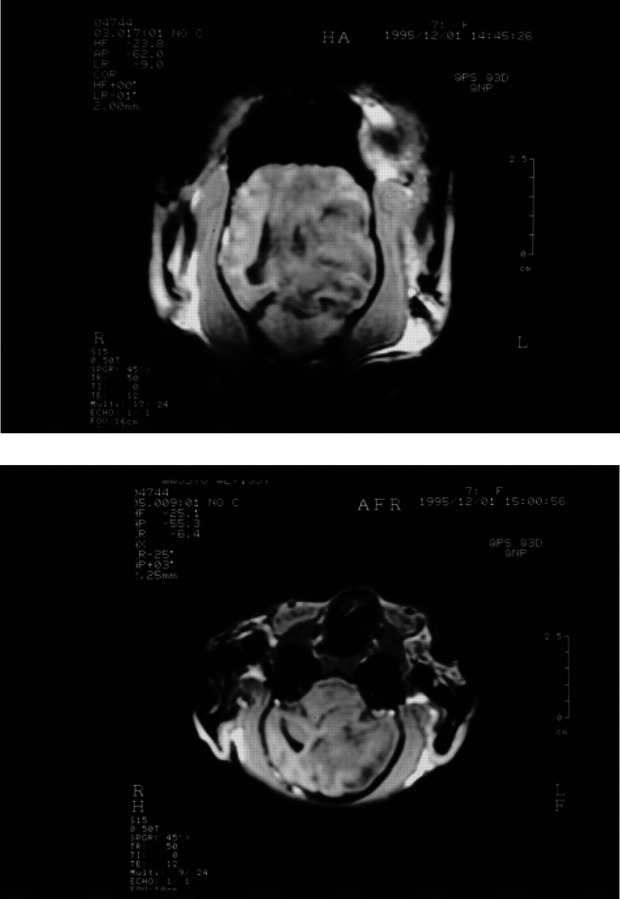
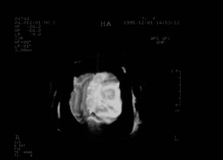
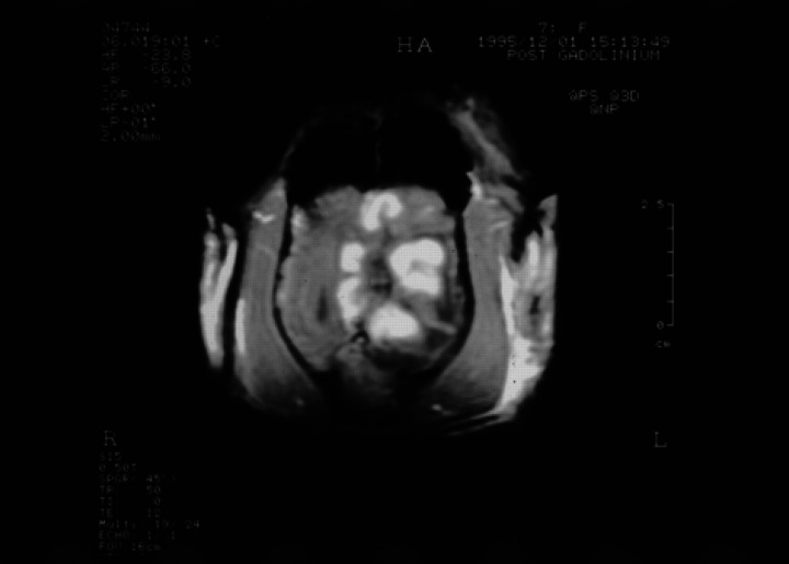
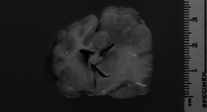
A 7-year-old cat was presented for seizures. Cerebrospinal fluid cytology and serology were consistent with a diagnosis of toxoplasmosis. The cat was treated with clindamycin but seizures continued and additional neurological signs developed over 6 months. A mass lesion was identified in the left cerebral hemisphere using magnetic resonance imaging (MRI). The lesion enhanced after gadolidium and a tumour was considered likely. Histologically, the lesion proved to be a cryptococcal granuloma and retrospective serology confirmed that the cat had cryptococcosis at its initial presentation. This report provides the first description in the veterinary literature of the MRI appearance of a cerebral cryptococcoma and emphasises the importance of performing cryptococcal antigen determination in cats with signs of intracranial disease.
Copyright 2000 European Society of Feline Medicine.
Figures
References
-
- Andreula CF, Burdi N, Carella A. (1993) CNS cryptococcosis in AIDS: Spectrum of MR findings. Journal of Computer Assisted Tomography 17, 438–441. - PubMed
-
- Dimech WJ. (1991) Diagnosis, identification and epidemiology of Cryptococcus neoformans infection. Australian Journal of Medical Laboratory Science 12, 13–21.
-
- Dubey JP. (1994) Toxoplasmosis and other coccidial infections. In: The Cat: Diseases and Clinical Management, (2nd edn). Sherding R. (ed). New York: Churchill Livingstone, pp 565–584.
-
- Dzendrowski TE, Himmelreich U, Dowd S, Allen C, Mountford C, Sorrell TC. (1999) An animal model for ex vivo discrimination of cerebral cryptococcoma from glioma. In: Programme and Abstracts of the 4th International Conference on Cryptococcus and Cryptococcosis, London, UK. London: The Royal Society, pp 185.
-
- Glass E, deLahunta A, Kent M, Kapatkin A, Joseph R. (1996) A cryptococcal granuloma in the brain of a cat causing focal signs. Progress in Veterinary Neurology 7, 141–144.
Publication types
MeSH terms
LinkOut - more resources
Full Text Sources
Miscellaneous
