. 2001 Dec;183(24):6991-8.
doi: 10.1128/JB.183.24.6991-6998.2001.

MxiM and MxiJ, base elements of the Mxi-Spa type III secretion system of Shigella, interact with and stabilize the MxiD secretin in the cell envelope

Affiliations

MxiM and MxiJ, base elements of the Mxi-Spa type III secretion system of Shigella, interact with and stabilize the MxiD secretin in the cell envelope

R Schuch et al. J Bacteriol. 2001 Dec.

Abstract

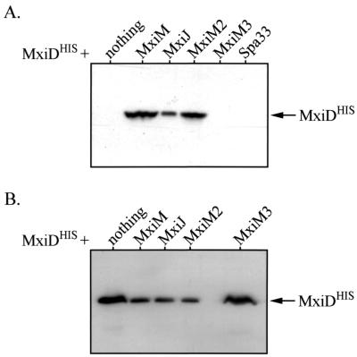
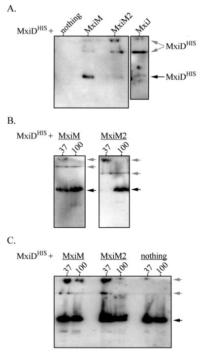
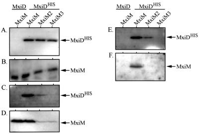
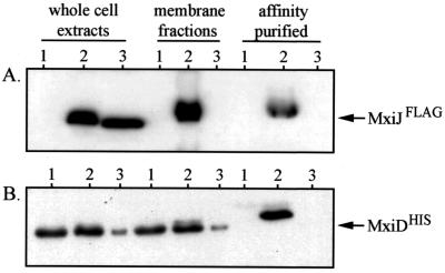
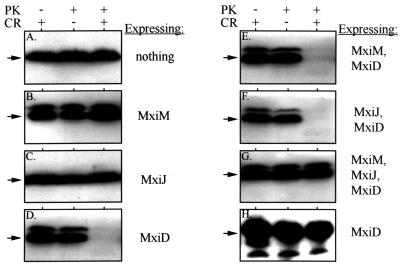
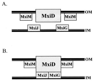
The type III secretion pathway is broadly distributed across many parasitic bacterial genera and serves as a mechanism for delivering effector proteins to eukaryotic cell surface and cytosolic targets. While the effectors, as well as the host responses elicited, differ among type III systems, they all utilize a conserved set of 9 to 11 proteins that together form a bacterial envelope-associated secretory organelle or needle complex. The general structure of the needle complex consists of a transenvelope base containing at least three ring-forming proteins (MxiD, MxiJ, and MxiG in Shigella) that is connected to a hollow needle-like extension that projects away from the cell surface. Several studies have shown that the initial steps in needle complex assembly require interactions among the base proteins, although specific details of this process remain unknown. Here we identify a role for another base element in Shigella, MxiM, in interactions with the major outer-membrane-associated ring-forming protein, MxiD. MxiM affects several features of MxiD, including its stability, envelope association, and assembly into homomultimeric structures. Interestingly, many of the effects were also elicited by the inner-membrane-associated base element, MxiJ. We confirmed that MxiM-MxiD and MxiJ-MxiD interactions occur in vivo in the cell envelope, and we present evidence that together these base elements can form a transmembrane structure which is likely an important intermediary in the process of needle complex assembly.


Figures

FIG. 1
FIG. 1
Analysis of MxiDHIS stability in the presence or absence of other Mxi-Spa proteins. Whole-cell protein extracts of various BS103 derivatives were separated by SDS-PAGE and analyzed by immunoblotting with anti-His antibodies. The position of MxiDHIS (∼62 kDa) is indicated by the arrows. (A) Induction of MxiDHIS (from low copy-number-vector pBAD33) in either the absence or presence of different Mxi-Spa proteins (expressed from pBluescript SK+). Protein from 1 × 10⁸ bacteria was examined in each case. (B) Induction of MxiDHIS (from high-copy-number vector pBAD18) in the absence or presence of different Mxi-Spa proteins (expressed from pBAD33). For lanes in which MxiDHIS was expressed with nothing or MxiM3, whole-cell extracts from 1 × 10⁶ bacteria were used. For the remaining lanes, protein from 1 × 10⁷ bacteria was used.
FIG. 2
FIG. 2
Formation of MxiDHIS homomultimers in the presence or absence of other Mxi-Spa proteins. Whole-cell protein extracts of various BS103 derivatives were separated by SDS-PAGE. The stacking and separating gels were transferred to polyvinylidene difluoride membranes and analyzed by immunoblotting with anti-His antibodies. The positions of MxiDHIS monomers (∼62 kDa) are indicated by black arrows, while the positions of the two MxiDHIS multimers (>200 kDa) are indicated by gray arrows. The lower multimer is at the interface between the stacking and separating gels. (A) Induction of MxiDHIS (from low-copy-number vector pBAD33) in either the presence or the absence of mxiM, mxiM2, or mxiJ (expressed from pBluescript SK+). In each case, protein from 2.5 × 10⁸ bacteria was boiled for 3 min and examined. (B) Resistance of MxiDHIS multimers to heat. MxiDHIS was induced (from pBAD33) in the presence of either MxiM or MxiM2 (expressed from pBluescript SK+), and samples were incubated at either 37°C (lanes 37) or 100°C (lanes 100). Protein from 3 × 10⁸ bacteria was examined in each case. (C) Induction of MxiDHIS (from high-copy-number vector pBAD18) in either the presence or the absence of MxiM or MxiM2 (expressed from pBAD33). Samples were incubated at either 37°C (lanes 37) or 100°C (lanes 100). Protein from either 3 × 10⁷ bacteria (for MxiM- and MxiM2-expressing strains) or 1 × 10⁶ bacteria (for the strain expressing only MxiDHIS) was used.
FIG. 3
FIG. 3
In vivo interaction between MxiM and MxiDHIS. BS103 derivatives expressing either MxiD or MxiDHIS (from pBAD18) in the presence of MxiM, MxiM2, or MxiM3 (expressed from pBAD33) were examined in coprecipitation analyses. The presence of MxiDHIS and MxiM (and its derivatives) was monitored in cultures prior to cross-linking with DSP (panels A and B, respectively), in membrane fractions from cross-linked cells (panels C and D, respectively), and after purification from membrane fractions using Ni-NTA beads (panels E and F, respectively). Cross-linked proteins were released prior to analysis by reducing DSP with 5% β-mercaptoethanol. The samples were then resolved on SDS–10% polyacrylamide gels and immunodetected with either anti-His or anti-MxiM antibodies. The arrows indicate the positions of MxiM (∼13 kDa) and MxiDHIS (∼62 kDa).
FIG. 4
FIG. 4
In vivo interaction between MxiJFLAG and MxiDHIS. BS103 derivatives expressing either MxiJ (lanes 1), MxiJFLAG (lanes 2), or MxiJ2FLAG (lanes 3) from pBAD24 in the presence of MxiDHIS (expressed from low-copy-number vector pBAD33) were examined in coprecipitation analyses. The presence of MxiJFLAG and MxiDHIS was monitored in whole-cell protein extracts of each strain, in the resulting membrane fractions of lysed cells, and in proteins affinity purified from the membranes by using an anti-FLAG affinity gel. No cross-linker was used in this experiment. The samples were resolved on SDS–10% polyacrylamide gels and immunodetected with either anti-FLAG or anti-MxiM antibodies. The arrows indicate the positions of MxiJFLAG (27 kDa) and MxiDHIS (∼62 kDa).
FIG. 5
FIG. 5
Susceptibility of periplasmic BlaM and cytoplasmic H-NS to extracellular protease. BS103 derivatives expressing either MxiM, MxiJ, or MxiD or various combinations of these proteins (indicated to the right of each panel) were incubated with proteinase K (PK) and/or Congo red (CR) for 20 min. Whole-cell protein extracts of each strain were resolved on SDS–10% polyacrylamide gels and examined by immunoblotting with either anti-BlaM (A to G) or anti-H-NS (H) antibodies. The arrow to the left of each panel indicates the position of BlaM (∼29 kDa) or H-NS (∼15 kDa). In these backgrounds, MxiD was expressed from the high-copy-number vector pBAD18, while MxiJ was expressed from pBAD33. MxiM was expressed from either pBAD33 (when it was used alone or with MxiD) or from pWSK129. Protein from 5 × 10⁸ bacteria was used in each lane of panels A to G; protein from 5 × 10⁷ bacteria was used in each lane of panel H. The minor higher-Mr band present in some panels probably corresponds to unprocessesed β-lactamase.
FIG. 6
FIG. 6
Interactions and localizations of Mxi-Spa base elements in the S. flexneri envelope. (A) Positions of MxiM, MxiD, and MxiJ. MxiM is anchored via a lipid moiety to the inner face of the OM (25), while the MxiD secretin is an integral OM protein (3). MxiJ is predicted to be anchored to the outer face of the IM (2, 14). (B) Interactions among MxiM, MxiD, and MxiJ predicted on the basis of our study. Interactions are indicated by rectangles that touch each other. MxiM and MxiD probably interact in the OM, while the MxiD-MxiJ interaction spans the periplasmic space.

References

    1. Allaoui A, Sansonetti P J, Ménard R, Barzu S, Mounier J, Phalipon A, Parsot C. MxiG, a membrane protein required for secretion of Shigella spp. Ipa invasins: involvement in entry into epithelial cells and in intercellular dissemination. Mol Microbiol. 1995;17:461–470. - PubMed
    1. Allaoui A, Sansonetti P J, Parsot C. MxiJ, a lipoprotein involved in secretion of Shigella Ipa invasins, is homologous to YscJ, a secretion factor of the Yersinia Yop proteins. J Bacteriol. 1992;174:7661–7669. - PMC - PubMed
    1. Allaoui A, Sansonetti P J, Parsot C. MxiD: an outer membrane protein necessary for the secretion of the Shigella flexneri Ipa invasins. Mol Microbiol. 1993;7:59–68. - PubMed
    1. Bahrani F K, Sansonetti P J, Parsot C. Secretion of Ipa proteins by Shigella flexneri: inducer molecules and kinetics of activation. Infect Immun. 1997;65:4005–4010. - PMC - PubMed
    1. Blocker A, Jouihri N, Larquet E, Gounon P, Ebel F, Parsot C, Sansonetti P, Allaoui A. Structure and composition of the Shigella flexneri ‘needle complex,’ a part of its type III secreton. Mol Microbiol. 2001;39:652–663. - PubMed

Publication types

MeSH terms

LinkOut - more resources