. 2001 Dec 1;21(23):9151-9.
doi: 10.1523/JNEUROSCI.21-23-09151.2001.

Distance-dependent increase in AMPA receptor number in the dendrites of adult hippocampal CA1 pyramidal neurons

Affiliations

Distance-dependent increase in AMPA receptor number in the dendrites of adult hippocampal CA1 pyramidal neurons

B K Andrasfalvy et al. J Neurosci. .

Abstract

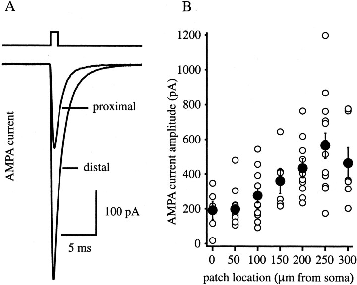
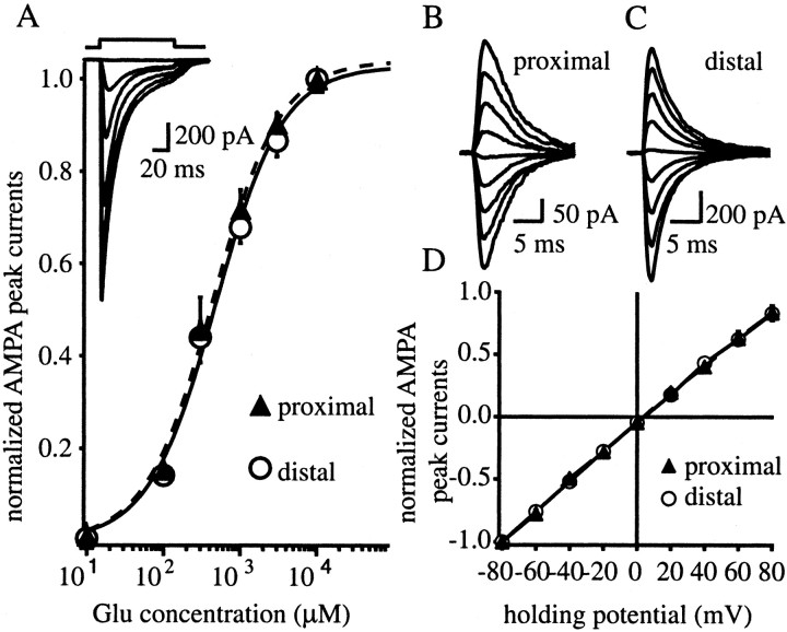
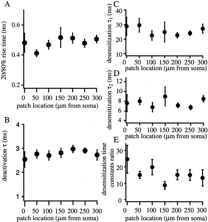
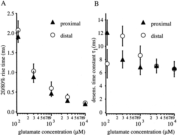
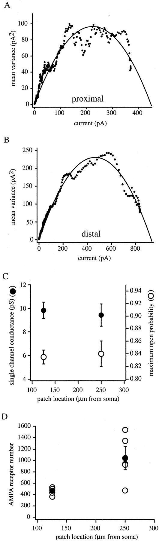
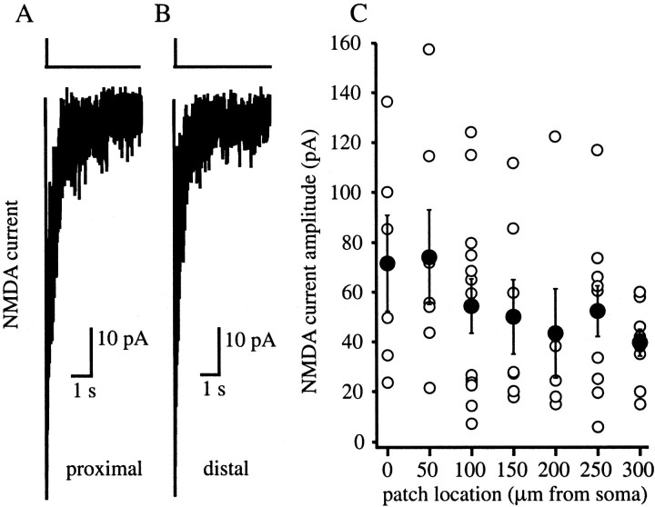
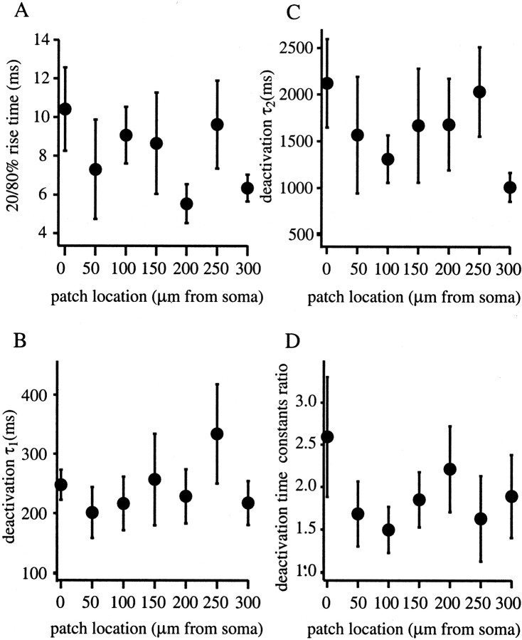
The Schaffer collateral pathway provides hippocampal CA1 pyramidal cells with a fairly homogeneous excitatory synaptic input that is spread out across several hundred micrometers of their apical dendritic arborizations. A progressive increase in synaptic conductance, with distance from the soma, has been reported to reduce the location dependence that should result from this arrangement. The excitatory synaptic contacts within this pathway primarily use AMPA- and NMDA-type glutamate receptors. To investigate the underlying mechanism of the increased distal excitatory postsynaptic conductance, we used outside-out patches and a fast application system to characterize the properties and distribution of synaptic glutamate receptors across the range of apical dendrites receiving Schaffer collateral input. We observed an approximately twofold increase in AMPA-mediated current amplitude (0.3-0.6 nA) in the range of CA1 apical dendrites that receive a uniform density of Schaffer collateral input (approximately 100-250 micrometer from soma). NMDA-mediated current amplitude, however, remained unchanged. We analyzed the current kinetics, agonist affinity, single-channel conductance, maximum open probability, and reversal potential of AMPA receptors and did not find any differences. Instead, the number of AMPA receptors present in our patches increased approximately twofold. These data suggest that an increase in the number of AMPA receptors present at distal synapses may play an important role in the distance-dependent scaling of Schaffer collateral synapses.


Figures

Fig. 1.
Fig. 1.
AMPA receptor-mediated glutamate currents in excised patches from different dendritic locations. A, Currents activated by 1 msec application of 1 mm glutamate to outside-out patches excised from either a proximal (∼50 μm from soma) or distal (∼250 μm from soma) dendritic location. Currents were recorded in the presence of 1 mmMg2+ and in the absence of glycine. Eachtrace is an average of five sweeps (Vh was −80 mV). B, AMPA receptor-mediated mean current amplitudes are plotted against the location of the excised patches from the soma (n = 69). Current amplitude increases approximately threefold across the range. Open circles are the peaks of single patches, andfilled circles are the means of these patches with SE bars.
Fig. 2.
Fig. 2.
Agonist affinity and voltage dependence of AMPA receptor-mediated glutamate current. A, Mean amplitude of AMPAR currents evoked by fast application of 10–104 μm glutamate with 100 msec pulses to proximal patches (∼50 μm from soma; n= 6; dashed line with filled triangles) and distal patches (∼250 μm from soma; n = 7;solid line with open circles). Currents were normalized to the response at 10 mm glutamate.B, C, AMPA currents evoked by the application of 1 mm glutamate for 1 msec (Vh was −80 to +80 mV, 20 mV steps) to proximal (B) and distal (C) excised patches. Each trace is a single sweep.D, Mean amplitude of AMPA currents for different holding potentials (Vh was −80 to +80 mV, 20 mV steps) during fast application of 1 mm glutamate for 1 msec to proximal (∼50 μm from soma; n = 5;dashed line with filled triangles) and distal (∼250 μm from soma; n = 6; solid line with open circles) patches. Currents were normalized to the response at −80 mV. In most cases, the size of thesymbols are bigger than the SE bars. In all experiments, 0.1–1 mm Mg2+ was added, with no additional glycine.
Fig. 3.
Fig. 3.
Kinetic properties of AMPAR-mediated glutamate currents versus location. A, Mean of 20–80% rise time of AMPA current evoked by 1 msec application of 1 mmglutamate on outside-out membrane patches excised from various locations of apical dendrites. B, Mean of deactivation time constants (τ) of the same AMPA currents evoked as inA. C, Mean of fast desensitization time constants (τ1) of AMPA currents evoked by 100 msec application of 1 mm glutamate to outside-out patches taken from various locations of apical dendrites. D, Mean of slow desensitization time constants (τ2) of the same currents as in C. E, Mean of the desensitization time constant ratio of AMPA currents shown inB and C. In all experiments, 0.1–1 mm Mg2+ was added with no additional glycine. In each figure, error bars represent the SEM (n = 69).
Fig. 4.
Fig. 4.
Concentration dependence of AMPA current rise and desensitization. A, Concentration dependence of AMPA current 20–80% rise time for a 100 msec application of various concentrations of glutamate (100–104μm). Data from proximal (∼50 μm from soma;n = 6; dashed line withfilled triangles) and distal (∼250 μm from soma;n = 6; solid line with open circles) patches are shown. Glutamate concentration is shown in log scale. B, Concentration dependence of the fast desensitization time constant (τ1) with 100 msec application of glutamate for proximal (∼50 μm from soma;n = 6; dashed line withfilled triangles) and distal (∼250 μm from soma;n = 6; solid line with open circles) patches. Glutamate concentration is shown in log scale.
Fig. 5.
Fig. 5.
Nonstationary fluctuation analysis of AMPA receptor-mediated glutamate current. Mean variance plotted as a function of the mean current for a proximal (A) and distal (B) patch. Solid lineis fit of the data by a parabolic equation (see Materials and Methods) that was used to determine single-channel conductance (γ), channel number (N), and maximum open probability (Po,max). Proximal patch from ∼125 μm from soma and distal patch ∼250 μm from soma.C, Single-channel conductance (left axis,filled circles) and maximum open probability (right axis, open circles) versus location of the patches. Currents were evoked by a 1 msec application of 10 mm glutamate with 1 mmMg2+ and in the absence of glycine.D, AMPA receptor number (N) versus patch location determined from the same patches as in C.Open circles are the N of single patches;filled circles are the mean of these patches with SE bars.
Fig. 6.
Fig. 6.
NMDA receptor-mediated glutamate currents in patches excised from different distances from soma. NMDA receptor-mediated currents activated by a 10 msec pulse of 1 mm glutamate to either proximal (A) (∼50 μm from soma) or distal (B) (∼250 μm from soma) patches show the same amplitudes. Currents were recorded in the presence of 10 μm glycine and in the absence of Mg2+. Each trace is an average of five sweeps (Vh was −80 mV).C, NMDA receptor-mediated mean current amplitudes are plotted against the location of the excised patches from soma on apical dendrite (n = 60). Open circles are the peaks of single patches; filled circles are the means of these patches with SE bars.
Fig. 7.
Fig. 7.
Kinetic properties of NMDAR-mediated glutamate currents versus location. A, Mean of NMDA current 20–80% rise time for currents evoked by 10 msec application of 1 mm glutamate to outside-out membrane patches excised from various locations of apical dendrites. B, Mean of fast deactivation time constants (τ1) for NMDA currents evoked as in A. C, Mean of slow deactivation time constants (τ2) of NMDA currents as in A and B. D, Mean of deactivation time constant ratio of the NMDA currents evoked as inB and C. Currents were recorded in the presence of 10 μm glycine and in the absence of Mg2+. Each point is an average of five sweeps (Vh was −80 mV).
Fig. 8.
Fig. 8.
MK-801 block and recovery of synaptically active NMDA receptors. A, Dendritic recording in whole-cell mode, during electrically evoked EPSCs (see Materials and Methods). EPSC before (dashed line) and after (solid line) 10 min bath application of 20 μm MK-801, during which synaptic stimulation was given every 20 sec. The transient component of the EPSCs (primarily carried by AMPA receptors) did not change, whereas the slow component (primarily carried by NMDA receptors) decreased significantly. Currents were recorded in the presence of 10 μm bicuculline, 1 mmMg2+, and 0 mm added glycine.B, NMDA-mediated current evoked by 10 msec pulse of 1 mm glutamate to outside-out patches excised near the stimulating electrode (see Materials and Methods). Currents were recorded in the presence of 10 μm glycine and in the absence of Mg2+. Firsttrace shows a small NMDA current (0 min) because of little relief from MK-801 block. Ten minutes later (10 min), a much bigger current was detected as recovery had proceeded. To speed the recovery from block, patches were depolarized every second minute from −80 to +20 mV, and three to four glutamate pulses were applied for 500 msec (see Results). Each trace is an individual sweep (Vh was −80 mV). C, NMDA receptor-mediated glutamate current evoked by 10 msec pulses of 1 mm glutamate current was integrated and normalized to the 0 min value and plotted against time. The charge increased rapidly with time during the relief of MK-801 block in the case of previous stimulation (open circles with solid line), whereas the control shows only a slight increase (filled circles with dashed line).D, NMDA charge recorded after 10 min divided by the charge of the first NMDA current (10 min/0 min) under control or test conditions. NMDA charge increased approximately ninefold in the stimulated cases (open bar; n = 5), whereas in control only twofold increase was observed (filled bar; n = 6). This suggests that the majority of the glutamate receptors in outside-out patches are synaptic.

References

    1. Alvarez FJ, Dewey DE, Harrington DA, Fyffe RE. Cell-type specific organization of glycine receptor clusters in the mammalian spinal cord. J Comp Neurol. 1997;379:50–70. - PubMed
    1. Andersen P, Silfvenius H, Sundberg SH, Sveen O. A comparison of distal and proximal dendritic synapses on CA1 pyramids in guinea-pig hippocampal slices in vitro. J Physiol (Lond) 1980;307:273–299. - PMC - PubMed
    1. Banke TG, Bowie D, Lee H-K, Huganir RL, Schousboe A, Traynelis SF. Control of GluR1 AMPA receptor function by cAMP-dependent protein kinase. J Neurosci. 2000;20:89–102. - PMC - PubMed
    1. Bannister NJ, Larkman AU. Dendritic morphology of CA1 pyramidal neurons from the rat hippocampus. II. Spine distribution. J Comp Neurol. 1995;360:161–171. - PubMed
    1. Benke TA, Luthi A, Isaac JT, Collingridge GL. Modulation of AMPA receptor unitary conductance by synaptic activity. Nature. 1998;393:793–797. - PubMed

Publication types

MeSH terms

Substances

LinkOut - more resources