. 2001 Dec 1;21(23):9214-23.
doi: 10.1523/JNEUROSCI.21-23-09214.2001.

The chemokine receptor CCR2 mediates the binding and internalization of monocyte chemoattractant protein-1 along brain microvessels

Affiliations

The chemokine receptor CCR2 mediates the binding and internalization of monocyte chemoattractant protein-1 along brain microvessels

K A Dzenko et al. J Neurosci. .

Abstract

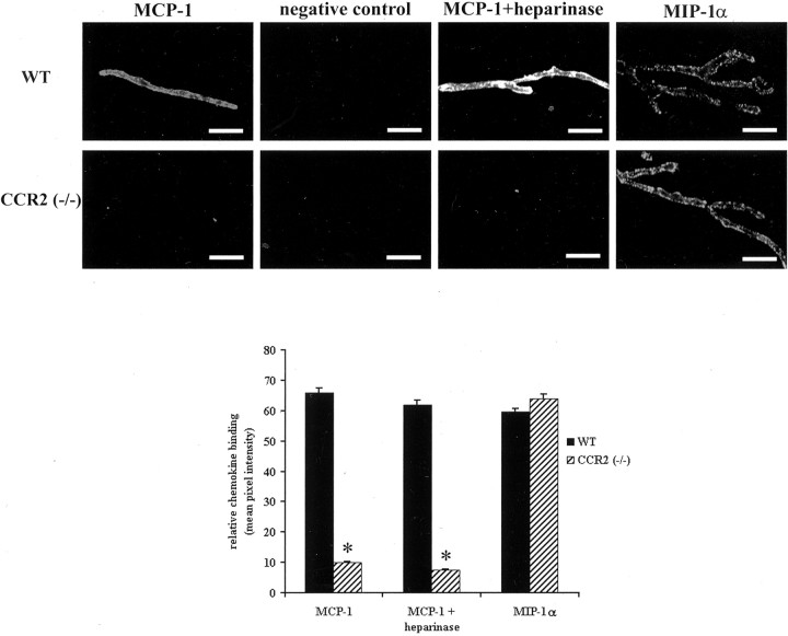
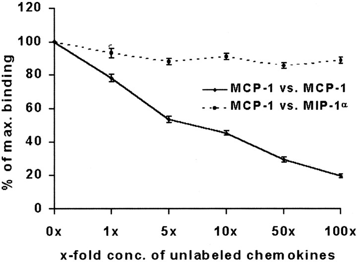
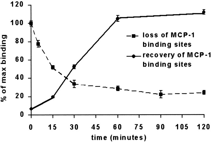
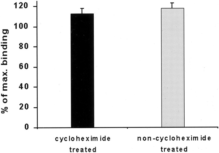
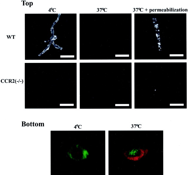
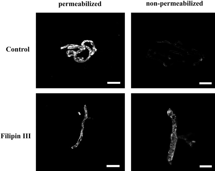
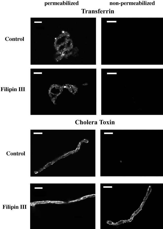
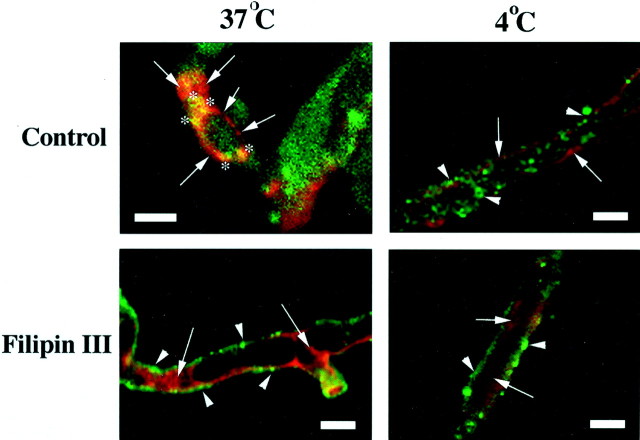
Previous results from this laboratory revealed the presence of high-affinity saturable binding sites for monocyte chemoattractant protein-1 (MCP-1) along human brain microvessels (Andjelkovic et al., 1999; Andjelkovic and Pachter, 2000), which suggested that CC chemokine receptor 2 (CCR2), the recognized receptor for this chemokine, was expressed by the brain microvascular endothelium. To test the role of CCR2 directly in mediating MCP-1 interactions with the brain microvasculature, we assessed MCP-1 binding activity in murine brain microvessels isolated from wild-type mice and from CCR2 (-/-) mice engineered to lack this receptor. Results demonstrate that MCP-1 binding is greatly attenuated in microvessels prepared from CCR2 (-/-) mice compared with wild-type controls. Moreover, microvessels from wild-type mice exhibited MCP-1-induced downmodulation in MCP-1 binding and a recovery of binding activity that was not dependent on de novo protein synthesis. Furthermore, MCP-1 was shown to be internalized within wild-type microvessels, but not within microvessels obtained from CCR2 (-/-) mice, additionally demonstrating that CCR2 is obligatory for MCP-1 endocytosis. Last, internalization of MCP-1, but not transferrin, was observed to be inhibited by disruption of caveolae. Internalized MCP-1 also colocalized at some sites with caveolin-1, a major protein of caveolae, implying that this chemokine is endocytosed, in part, via nonclathrin-coated vesicles. These results prompt consideration that MCP-1 signals may be relayed across the blood-brain barrier by highly specialized interactions of this chemokine with its cognate receptor, CCR2, along brain microvascular endothelial cells.


Figures

Fig. 1.
Fig. 1.
Biotinylated chemokine binding along murine brain microvessels. Top, Representative examples of fluorescent detection of chemokines attached to the brain microvascular surface. Detection of biot.-rmMCP-1 and biot.-rmMIP-1α binding was performed at 4°C as described in Materials and Methods. Diminished binding of biot.-rmMCP-1 to microvessels from CCR2 (−/−) mice, compared with those from wild-type mice (WT), is readily apparent. No such difference between these respective microvessel populations is observed for biot.-rmMIP-1α binding, and heparinase treatment failed to abrogate biot.-rmMCP-1 binding. Scale bar, 50 μm. Bottom, Quantitative analysis of biotinylated chemokine binding. Mean pixel intensities ± SEM, reflecting relative chemokine binding, were determined as described in Materials and Methods and were corrected for background noise by subtraction of intensity values associated with negative controls. Values represent those determined from at least three different experiments. *p < 0.001 when contrasted with corresponding wild-type value.
Fig. 2.
Fig. 2.
Competition of biotinylated MCP-1 binding along murine brain microvessels. Competition studies were performed at 4°C with a constant concentration of biot.-rmMCP-1 and increasing concentrations of unlabeled chemokines (indicated bysymbols). Binding was quantitated as described in Materials and Methods and is reported as the percentage ± SEM of maximal binding achieved in the absence of inhibitor ligand. Values represent those determined from at least three different experiments.
Fig. 3.
Fig. 3.
Loss and recovery of MCP-1 binding sites on murine brain microvessels after ligand exposure. Microvessels were reacted with unlabeled MCP-1 at 37°C for 2 hr (recovery) or for varying periods of time (loss). After exposure to unlabeled chemokine the microvessels were washed in PBS and either were exposed immediately to biot.-rmMCP-1 for 2 hr at 4°C (loss) or were incubated at 37°C for varying periods of time and then exposed to biot.-rmMCP-1 (recovery). After reaction with biot.-rmMCP-1 the samples were subjected to the standard binding assay conditions, and binding intensity along the microvessels was analyzed as described in Materials and Methods. The extent of both loss and recovery of biot.-rmMCP-1 binding is indicated as the percentage ± SEM of maximal binding obtained in the absence of any previous exposure of the microvessels to unlabeled MCP-1. Values represent those determined from at least three different experiments.
Fig. 4.
Fig. 4.
Effect of protein synthesis inhibition on the recovery of MCP-1 binding sites along murine brain microvessels. Microvessels were exposed to unlabeled MCP-1 as described in Figure 3, except that in the last 30 min the protein synthesis inhibitor cycloheximide (10 μg/ml) either was added to the incubation mixture or was not. After chemokine exposure the microvessels treated with cycloheximide were allowed to recover for 90 min in the continued presence of protein synthesis inhibitor while the control samples continued their recovery in the absence of cycloheximide. Binding was quantitated as described in Materials and Methods and is reported as the percentage ± SEM of maximal binding obtained in the absence of any previous exposure of the microvessels to unlabeled MCP-1. Values represent those determined from three different experiments.
Fig. 5.
Fig. 5.
Internalization of MCP-1 binding sites along murine brain microvessels. Top, Microvessels were reacted with biot.-rmMCP-1 at 37°C, fixed with 4% paraformaldehyde, and then either reacted directly with avidin-fluorescein (nonpermeabilized) or permeabilized with Tween 20 before reaction with avidin-fluorescein, as described in Materials and Methods. Compared with Figure 1, wild-type microvessels (WT) that were reacted with labeled chemokine at 37°C and that were not permeabilized reveal a greatly attenuated signal. Permeabilization, however, restores the detection of biot.-rmMCP-1, suggesting that the labeled chemokine had been internalized within wild-type microvessels at the elevated temperature. Microvessels from CCR2 (−/−) mice, in contrast, demonstrated only a weak signal regardless of whether or not they had been permeabilized, also implying that they do not internalize MCP-1. Scale bar, 50 μm. Bottom, Three-dimensional renderings of microvessels from wild-type mice depicting the distribution of biot.-rmMCP-1 relative to the endothelial plasma membrane. In accordance with the procedures that were described in Materials and Methods, the microvessels were reacted with biot.-rmMCP-1 at either 4 or 37°C and then were fixed and permeabilized. Next the samples were stained consecutively with avidin-fluorescein to reveal chemokine localization (green) and then with rho.-WGA to indicate endothelial plasma membrane (red). Samples were subject to confocal microscopy and three-dimensional rendering as described in Materials and Methods, and the images are oriented so that viewer is looking “on face” toward the microvascular lumen. Chemokine staining clearly lies external to the plasma membrane at 4°C and internal to it at 37°C (some chemokine staining actually might reflect complete transit into the lumen at the elevated temperature).
Fig. 6.
Fig. 6.
Effect of filipin III on internalization of MCP-1 along murine brain microvessels. To gauge whether MCP-1 internalization may be mediated by caveolae, we exposed the microvessels to the caveolae-disrupting agent filipin III before incubation with biot.-rmMCP-1 at 37°C. Contrary to nontreated controls, the microvessels pretreated with filipin III did not require permeabilization to enable the detection of labeled chemokine, suggesting that biot.-rmMCP-1 remained on the cell surface as a consequence of caveolar disruption. Scale bar, 50 μm.
Fig. 7.
Fig. 7.
Effect of filipin III on internalization of transferrin and cholera toxin along murine brain microvessels. Microvessels received (±) filipin III treatment, were exposed to biot.-transferrin or biot.-cholera toxin, and then were processed as described in Figure 6. Both control and filipin III-treated microvessels required permeabilization to detect biot.-transferrin, indicating that the process of transferrin internalization proceeded despite disruption of the caveolae. In contrast, biot.-cholera toxin could be observed along the surface of filipin III-treated microvessels both with and without permeabilization, reflecting filipin III-mediated interference with the internalization of this ligand. Scale bar, 50 μm.
Fig. 8.
Fig. 8.
Colocalization of internalized MCP-1 with caveolin-1. Microvessels were pretreated (±) with filipin III. Then the samples were exposed to biot.-rmMCP-1 at 4 or 37°C, fixed/permeabilized, and stained to reveal labeled chemokine (green) and caveolin-1 (red) localization, as described in Materials and Methods. Then confocal images were obtained at a level approximately midway through the interior of the microvascular samples, revealing the relative distribution patterns of labeled chemokine and caveolin-1. In the control sample exposed to biot.-rmMCP-1 at 37°C, caveolin-1 staining can be seen concentrated around the periphery of the microvascular segment (arrows), with chemokine apparently present diffusely in the cytoplasm. Areas of yellow fluorescence (asterisks) indicate presumed sites of biot.-rmMCP-1/caveolin-1 colocalization. In the filipin III-treated sample exposed to chemokine at 37°C, no sites of colocalization are detected readily, and biot.-rmMCP-1 appears to be confined to the membrane surface (arrowheads), with caveolin-1 expression heightened in some cytoplasmic locales (arrows). Microvessels exposed to chemokine at 4°C (±) filipin III pretreatment also fail to show any areas of biot.-rmMCP-1/caveolin-1 colocalization. These samples also do not demonstrate as strong a cytoplasmic distribution of labeled chemokine as that observed in the control sample at 37°C but seemingly manifest a more peripheral chemokine staining (arrowheads), possibly restricted to the membrane surface. As with the samples exposed to chemokine at 37°C, caveolin-1 distribution appears to be concentrated along the periphery of the microvessel in the control (arrowheads) but is dispersed more cytoplasmically in the filipin III-treated sample. Arrows, Caveolin-1;arrowheads, biot.-rmMCP-1; asterisks, biot.-rmMCP-1/caveolin-1 colocalization. Scale bar, 10 μm.

Similar articles

Cited by

References

    1. Abbott NJ, Romero IA. Transporting therapeutics across the blood–brain barrier. Mol Med Today. 1996;2:106–113. - PubMed
    1. Amara A, Gall SL, Schwartz O, Salamero J, Montes M, Loetscher P, Baggliolini M, Virelizier JL, Arenzana-Seisdedos F. HIV coreceptor downregulation as antiviral principle: SDF-1α-dependent internalization of the chemokine receptor CXCR4 contributes to inhibition of HIV replication. J Exp Med. 1997;186:139–146. - PMC - PubMed
    1. Anderson RGW. The caveolae membrane system. Annu Rev Biochem. 1998;67:199–225. - PubMed
    1. Andjelkovic AV, Pachter JS. Characterization of binding sites for chemokines MCP-1 and MIP-1α on human brain microvessels. J Neurochem. 2000;75:1898–1906. - PubMed
    1. Andjelkovic AV, Spencer DD, Pachter JS. Visualization of chemokine binding sites on human brain microvessels. J Cell Biol. 1999;145:403–412. - PMC - PubMed

Publication types

MeSH terms