. 2001 Dec 1;21(23):9246-54.
doi: 10.1523/JNEUROSCI.21-23-09246.2001.

Rats expressing human cytosolic copper-zinc superoxide dismutase transgenes with amyotrophic lateral sclerosis: associated mutations develop motor neuron disease

Affiliations

Rats expressing human cytosolic copper-zinc superoxide dismutase transgenes with amyotrophic lateral sclerosis: associated mutations develop motor neuron disease

M Nagai et al. J Neurosci. .

Abstract

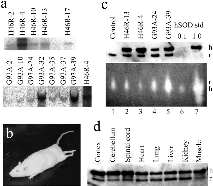
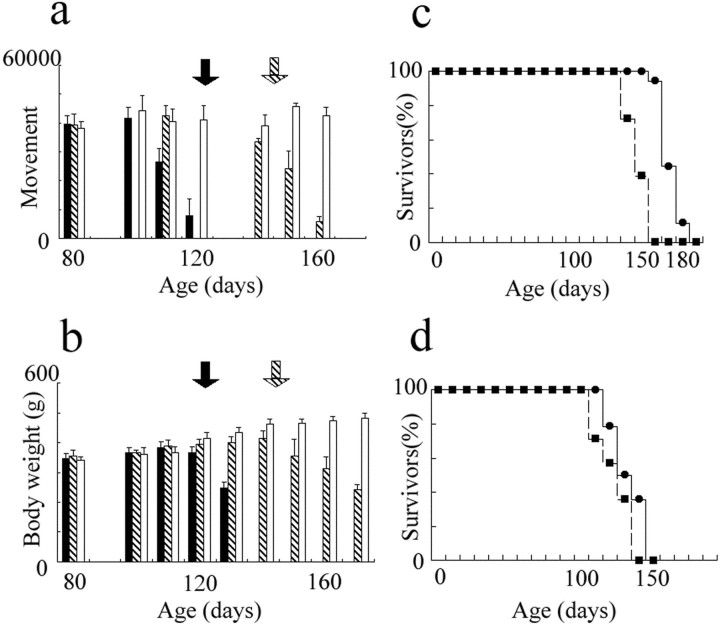
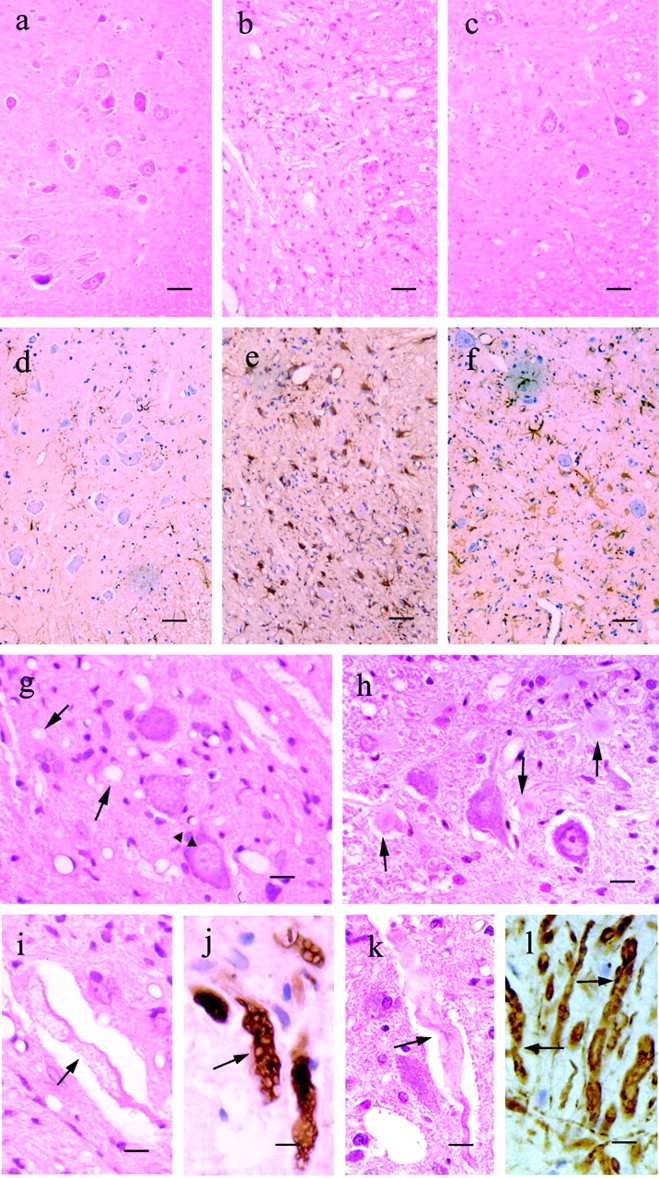
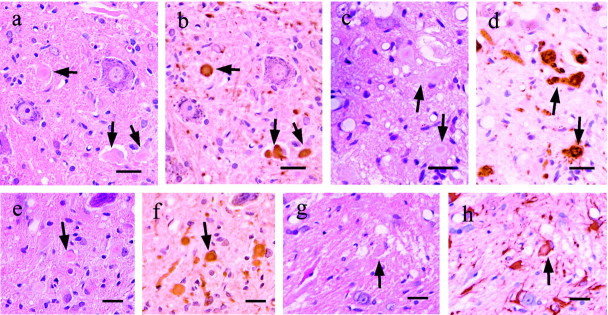
Some cases of familial amyotrophic lateral sclerosis (ALS) are caused by mutations in the gene encoding cytosolic, copper-zinc superoxide dismutase (SOD1). We report here that rats that express a human SOD1 transgene with two different ALS-associated mutations (G93A and H46R) develop striking motor neuron degeneration and paralysis. As in the human disease and transgenic ALS mice, pathological analysis demonstrates selective loss of motor neurons in the spinal cords of these transgenic rats. In spinal cord tissues, this is accompanied by activation of apoptotic genes known to be activated by mutant SOD1 protein in vitro and in vivo. These animals provide additional support for the proposition that motor neuron death in SOD1-related ALS reflects one or more acquired, neurotoxic properties of the mutant SOD1 protein. The larger size of this rat model as compared with the ALS mice will facilitate studies involving manipulations of spinal fluid (implantation of intrathecal catheters for chronic therapeutic studies; CSF sampling) and spinal cord (e.g., direct administration of viral- and cell-mediated therapies).


Figures

Fig. 1.
Fig. 1.
Analysis of human SOD1 transgene (H46R or G93A) copy number, protein expression, and SOD1 activity. a, Southern analysis of the human SOD1 gene in transgenic rats as determined by tail DNA blots. Five transgenic lines were established with the H46R mutation (top row), and seven transgenic lines were established with the G93A mutation (bottom row). The H46R-4 blot is shown in each row to allow comparison of the H46R and G93A results. b, An affected transgenic rat from the H46R-4 line demonstrates hindlimb weakness and abnormal posturing with segmental spasticity of the tail. c,Top panel, Quantitative immunoblotting of 4 μg of total protein extracts of spinal cord from nontransgenic littermate control, H46R lines (H46R-4, H46R-13), and G93A lines (G93A-24, G93A-39) using sheep polyclonal antibodies that recognize a common epitope shared between human (h) and rat (r) SOD1. Two 10-fold dilutions of purified human erythrocyte SOD1 (0.1, 1.0 U) were immunoblotted in parallel to provide standards for quantitation. Bottom panel, SOD1 enzymatic activity in spinal cord extracts (20 μg of protein) from the same nontransgenic littermate control or transgenic rats determined on native gels. Note that the electrophoretic migration of rat SOD1 (r) differs from that of human erythrocyte SOD1 (h). d, Total protein (4 μg) from various tissues from 2-month-old transgenic H46R-4 rats was immunoblotted with the same sheep polyclonal antibody as inc, recognizing human and rat SOD1.
Fig. 2.
Fig. 2.
Phenotypic markers of disease progression in the transgenic (H46R-4, G93A-39) ALS rats. a, Spontaneous walking movements decreased by 110 and 150 d for the G93A-39 and H46R-4 rats, respectively, as measured using an Automap II apparatus.b, Body weights for these two lines of rats began to fall at approximately the same age as onset of clinically apparent weakness (∼123 d for the G93A-39 line and ∼145 d for the H46R-4 line). c, Kaplan-Meier curves illustrating the ages of onset (mean 145 d) and death (mean 169 d) for the H46R-4 rats. d, Kaplan-Meier curves illustrating the ages of onset (mean 123 d) and death (mean 131 d) for the G93A-39 rats. In a and b, solid bars = G93A-39 transgenic rats, hatched bars = H46R-4 transgenic rats, and open bars = nontransgenic littermate control rats. The ages of the first appearance of clinical weakness are indicated by thesolid (G93A-39) and hatched (H46R-4)arrows. In c and d, thedashed lines with black square data points designate the onset curves (percentage without weakness), whereas the solid lines with black dots designate the survival curves (percentage surviving).
Fig. 3.
Fig. 3.
Major histopathological findings in the G93A-39 and H46R-4 transgenic rats. af, Ventral horns of the lumbar spinal cord from a 6-month-old normal littermate(a, d), a G93A-39 transgenic rat at 4.5 months (b, e), and an H46R-4 transgenic rat at 6.0 months (c, f). Sections were stained with hematoxylin and eosin (ac) and immunostained using GFAP (df). g, h, Higher magnification views of the neuropil in the ventral horn of the lumbar spinal cord in G93A-39 (g) and H46R-4 (h) transgenic rats. g, Conspicuous vacuoles in the neuropil (arrows) and perikarya of motor neuron (arrowheads) are indicated in the G93A-39 transgenic rat section. h, Lewy body-like inclusions are readily apparent (arrows) in the neuropil of the H46R-4 transgenic rat. i, Axonal swelling and vacuolation in the G93A-39 transgenic rat. j, Phosphorylated neurofilament is identified using antibody SMI-31 in the G93A-39 rats. k, Swollen and tortuous ventral axon in H46R-4. l, Phosphorylated neurofilaments are also identified with antibody SMI-31 in the H46R-4 rats. Inil, the arrows designate axons. Scale bars: af, 50 μm;g, h, 20 μm;il, 10 μm.
Fig. 4.
Fig. 4.
Intracellular inclusions in the 6-month-old H46R-4 transgenic rats. ad, Lewy body-like cytoplasmic inclusions (arrows) in neuropil and in motor neurons. Inclusions consist of a pale periphery and dense central core as stained by hematoxylin and eosin (a,c). Destaining (a, c) and restaining (b, d) of the same section with anti-human SOD1 antibody (b) and anti-ubiquitin antibody (d) demonstrated that these inclusions are immunoreactive for human SOD1 and ubiquitin.eh, Lewy body-like cytoplasmic inclusions in astrocytes. Inclusions stained by hematoxylin and eosin (e, g) have clear periphery and dense core like those in the neurons and the neuropil. Destaining (e, f) and restaining (g, h) of the same section revealed that inclusions are immunostained by the anti-human SOD1 antibody (f) but not by anti-GFAP antibodies (h), although the cell itself is GFAP-positive (h). Scale bars, 40 μm.
Fig. 5.
Fig. 5.
Amino acid levels in CSF from 2-month-old and end stage G93A-39 transgenic rats. a, Mean glutamine concentrations in CSF from 2-month-old and end stage transgenic rats and control littermates. *p < 0.05.b, Mean concentrations of asparagine (solid bar), aspartate (shaded bar), glutamate (hatched bar), and glycine (open bar) in CSF from 2-month-old and end stage transgenic rats and control littermates. The error bars denote the SD.
Fig. 6.
Fig. 6.
Caspase-1 and -3 are sequentially activated in the spinal cord of ALS transgenic rats. a, Caspase-1-like activity was measured as described and is reported as fluorescence emitted by the free AMC after cleavage of the caspase-1 substrate YVAD-AMC. b, DEVD-AMC cleavage was measured to determine caspase-3 activity in spinal cord lysates of the ALS rats and their littermate controls. Caspase-3 activity is expressed as fluorescence emitted from the free fluorogenic group AMC. Data are the mean ± SD for experiments assayed in duplicate. Asterisks(*p < 0.05; **p < 0.01) indicate significant differences with respect to the control groups.Squares and diamondsindicate data derived from H46R-4 and control rats, respectively. Ages are in months.

References

    1. Aoki M, Ogasawara M, Matsubara Y, Narisawa K, Nakamura S, Itoyama Y, Abe K. Mild ALS in Japan associated with novel SOD mutation. Nat Genet. 1993;5:323–324. - PubMed
    1. Aoki M, Ogasawara M, Matsubara Y, Narisawa K, Nakamura S, Itoyama Y, Abe K. Familial amyotrophic lateral sclerosis (ALS) in Japan associated with H46R mutation in Cu/Zn superoxide dismutase gene: a possible new subtype of familial ALS. J Neurol Sci. 1994;126:77–83. - PubMed
    1. Beauchamp C, Fridovich I. Superoxide dismutase: improved assays and an assay applicable to acrylamide gels. Anal Biochem. 1971;44:276–287. - PubMed
    1. Borchelt DR, Lee MK, Slunt HS, Guarnieri M, Xu ZS, Wong PC, Brown RHJ, Price DL, Sisodia SS, Cleveland DW. Superoxide dismutase 1 with mutations linked to familial amyotrophic lateral sclerosis possesses significant activity. Proc Natl Acad Sci USA. 1994;91:8292–8296. - PMC - PubMed
    1. Brown RHJ. Amyotrophic lateral sclerosis: recent insights from genetics and transgenic mice. Cell. 1995;80:687–692. - PubMed

Publication types

MeSH terms