. 2001 Dec 1;21(23):9355-66.
doi: 10.1523/JNEUROSCI.21-23-09355.2001.

Functional interactions between tumor and peripheral nerve: morphology, algogen identification, and behavioral characterization of a new murine model of cancer pain

Affiliations

Functional interactions between tumor and peripheral nerve: morphology, algogen identification, and behavioral characterization of a new murine model of cancer pain

P W Wacnik et al. J Neurosci. .

Abstract

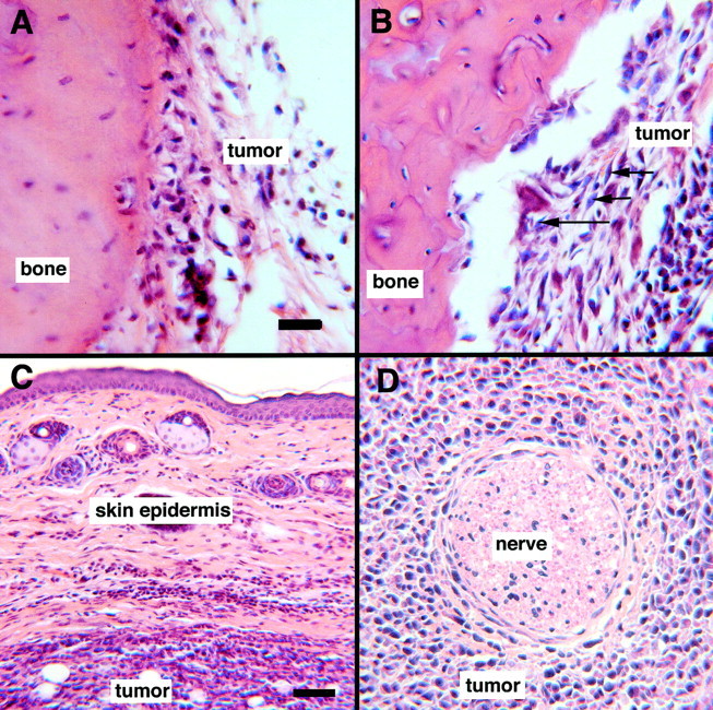
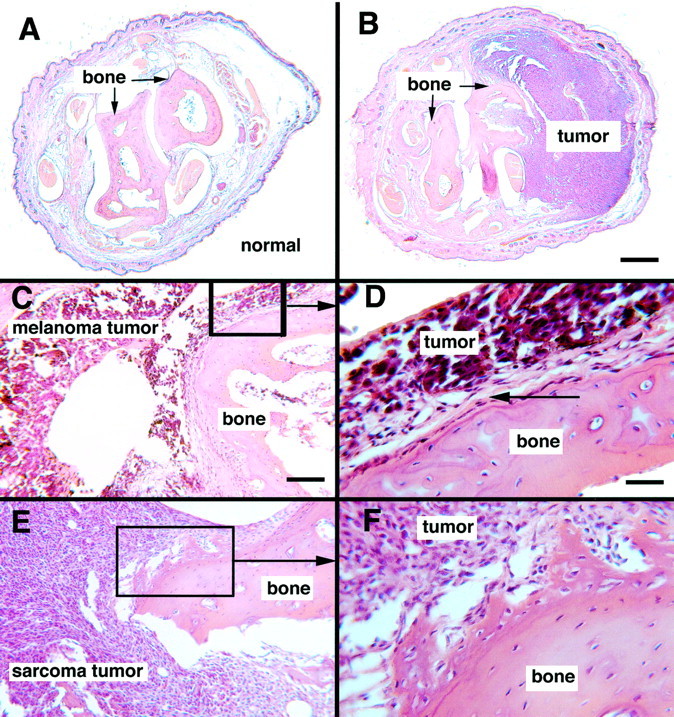
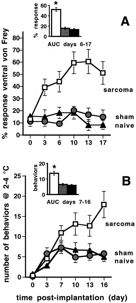
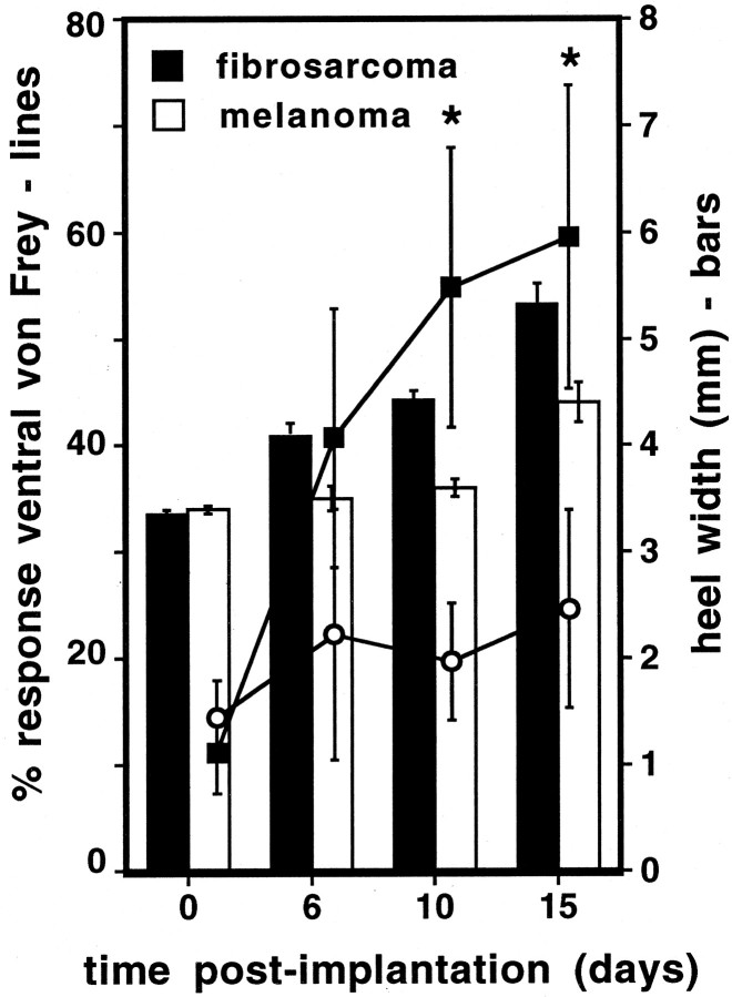
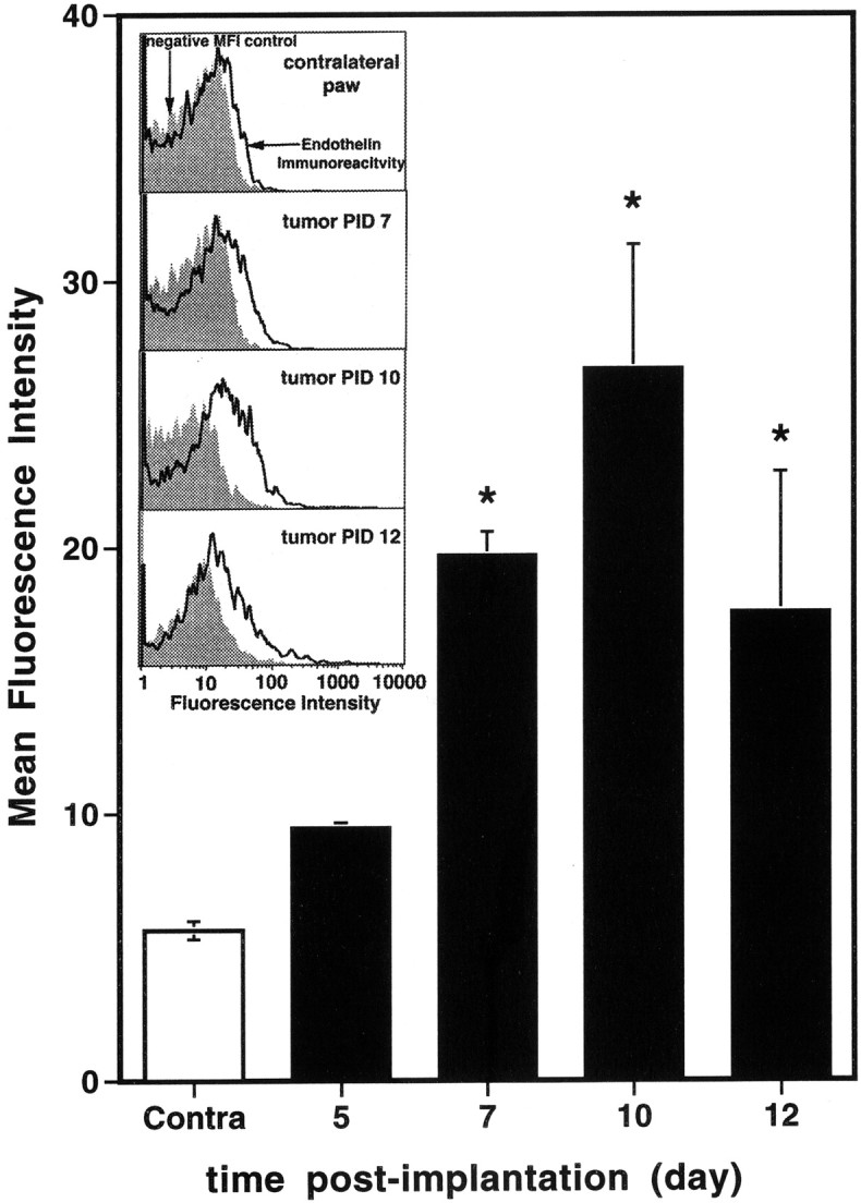
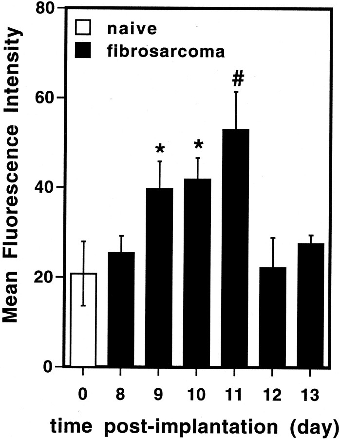
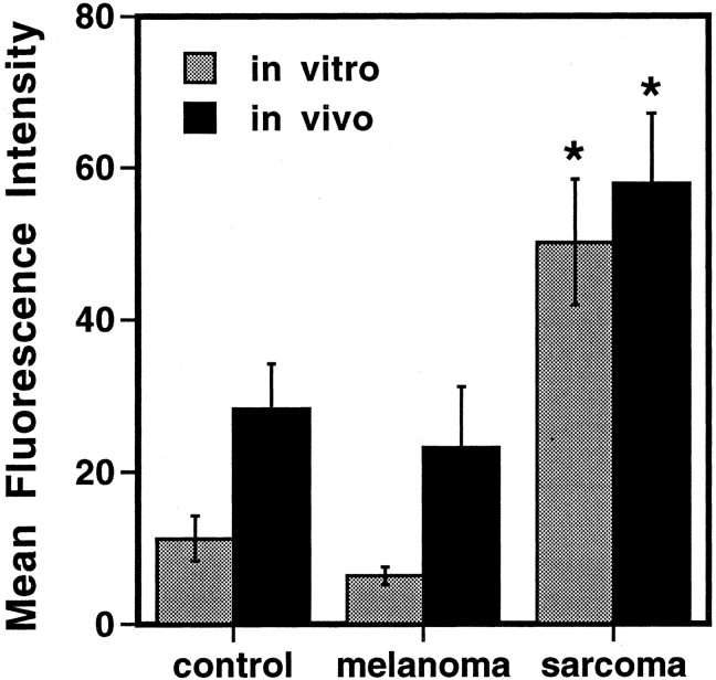
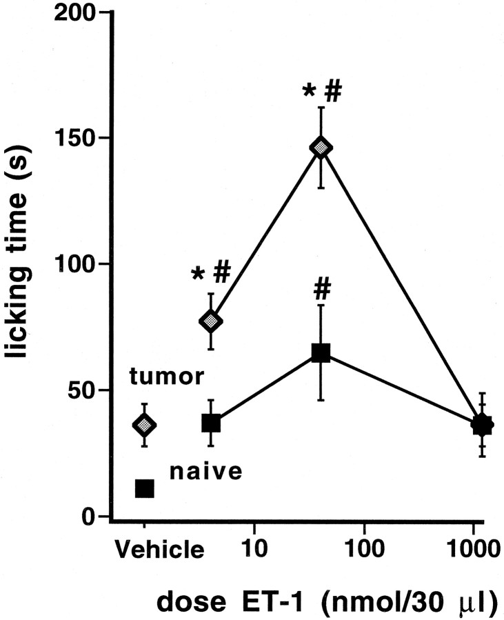
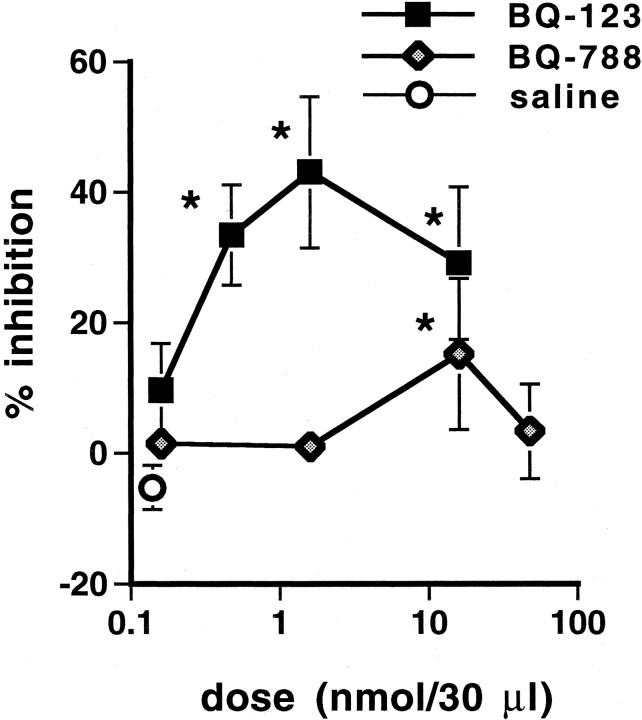
This paper describes a model of tumor-induced bone destruction and hyperalgesia produced by implantation of fibrosarcoma cells into the mouse calcaneus bone. Histological examination indicates that tumor cells adhere to the bone edge as early as post-implantation day (PID) 3, but osteolysis does not begin until PID 6, correlating with the development of hyperalgesia. C3H/He mice exhibit a reproducible hyperalgesia to mechanical and cold stimuli between PID 6 and 16. These behaviors are present but significantly reduced with subcutaneous implantation that does not involve bone. Systemic administration of morphine (ED(50) 9.0 mg/kg) dose-dependently attenuated the mechanical hyperalgesia. In contrast, bone destruction and hypersensitivity were not evident in mice implanted with melanoma tumors or a paraffin mass of similar size. A novel microperfusion technique was used to identify elevated levels of the putative algogen endothelin (ET) in perfusates collected from the tumor sites of hyperalgesic mice between PID 7 and 12. Increased ET was evident in microperfusates from fibrosarcoma tumor-implanted mice but not from melanoma tumor-implanted mice, which are not hyperalgesic. Intraplantar injection of ET-1 in naive and, to a greater extent, fibrosarcoma tumor-bearing mice produced spontaneous pain behaviors, suggesting that ET-1 activates primary afferent fibers. Intraplantar but not systemic injection of the ET-A receptor antagonist BQ-123 partially blocked tumor-associated mechanical hyperalgesia, indicating that ET-1 contributes to tumor-induced nociception. This model provides a unique approach for quantifying the behavioral, biochemical, and electrophysiological consequences of tumor-nerve interactions.


Figures

Fig. 1.
Fig. 1.
Photomicrographs of hematoxylin and eosin-stained fibrosarcoma tumor sections at different PID time points.A, PID 3 fibrosarcoma: tumor cells are closely adhered to bone surface, but the bone edge is intact. B, PID 6 fibrosarcoma: bone edge is irregular, indicative of osteolysis.Arrows indicate individual spindle-shaped fibrosarcoma cells. C, Skin overlying tumor at PID 9: note the lack of skin invasion by tumor cells and normal skin morphology.D, PID 12 fibrosarcoma: intact nerve bundle surrounded by tumor cells. There is no evidence of nerve invasion by tumor cells or of nerve degeneration at this time point. Scale bars (shown inA): A, B,D, 20 μm; C, 60 μm.
Fig. 2.
Fig. 2.
Photomicrographs of hematoxylin and eosin-stained sections of a normal and tumor-bearing mouse heel. A, Cross section of normal mouse heel. B, Cross section of a comparable area at PID 9 of fibrosarcoma cells.CF, Comparison of fibrosarcoma tumor and control melanoma tumor morphology. C, PID 9 melanoma tumor. D, Enlargement of boxed area inC: note the regular bone edge and layer of periosteum (arrow) separating bone and tumor cells.E, PID 9 fibrosarcoma tumor. F, Enlargement of boxed area in E: note irregular bone edge and invasion of tumor cells into bone, indicating osteolysis. Scale bars: (shown in B) A,B, 500 μm; (shown in C)C, E, 100 μm; (shown inD) D, F, 30 μm.
Fig. 3.
Fig. 3.
A, Mechanical hyperalgesia after development of the fibrosarcoma tumor. The withdrawal responses evoked by a normally non-noxious von Frey monofilament (3.4 mN bending force) increased significantly as early as PID 3. The percentage response was calculated as the number of positive responses divided by 6 (the total number of stimuli per paw) multiplied by 100. The mean and SEM were calculated for each group for PID 6–15, and the data were analyzed for significance (∗) using ANOVA (p < 0.01) with Bonferroni post hoc tests. B, Cold hypersensitivity was detected after the development of the fibrosarcoma tumor and measured as a significant increase in the number of nociceptive behaviors over a 4 min period on a 2–4°C cold plate. The mean and SEM were calculated for each group for PID 7–16, and the data were analyzed for significance using ANOVA (∗ indicates significance;p < 0.01) with Bonferroni post hoctests (n = 14 tumor; n = 9 sham; n = 10 naive).
Fig. 4.
Fig. 4.
B6C3f1/cr mice implanted with fibrosarcoma cells, but not melanoma or saline (sham, data not shown), showed significant mechanical hyperalgesia in response to plantar stimulation with a normally non-noxious 1.6 mN von Frey monofilament. Lines(left axis) show increasing hyperalgesia as the fibrosarcoma tumor grew, with greater hyperalgesia evident on PID 10 and 15 than both the melanoma-implanted and sham mice (data not shown).Bars (right axis) show the corresponding progression of heel width for both fibrosarcoma and melanoma as measured on PID 1–15 (PID 1–5 not shown) with a micrometer; note that the two tumor types had equivalent size at PID 15 (melanoma, 4.4 mm ± 0.19) and PID 10 (fibrosarcoma, 4.43 mm ± 0.20) (n ≥ 7). The mean and SEM are shown for each experimental group. Fibrosarcoma tumor, melanoma tumor, and sham groups were compared by ANOVA followed by a Fisher's post hoc test where ∗ indicates significant differences from control (p < 0.05).
Fig. 5.
Fig. 5.
Homogenates of harvested tumors contain increased levels of ET on PID 7, 10, and 12 as compared with homogenates from the contralateral limb. Tumors were harvested and homogenized; the supernatant was analyzed by flow cytometry for ET. Group size is four to five mice at each time point; *p < 0.05.Inset indicates representative flow cytometry histograms from tumor homogenates analyzed by MBISA. Tumor homogenate proteins were adsorbed to 4 μm latex microbeads, and the beads were labeled with anti-ET antisera (open histograms). Nonspecific antibody labeling was determined using negative control NRS (negative MFI control, filled histograms). Bead staining was assessed by flow cytometry and from these data; the MFI was calculated from the histograms and subjected to statistical analyses.
Fig. 6.
Fig. 6.
Fibrosarcoma tumors secrete increased levels of ET on PID 9, 10, and 11. Extracellular fluid from fibrosarcoma tumor sites was sampled by microperfusion on PID 8–13 and compared with microperfusates from the hindlimb of naive mice. The relative level of ET was determined by flow cytometry. Group size is four to six mice per time point; ∗ indicates p < 0.05 compared with naive; # indicates p < 0.01 compared with naive.
Fig. 7.
Fig. 7.
Fibrosarcoma cells in vitro(gray bars) and in vivo(black bars) secrete more ET than melanoma cells. Serum-free medium incubated with tumor cells for 24 hr was analyzed in triplicate for the presence of ET by flow cytometry. Levels in conditioned media were compared with those in cell-free media (control). For in vivo analysis, mice implanted with fibrosarcoma or melanoma tumors and naive mice (control) underwent microperfusion on PID 8. The relative levels of ET were determined by flow cytometry. Group size is three to four mice per condition at each time point; ∗ indicatesp < 0.05 compared with control and melanoma samples.
Fig. 8.
Fig. 8.
The duration of nocifensive behaviors in seconds (s) was accumulated over a 10 min period after ET-1 (4.0 pmol–1.2 nmol/30 μl) or saline (30 μl) injection into the fibrosarcoma tumor site of C3H/He mice at PID 10 or into the heel of naive mice. Data are presented as mean and SEM, analyzed by ANOVA, and further tested for differences from respective saline–sham control (∗) and between-treatment groups (#) with the Fisher'spost hoc test; ∗ and # indicate statistical significance; p < 0.05 (n ≥ 10).
Fig. 9.
Fig. 9.
The ET-A receptor antagonist BQ-123 (0.16–16 nmol/30 μl) injected into the tumor site of hyperalgesic C3H/He mice with fibrosarcoma tumors on PID 10 produced dose-dependent attenuation of mechanical hyperalgesia (45 min post-drug; n ≥ 9; filled squares). Injection of the ET-B receptor antagonist BQ-788 (0.16–48 nmol/30 μl) on PID 12 attenuated mechanical hyperalgesia only at the highest dose tested (45 min post-drug; n ≥ 9; shaded diamonds). Saline, similarly injected into the tumor site of PID 10 and 12 hyperalgesic C3H/He mice, was inactive (n = 20;open circles). Data are presented as mean and SEM, analyzed by ANOVA, and further tested for differences from saline–vehicle control (∗) with the Fisher's post hoc test; ∗ indicates statistical significance;p < 0.05.

References

    1. Asham EH, Loizidou M, Taylor I. Endothelin-1 and tumour development. Eur J Surg Oncol. 1998;24:57–60. - PubMed
    1. Banning A, Sjogren P, Henriksen H. Treatment outcome in a multidisciplinary cancer pain clinic. Pain. 1991;47:129–134. - PubMed
    1. Cain DM, Khasabov SG, Simone DA. Response properties of mechanoreceptors and nociceptors in mouse glabrous skin: an in vivo study. J Neurophysiol. 2001a;85:1561–1574. - PubMed
    1. Cain DM, Wacnik PW, Turner M, Wendelschafer-Crabb G, Kennedy WR, Wilcox GL, Simone DA. Functional interactions between tumor and peripheral nerve: changes in excitability and morphology of primary afferent fibers in a murine model of cancer pain. J Neurosci. 2001b;21:9367–9376. - PMC - PubMed
    1. Caraceni A, Portenoy RK. An international survey of cancer pain characteristics and syndromes. IASP Task Force on Cancer Pain. International Association for the Study of Pain. Pain. 1999;82:263–274. - PubMed

Publication types

MeSH terms