. 2001 Dec 1;21(23):9367-76.
doi: 10.1523/JNEUROSCI.21-23-09367.2001.

Functional interactions between tumor and peripheral nerve: changes in excitability and morphology of primary afferent fibers in a murine model of cancer pain

Affiliations

Functional interactions between tumor and peripheral nerve: changes in excitability and morphology of primary afferent fibers in a murine model of cancer pain

D M Cain et al. J Neurosci. .

Abstract

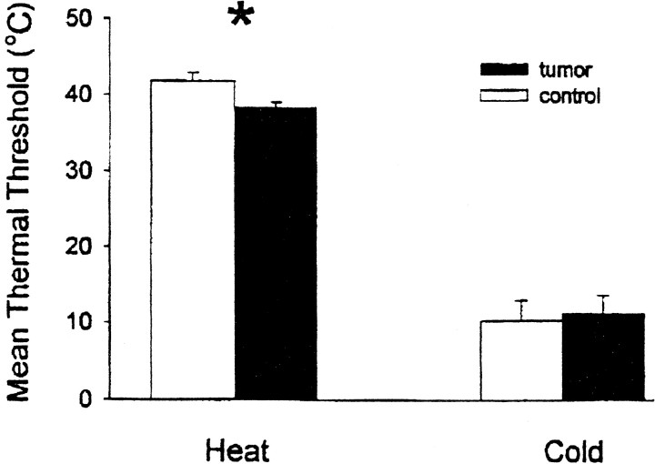
We used a murine model to investigate functional interactions between tumors and peripheral nerves that may contribute to pain associated with cancer. Implantation of fibrosarcoma cells in and around the calcaneus bone produced mechanical hyperalgesia of the ipsilateral paw. Electrophysiological recordings from primary afferent fibers in control and hyperalgesic mice with tumor revealed the development of spontaneous activity (0.2-3.4 Hz) in 34% of cutaneous C-fibers adjacent to the tumor (9-17 d after implantation). C-fibers in tumor-bearing mice exhibited a mean decrease in heat threshold of 3.5 +/- 0.10 degrees C. We also examined innervation of the skin overlying the tumor. Epidermal nerve fibers (ENFs) were immunostained for protein gene product 9.5, imaged using confocal microscopy, and analyzed in terms of number of fibers per millimeter of epidermal length and branching (number of nodes per fiber). Divergent morphological changes were linked to tumor progression. Although branching of ENFs increased significantly relative to control values, in later stages (16-24 d after implantation) of tumor growth a sharp decrease in the number of ENFs was observed. This decay of epidermal innervation of skin over the tumor coincided temporally with gradual loss of electrophysiological activity in tumor-bearing mice. The development of spontaneous activity and sensitization to heat in C-fibers and increased innervation of cutaneous structures within the first 2 weeks of tumor growth suggest activation and sensitization of a proportion of C-fibers. The decrease in the number of ENFs observed in later stages of tumor development implicates neuropathic involvement in this model of cancer pain.


Figures

Fig. 1.
Fig. 1.
Representative examples of spontaneous activity and conduction latency of three C-fibers recorded in skin overlying a fibrosarcoma tumor. A, Top, Spontaneous activity of this nociceptor occurred generally at a steady rate of ∼1 Hz. A, Bottom, Three superimposed oscilloscope traces showing constant conduction latency evoked by electrical stimulation at the RF. B, A second C-fiber with a slower (0.15 Hz) rate of spontaneous activity.C, Spontaneous activity of this nociceptor occurred in bursts of three to five action potentials.Arrows indicate stimulus artifact. The 10 sec calibration below the spontaneous activity in C applies to all three traces of ongoing activity. Calibrations of individual conduction latencies are shown under the overlaid traces of constant conduction velocity.
Fig. 2.
Fig. 2.
Mean (±SE) response threshold for heat and cold in control (unfilled columns) and in mice with fibrosarcoma tumors (filled columns). The mean heat threshold of C-fibers in control mice was 41.8 ± 1.09°C compared with 38.3 ± 10.72°C in mice with tumor (p = 0.011). ∗ indicates a significant difference from control. The cold threshold of C-fibers in mice with tumor was 11.2 ± 2.4°C compared with 10.2 ± 2.67°C in control mice.
Fig. 3.
Fig. 3.
Stimulus response curves for C-fibers in control mice (○) and mice with tumor (●) indicating mean (±SE) number of impulses in response to thermal stimuli. A, C-fibers discharged a greater number of impulses to heat stimuli 37–47°C in mice with tumor than in control mice. B, No significant difference was observed in C-fiber discharge to cold stimuli between control mice and mice with tumor.
Fig. 4.
Fig. 4.
Mean (±SE) thermal threshold in control mice (black) and nonspontaneously active and spontaneously active C-fibers in mice with tumor. The asterisk(left columns) indicates a significant difference in mean heat thresholds between control and mice with tumor, but thresholds of C-fibers in tumor mice with spontaneous activity did not differ from those without it. Right columns, Neither the presence of tumor nor ongoing activity appeared to have an effect on cold thresholds of C-fibers.
Fig. 5.
Fig. 5.
Effect of tumor growth on morphology of ENFs.Left panels, Confocal images of glabrous skin biopsies indicating ENFs (green) and basement membrane (BM) and blood vessels (red) from a control mouse (top), a mouse with tumor 10 d after implantation of tumor-inducing cells (PID 10,middle), and a mouse with tumor 24 d after implantation (PID 24, bottom). ENFs in control mouse (top left) show normal innervation with relatively little branching. Within 2 weeks of implantation, increased fiber branching is evident (middle left). After >3 weeks of tumor progression, extensive atrophy results in loss of most ENFs. The scale bar applies to each of the panels. Right three panels, Neurolucida tracings corresponding to the composite image stack of ENFs on the corresponding left panels. Each image stack consists of a Z-series acquired in 1 mm increments throughout the thickness of the section. Note that the standard criterion for quantifying the number of fibers restricted tracings to continuous fibers passing through the basement membrane. Fiber fragments such as some of those seen in the bottom left image, for example, were not quantified. Straight lines approximating the basement membrane underneath ENF tracings (right panels) were used to determine the epidermal length of the image stack for calculation of number of fibers per millimeter of epidermis. Different branches of individual fibers are illustrated by different colors.
Fig. 6.
Fig. 6.
A, Effect of tumor growth on branching of ENFs and on epidermal innervation. Comparison of mean (±SE) branching (nodes per fiber) observed in biopsy images from control mice (0.14 nodes per fiber ± 0.02; n= 60) and mice with tumor (0.27 ± 0.03; n = 117). Significant difference is indicated by asterisk.B, The mean number of fibers per millimeter length of epidermis. The histogram shows a significant (asterisk) decline in the number of ENFs per millimeter of epidermis observed in biopsies from mice with tumor.
Fig. 7.
Fig. 7.
For control mice and mice with tumor, the proportions of ENFs having no branching, one node per fiber, or two or more nodes per fiber are compared. Fibers having no branching were significantly more abundant in control mice. Conversely, ENFs exhibiting one or more than or equal to two branch points were significantly more prevalent in mice with tumor.Asterisks indicate significant differences among groups.

References

    1. Ali Z, Ringkamp M, Hartke TV, Chien HF, Flavahan NA, Campbell JN, Meyer RA. Uninjured C-fiber nociceptors develop spontaneous activity and alpha-adrenergic sensitivity following L6 spinal nerve ligation in monkey. J Neurophysiol. 1999;81:455–466. - PubMed
    1. Allen BJ, Rogers SD, Ghilardi JR, Menning PM, Kuskowski MA, Basbaum AI, Simone DA, Mantyh PW. Noxious cutaneous thermal stimuli induce a graded release of endogenous substance P in the spinal cord: imaging peptide action in vivo. J Neurosci. 1997;17:5921–5927. - PMC - PubMed
    1. Allen BJ, Li J, Menning PM, Rogers SD, Ghilardi J, Mantyh PW, Simone DA. Primary afferent fibers that contribute to increased substance P receptor internalization in the spinal cord after injury. J Neurophysiol. 1999;81:1379–1390. - PubMed
    1. Andrew D, Greenspan JD. Mechanical and heat sensitization of cutaneous nociceptors after peripheral inflammation in the rat. J Neurophysiol. 1999;82:2649–2656. - PubMed
    1. Baumann TK, Simone DA, Shain CN, Lamotte RH. Neurogenic hyperalgesia: the search for the primary cutaneous afferent fibers that contribute to capsaicin-induced pain and hyperalgesia. J Neurophysiol. 1991;66:212–227. - PubMed

Publication types

MeSH terms