Clinical Trial
. 2001 Dec 1;21(23):9430-7.
doi: 10.1523/JNEUROSCI.21-23-09430.2001.

Functions of the medial frontal cortex in the processing of conflict and errors

Affiliations
Clinical Trial

Functions of the medial frontal cortex in the processing of conflict and errors

W J Gehring et al. J Neurosci. .

Abstract

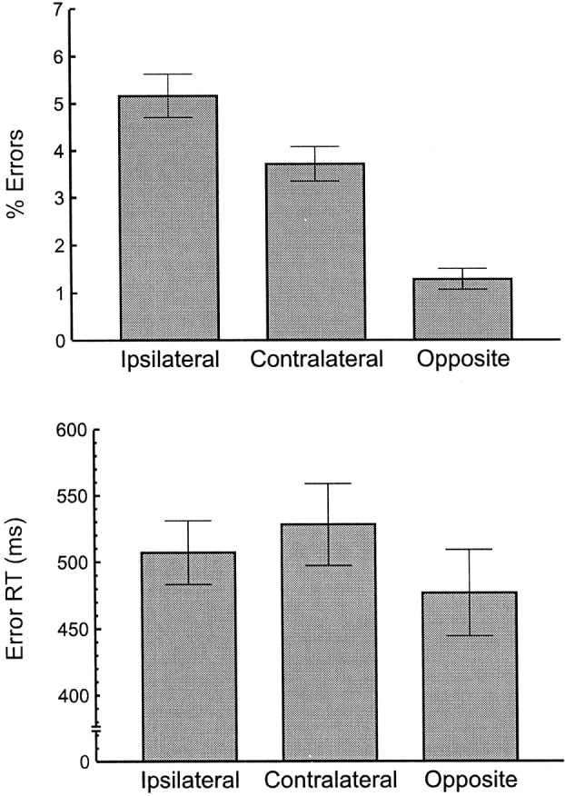
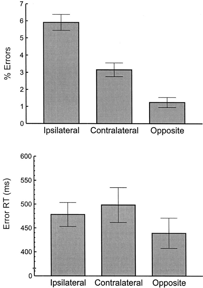
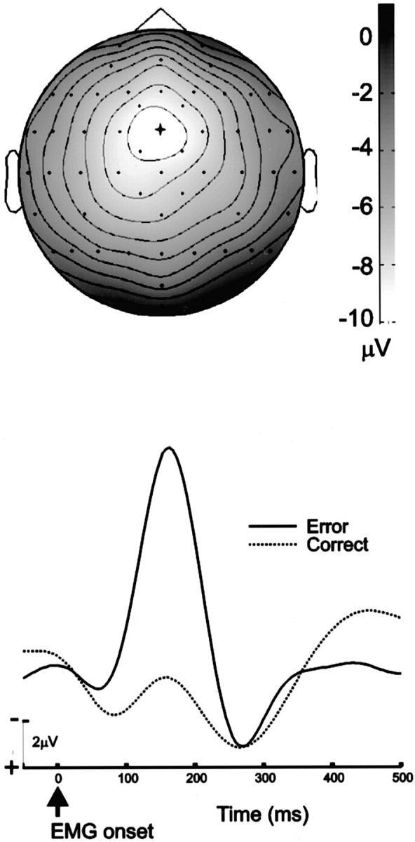
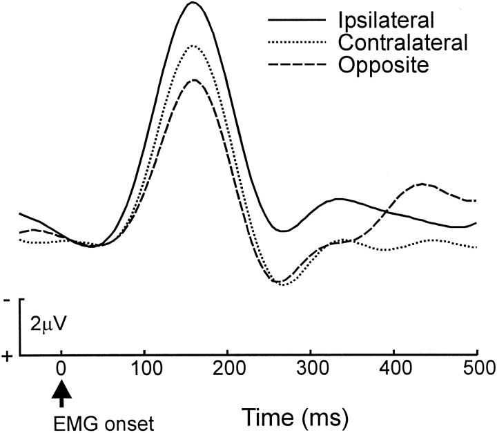
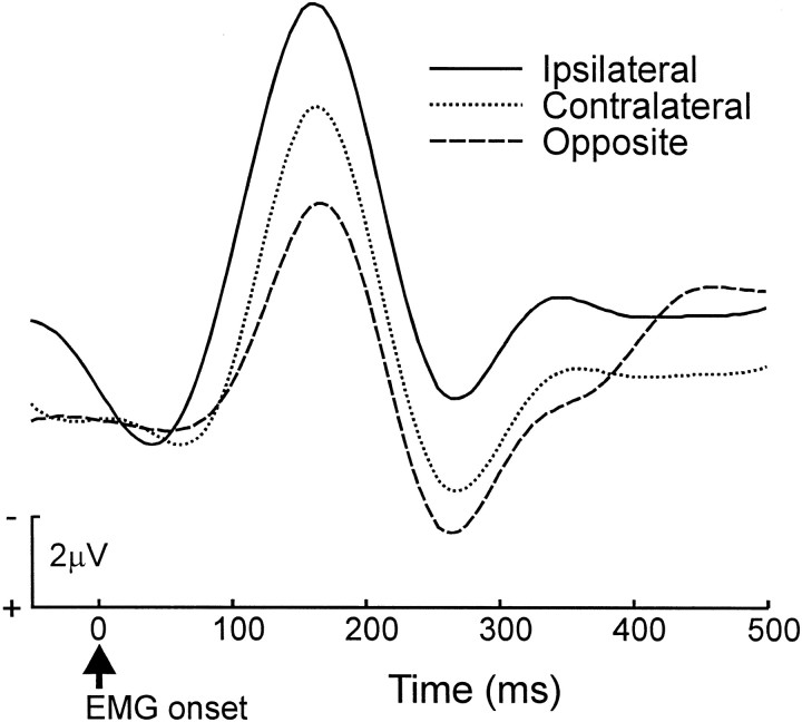
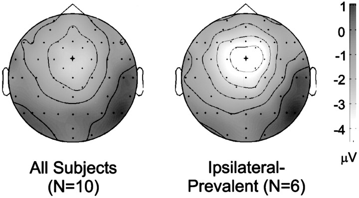
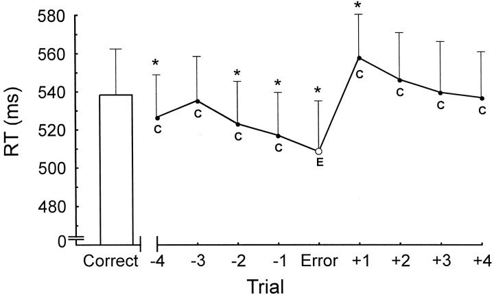
A principal function of the medial frontal cortex, in particular the anterior cingulate cortex (ACC), is to monitor action. The error-related negativity (ERN, or N(E)), an event-related brain potential, reflects medial frontal action-monitoring processes. Specifically, the error-detection theory of the ERN states that the ERN reflects ACC processing that is directly related to detecting the error. This theory predicts that ERN and ACC activity should increase directly with the dissimilarity of the error from the correct response, with similarity defined with respect to the common movement features of the responses. In contrast, the conflict-detection theory claims that ACC and ERN activity represent the detection of response conflict. This theory predicts that the activity should increase directly with the similarity of the error and the correct response. To test these theories, we investigated the effects of response similarity and conflict on the ERN, using a task that involved hand and foot movements. ERN activity was largest under conditions of high response conflict, where the error was similar to the correct response. This finding favors the conflict-detection theory over the error-detection theory, although the ERN was not associated with posterror slowing, as predicted by proponents of both theories. Discrepancies between our results and those of past studies may stem from the use in previous studies of four-finger response tasks which are subject to unique physiological and biomechanical constraints. We conclude that the ERN reflects medial frontal activity involved in the detection or affective processing of response conflict.


Figures

Fig. 1.
Fig. 1.
Response conflict was greatest for error responses that were similar to the correct response. Percentage of errors (top) and mean RT (bottom) are shown as a function of error type for all 10 participants.Ipsilateral denotes errors on the same side of the body as the correct response. Contralateral indicates an error committed on the incorrect side of the body, using the correct limb. Opposite indicates an error for which the limb and side were both incorrect. Error bars are ±1 SE.
Fig. 2.
Fig. 2.
A subgroup of six participants showing the greatest tendency to make ipsilateral errors. Percentage of errors (top) and mean RT (bottom) are shown as a function of error type for participants showing more ipsilateral errors than contralateral errors (ipsilateral-prevalent participants). Error types are described in Figure 1. Error bars are ±1 SE.
Fig. 3.
Fig. 3.
Grand average ERN scalp topography and waveform.Top, A topographic map of the ERN peak (error-correct difference) at 155–165 msec after the onset of EMG activity.Lighter colors represent regions of greater negativity;isocontour lines represent increments of 1 μV.Dots represent electrode locations. The view shows the top of the head, with the nose pointing upward. The scalp maximum of the ERN occurs at the FCz electrode, indicated by thecross. Bottom, the grand average ERN waveform at FCz. The ERN is evident as a negative-polarity peak at 165 msec after EMG onset.
Fig. 4.
Fig. 4.
The amplitude of the ERN was greatest in the high-conflict, ipsilateral error condition. Error-trial ERN waveforms for all 10 participants are plotted as a function of error type. EMG onset occurs at time 0. The waveform is from the frontocentral electrode FCz. Error types are described in Figure1.
Fig. 5.
Fig. 5.
The effects of response conflict on the ERN waveform were greatest for participants whose behavior was disrupted most by response conflict. Error-trial ERN waveforms for ipsilateral-prevalent participants on error trials are plotted as a function of error type. EMG onset occurs at time 0. Error types are described in Figure 1.
Fig. 6.
Fig. 6.
Topographic maps confirm that the event-related potential conflict effects resulted from changes in the cortical generator of the ERN. Maps represent the difference between the ipsilateral errors and opposite errors at 155–165 msec after the onset of EMG activity. Lighter colors represent regions of greater negativity; isocontour lines represent increments of 1 μV. Dots represent electrode locations. The view shows the top of the head, with the nose pointing upward. The scalp maximum of the conflict effect occurs at the FCz electrode, the site of the ERN, indicated by the cross.Left, Map from all 10 participants;right, map from the ipsilateral-prevalent group.
Fig. 7.
Fig. 7.
Reaction times on correct trials (C) before and after the error (E) show that participants increased their speed before the error and then slowed responses after the error. Error bar indicates mean correct reaction time for all trials except those that occur immediately after errors. The reaction times in theline represent four correct trials before the error (−4) to four correct trials after the error (+4). Error bars are +1 SE. Values differing from the mean correct reaction time on theleft are indicated by asterisks(t(9) = p < 0.05; one-tailed t test). From left toright, the t values are 2.25, 1.25, 3.12, 4.42, 3.02, −2.07, −0.65, 0.50, and 1.21.
Fig. 8.
Fig. 8.
The degree of ERN activity was unrelated to the amount of posterror slowing. Left, Trials that were identified as associated with slowing after the error or with no slowing, where the two sets of trials had equivalent error-trial reaction times. Those trials formed the basis of the analysis on theright, which compares the ERN that preceded posterror slowing with the ERN that was not associated with slowing.

References

    1. American Electroencephalographic Society. American Electroencephalographic Society guidelines for standard electrode position nomenclature. J Clin Neurophysiol. 1991;8:200–202. - PubMed
    1. Beck AT. An inventory for measuring depression. Arch Gen Psychiatry. 1961;4:561–571. - PubMed
    1. Bernstein PS, Scheffers MK, Coles MGH. Where did I go wrong? A psychophysiological analysis of error-detection. J Exp Psychol Hum Percept Perform. 1995;21:1312–1322. - PubMed
    1. Blythe KW. Ipsilateral confusion in 2-choice and 4-choice responses with the hands and feet. Nature. 1963;199:1312. - PubMed
    1. Botvinick MM, Braver TS, Barch DM, Carter CS, Cohen JD. Conflict monitoring and cognitive control. Psychol Rev. 2001;108:624–652. - PubMed

Publication types

MeSH terms

LinkOut - more resources