. 2001 Dec 1;21(23):9506-18.
doi: 10.1523/JNEUROSCI.21-23-09506.2001.

Distribution of CB1 cannabinoid receptors in the amygdala and their role in the control of GABAergic transmission

Affiliations

Distribution of CB1 cannabinoid receptors in the amygdala and their role in the control of GABAergic transmission

I Katona et al. J Neurosci. .

Abstract

Cannabinoids are the most popular illicit drugs used for recreational purposes worldwide. However, the neurobiological substrate of their mood-altering capacity has not been elucidated so far. Here we report that CB1 cannabinoid receptors are expressed at high levels in certain amygdala nuclei, especially in the lateral and basal nuclei, but are absent in other nuclei (e.g., in the central nucleus and in the medial nucleus). Expression of the CB1 protein was restricted to a distinct subpopulation of GABAergic interneurons corresponding to large cholecystokinin-positive cells. Detailed electron microscopic investigation revealed that CB1 receptors are located presynaptically on cholecystokinin-positive axon terminals, which establish symmetrical GABAergic synapses with their postsynaptic targets. The physiological consequence of this particular anatomical localization was investigated by whole-cell patch-clamp recordings in principal cells of the lateral and basal nuclei. CB1 receptor agonists WIN 55,212-2 and CP 55,940 reduced the amplitude of GABA(A) receptor-mediated evoked and spontaneous IPSCs, whereas the action potential-independent miniature IPSCs were not significantly affected. In contrast, CB1 receptor agonists were ineffective in changing the amplitude of IPSCs in the rat central nucleus and in the basal nucleus of CB1 knock-out mice. These results suggest that cannabinoids target specific elements in neuronal networks of given amygdala nuclei, where they presynaptically modulate GABAergic synaptic transmission. We propose that these anatomical and physiological features, characteristic of CB1 receptors in several forebrain regions, represent the neuronal substrate for endocannabinoids involved in retrograde synaptic signaling and may explain some of the emotionally relevant behavioral effects of cannabinoid exposure.


Figures

Fig. 1.
Fig. 1.
Regional localization of CB1 cannabinoid receptors in the rodent amygdala I. A, Low-power light micrograph of CB1 receptor immunostaining reveals selective distribution of CB1 receptors in certain amygdala nuclei of the rat. Whereas the basolateral complex of the amygdala (BLA) shows very strong immunoreactivity, the central nucleus (Ce) is immunonegative for CB1 receptors. The micrograph was taken at bregma −2.5. B, At high magnification, dense CB1 receptor-immunoreactive axonal meshwork is visible only in the basolateral complex but not in the central nucleus. C,Immunostaining for CB1 receptor in CB1−/− mice gives rise to no staining at all, which confirms the selectivity of the antibody for CB1 receptors. D, In contrast, CB1+/+ mice have identical CB1 receptor localization pattern to rats. CB1 receptor-immunopositive axons clearly delineate the border between the basolateral complex and the central nucleus as in rats. Note the lack of dendritic labeling of CB1-immunoreactive neurons (arrowheads).BLA, Basolateral complex of the amygdala;Ce, central nucleus of the amygdala; ic, internal capsule. Scale bars: A, 500 μm;B–D, 100 μm.
Fig. 2.
Fig. 2.
Regional localization of CB1 cannabinoid receptors in the rodent amygdala II. Four rostrocaudal levels are presented showing the characteristic distribution pattern of CB1 receptors in the rat amygdala. A, At the frontal level, the nucleus of the lateral olfactory tract (NLOT) is highly immunostained for CB1 receptors, whereas the anterior cortical nucleus (Coa) contains only a moderate density of axons. Thewhite arrow indicates a CB1-positive cell body within the NLOT. The micrograph was taken at bregma −1.3. B, Although the medial nucleus (M) shows no labeling for CB1, the bed nucleus of the accessory olfactory tract (BAOT) contains moderate number of CB1-immunopositive axons and occasionally cell bodies as well (arrow). The micrograph was taken at bregma −2.1.C, One of the most strongly labeled nucleus for CB1 receptor is the basal nucleus and its magnocellular division (Bmc). In comparison, only few axons are visible in the caudal end of the anterior cortical nucleus (Coa), and there is a lack of CB1 immunostaining in the medial (M) and in the intercalated nucleus (I). The micrograph was taken at bregma −2.9. D, More caudally, the strongest CB1 immunoreactivity is visible in the periamygdaloid cortex (PAC) and in the accessory basal nucleus magnocellular division (ABmc). The picture was taken at bregma −3.4. Scale bars: A, 200 μm; B–D, 50 μm.
Fig. 3.
Fig. 3.
CB1 receptor is expressed by a selective subpopulation of cholecystokinin-immunoreactive interneurons in the basolateral complex of the amygdala. A,Immunofluorescence staining for cholecystokinin (CCK) in the basal nucleus reveals two types of CCK-immunoreactive interneurons. The arrow depicts a so-called large CCK-positive cell, whereas the arrowheadpoints to a small CCK-immunoreactive neuron. B,Double-immunofluorescence staining demonstrates that the large CCK-positive cell expresses CB1 receptor (arrow), in contrast to the small CCK-immunoreactive neuron (arrowhead), which is negative for CB1.C–D, A parvalbumin (PV)-immunoreactive interneuron in the basal nucleus (C, arrow) is also negative for CB1 receptor (D, arrow), and CB1 receptor-immunoreactive cells do not contain parvalbumin, as indicated by arrowheads inC and D. CCK, Cholecystokinin; CB1, CB1 cannabinoid receptor;PV, parvalbumin. Scale bars, 50 μm.
Fig. 4.
Fig. 4.
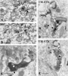
Distribution of CB1 cannabinoid receptors in the cell body is restricted to intracellular membrane compartments in the basal nucleus of the amygdala. A, Immunogold particles (small arrows), representing the localization of CB1 receptor protein, are always attached to the rough endoplasmic reticulum (RER) or to the Golgi apparatus (G) in the cell body but never to the plasmamembrane (arrowheads). B, CB1 receptors are often found on the surface of multivesicular bodies (MVB) or transport vesicles, indicating that the antibody recognizes CB1 protein that is packaged for transport to the axon terminals or is to be degraded. Scale bars: A, 1 μm; B, 0.5 μm.
Fig. 5.
Fig. 5.
Presynaptic localization of CB1 cannabinoid receptors in the amygdala. A, B, Serial sections cut from a CB1-immunoreactive axon terminal (labeled by anasterisk) forming a symmetrical synapse (thick arrow) on a cell body in the basal nucleus of the amygdala. Note that gold particle labeling is restricted to the inner surface of the bouton, where the intracellular C terminus epitope of CB1 is located. C, High-power electron micrograph from the basal nucleus depicts that a CB1-immunoreactive bouton (white asterisk) forms a symmetrical synapse with its postsynaptic target. In this experiment, immunoperoxidase procedure was used taking advantage of its higher sensitivity. The black reaction product within the axon terminal demonstrates the CB1 immunopositivity of the bouton. In contrast, the complete lack of staining in an axon terminal (b), forming asymmetrical synapse, suggests that glutamatergic axons do not contain CB1 receptors. D, E,Combined immunogold-immunoperoxidase double staining for CB1 receptor (gold particles labeled by small arrows) and CCK (the DAB end product of immunoperoxidase reaction is depicted byasterisk) confirms that the axon terminals of CCK-containing interneurons in the basal nucleus bear presynaptic CB1 receptors. Compare the CB1/CCK double-immunopositive bouton forming a symmetrical synapse (thick arrow) with the double-immunonegative axon terminals (b1, b2), which give asymmetrical synapses (arrowheads) onto the same dendritic shaft. Scale bars: A–E, 0.5 μm.
Fig. 6.
Fig. 6.
A synthetic cannabinoid agonist, WIN55,212–2, suppresses IPSCs in the basolateral complex of amygdala but not in the central nucleus. A, In rat, bath application of WIN55,212–2 (1 μm) causes a 50% reduction in the amplitude of monosynaptic IPSCs (eIPSCs) evoked in the basal nucleus (plot on the left) but not in the central nucleus of amygdala (plot on the right). Whole-cell patch-clamp recordings were obtained from spiny principal cells. The CB1 receptor antagonist SR141716A (1 μm) reverses the decrement of eIPSC amplitude. B, The amplitude of eIPSCs is suppressed by CB1 receptor activation in the lateral nucleus of wild type (CB1+/+) but not in CB1−/− knock-out mice. IPSCs were evoked by focal microstimulation delivered via a patch pipette placed into the close vicinity of the cell. All data points on the plots represent a mean ± SEM of six consecutive events. Inserts are average records of 6–10 consecutive IPSCs taken at the labeled time points. Stimulus artifacts were removed for clarity. Calibration: 10 msec, 100 pA.
Fig. 7.
Fig. 7.
Action potential-driven IPSCs, but not Ca2+-influx-independent miniature events, are sensitive for CB1 receptor activation in the basal nucleus of the amygdala. A, Spontaneous, action potential-dependent IPSCs (sIPSCs) are suppressed by bath application of the CB1 receptor agonist WIN55,212–2 (1 μm), as seen in the raw traces.B, Cumulative probability distributions of peak conductances and interevent intervals of sIPSCs are shown before (solid line;n = 231) and after (dotted line;n = 88) the application of 1 μm WIN55,212–2. The CB1 receptor agonist decreases the conductance and increases the interevent intervals (i.e., decreases the frequency) of spontaneous IPSCs (control, n = 352; WIN, n = 106). Averages of sIPSCs are shown in C. When averaged events are scaled to the same peak value (on the right), no changes in the IPSC kinetics can be observed after WIN55,212–2 application. D, In the presence of TTX (1 μm) and Cd2+ (200 μm), mIPSCs are unaltered after the bath application of 1 μmWIN55,212–2, as shown on representative records. E,Application of TTX and Cd2+ significantly reduces both the conductance and the frequency (i.e., increases interevent intervals) of spontaneous IPSCs. The addition of CB1 receptor agonist causes no further changes in miniature IPSCs, as seen on the cumulative distribution plots of the conductance (control, solid line,n = 301; TTX + Cd2+, dashed line,n = 96; WIN, dotted line,n = 76) and on the cumulative distribution plots of the interevent intervals (control, solid line,n = 502; TTX + Cd2+,dashed line,n = 105; WIN,dotted line,n = 89).F, Averaged IPSCs for spontaneous IPSCs and miniature IPSCs are superimposed. The peak scaled mIPSCs (on theright) show no alterations in the kinetic parameters. Calibration: A, D, 100 msec, 100 pA;C, F, 5 msec, 10 pA.

References

    1. Abood ME, Martin BR. Neurobiology of marijuana abuse. Trends Pharmacol Sci. 1992;13:201–206. - PubMed
    1. Acsády L, Katona I, Martinez-Guijarro FJ, Buzsáki G, Freund TF. Unusual target selectivity of perisomatic inhibitory cells in the hilar region of the rat hippocampus. J Neurosci. 2000;20:6907–6919. - PMC - PubMed
    1. Cador M, Robbins TW, Everitt BJ. Involvement of the amygdala in stimulus-reward associations: interaction with the ventral striatum. Neuroscience. 1989;30:77–86. - PubMed
    1. Chan PK, Chan SC, Yung WH. Presynaptic inhibition of GABAergic inputs to rat substantia nigra pars reticulata neurones by a cannabinoid agonist. NeuroReport. 1998;9:671–675. - PubMed
    1. Chen JP, Paredes W, Li J, Smith D, Lowinson J, Gardner EL. Delta 9-tetrahydrocannabinol produces naloxone-blockable enhancement of presynaptic basal dopamine efflux in nucleus accumbens of conscious, freely-moving rats as measured by intracerebral microdialysis. Psychopharmacology (Berl) 1990;102:156–162. - PubMed

Publication types

MeSH terms

Substances

LinkOut - more resources