. 2001 Dec 4;98(25):14268-73.
doi: 10.1073/pnas.241429098. Epub 2001 Nov 20.

Structure of a quinohemoprotein amine dehydrogenase with an uncommon redox cofactor and highly unusual crosslinking

Affiliations

Structure of a quinohemoprotein amine dehydrogenase with an uncommon redox cofactor and highly unusual crosslinking

S Datta et al. Proc Natl Acad Sci U S A. .

Abstract

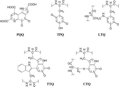
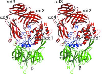
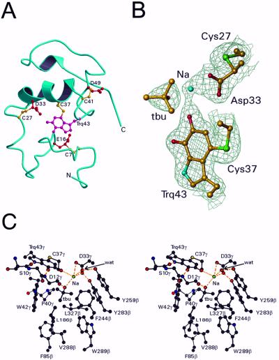
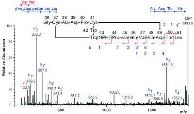
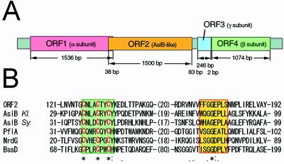
The crystal structure of the heterotrimeric quinohemoprotein amine dehydrogenase from Paracoccus denitrificans has been determined at 2.05-A resolution. Within an 82-residue subunit is contained an unusual redox cofactor, cysteine tryptophylquinone (CTQ), consisting of an orthoquinone-modified tryptophan side chain covalently linked to a nearby cysteine side chain. The subunit is surrounded on three sides by a 489-residue, four-domain subunit that includes a diheme cytochrome c. Both subunits sit on the surface of a third subunit, a 337-residue seven-bladed beta-propeller that forms part of the enzyme active site. The small catalytic subunit is internally crosslinked by three highly unusual covalent cysteine to aspartic or glutamic acid thioether linkages in addition to the cofactor crossbridge. The catalytic function of the enzyme as well as the biosynthesis of the unusual catalytic subunit is discussed.


Figures

Figure 1
Figure 1
Chemical structures of the quinone cofactors PQQ, TPQ, lysine tyrosylquinone (LTQ), and TTQ. The structure of the newly identified cofactor, cysteine tryptophylquinone (CTQ), is also shown.
Figure 2
Figure 2
Stereo ribbon diagram of the full QHNDH heterotrimer. The subunits α, β, and γ and their N and C termini are shown in red, green, and blue, respectively. The four domains of the α subunit (αd1, αd2, αd3, and αd4) and the β and γ subunits are labeled in black. The two heme groups in subunit α and the CTQ cofactor in the γ subunit are also shown. This and subsequent diagrams, except where stated, were prepared by using MOLSCRIPT (41) and rendered by using RASTER3D (42).
Figure 3
Figure 3
Structure of the γ subunit and active site of QHNDH. (A) The backbone ribbon is blue, the crosslinked side chains of Cys are yellow, and the crosslinked side chains of tryptophylquinone (Trq), Glu, and Asp are red. (B) Electron density around CTQ. Also shown are the putative active site base Asp-33γ, its covalently linked Cys-27γ, the molecule of t-butyl alcohol (tbu) and the sodium ion (Na). Only Cα and side chain atoms are included. The map was computed with coefficients (2FoFc) exp(−iα) where Fc and α, the calculated structure factors and phase angles, were derived from the final refined model. The contours are drawn at 1.2 σ, where σ is the rms value of the electron density. This diagram was prepared by using TURBO-FRODO (18). (C) Stereoview of the active site of QHNDH. See text for details.
Figure 4
Figure 4
ESI-MS/MS spectrum of the NPH-quinone peptide derived from the γ subunit. Fragmentation occurred for the most part along the backbone of a C-terminal portion of the peptide. Amino acids shown in three-letter code denote immonium ions assigned by SEQMS (24). Arrows below the amino acids show the sequences from the N and C termini based on y"m and bl ions, respectively, where m and l denote positions counted from the C and N termini that were produced by cleavage of peptide bonds during MS/MS. The predicted structure of the peptide is shown in the middle with possible cleavage sites.
Figure 5
Figure 5
Gene structure of QHNDH and local homology alignment of ORF2 protein with Radical S-adenosylmethionine (SAM) proteins. (A) Tandem arrangement of four ORFs coding for subunit α, the hypothetical [Fe—S] protein, subunit γ, and subunit β, respectively (from left to right). Nucleotide numbers of each ORF and of intervening noncoding regions are also indicated. (B) Multiple alignment of an N-terminal region of ORF2 protein of P. denitrificans with Radical SAM Proteins registered in GenBank [AslB of Klebsiella (Kl.), gi/114710; AslB of Synechocystis sp. (Sy.), gi/1652765; PflA (pyruvate formate-lyase activating enzyme), gi/6093720; NrdG (anaerobic ribonucleotide reductase activating protein), gi/730195; BssD (benzylsuccinate synthase activating enzyme), gi/3184129]. Aligned residue numbers are shown on both sides of each sequence, and invariant (*), highly (:), and weakly (.) conserved residues are indicated in the bottom line. Cys residues in the [Fe—S]-binding motifs (green box) are shown in red letters, and the putative SAM-binding Gly-rich motif is boxed in yellow.

Comment in

References

    1. Anthony C. Biochem J. 1996;320:697–711. - PMC - PubMed
    1. Janes S M, Mu D, Wemmer D, Smith A J, Kaur S, Maltby D, Burlingame A L, Klinman J P. Science. 1990;248:981–987. - PubMed
    1. Wang S X, Mure M, Medzihradszky K F, Burlingame A L, Brown D E, Dooley D M, Smith A J, Kagan H M, Klinman J P. Science. 1996;273:1078–1084. - PubMed
    1. McIntire W S, Wemmer D E, Chistoserdov A, Lidstrom M E. Science. 1991;252:817–824. - PubMed
    1. Klinman J P, Mu D. Annu Rev Biochem. 1994;63:299–344. - PubMed

Publication types

MeSH terms

Substances

Associated data

LinkOut - more resources