. 2001 Dec 4;98(25):14404-9.
doi: 10.1073/pnas.251548398. Epub 2001 Nov 20.

Peripheral myelin protein 22 is a constituent of intercellular junctions in epithelia

Affiliations

Peripheral myelin protein 22 is a constituent of intercellular junctions in epithelia

L Notterpek et al. Proc Natl Acad Sci U S A. .

Abstract

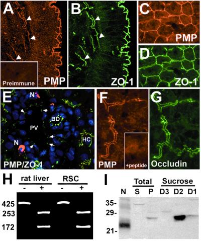
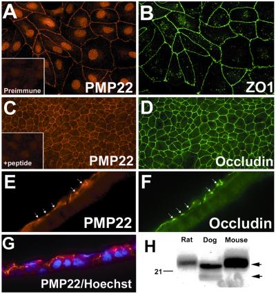
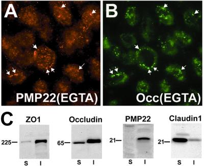
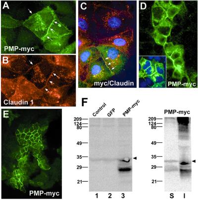
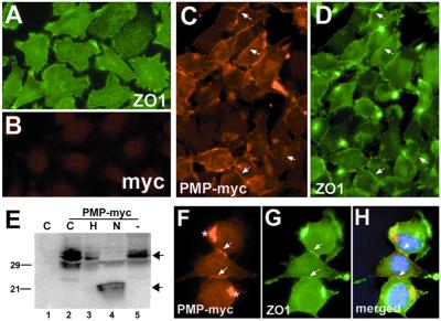
Alterations in peripheral myelin protein 22 (PMP22) gene expression are associated with a host of heritable demyelinating peripheral neuropathies, yet the function of the protein remains unknown. PMP22 expression is highest in myelinating Schwann cells of peripheral nerves; however, significant levels of PMP22 mRNAs can be detected in a variety of non-neural tissue, including epithelia. To date, PMP22 protein expression and localization in non-neural tissues have not been studied in detail. In adult rat liver and intestine, and cultured epithelial cells, we detected PMP22-like immunoreactivity associated with markers of the tight junctional complex, including zonula occludens 1 (ZO-1) and occludin. Upon disruption of intercellular contacts, PMP22 was internalized into vesicles that were immunoreactive for both anti-occludin and anti-PMP22 antibodies. Nonionic detergent extraction of cultured epithelial cells did not solubilize PMP22, as the majority of the protein remained in the detergent insoluble fraction, as did ZO-1 and occludin. We also observed the targeting of exogenous myc-tagged PMP22 to apical cell junctions in polarized epithelia and to anti-ZO-1 antibody immunoreactive cell contacts of L fibroblasts. These studies support a role for PMP22 at intercellular junctions of epithelia and may indicate a similar function in myelinating Schwann cells. Furthermore, our findings could provide an explanation for certain phenotypes of PMP22 neuropathy mice that cannot be accounted for by dysmyelination.


Figures

Figure 1
Figure 1
PMP22 is coexpressed with ZO-1 and occludin in colon epithelium and bile canaliculi. Frozen sections of normal adult rat colon (A and B) and liver (CG) were coimmunostained with polyclonal anti-PMP22 (A, C, E, and F) and monoclonal anti-ZO-1 (B, D, and E) or occludin (G) antibodies. (A and B) Confocal images showing the presence of PMP22 at the surface epithelium of the mucosa and in submucosal vasculature (arrowheads). (E) A high-resolution thin section of adult rat liver stained with anti-PMP22 (red), anti-ZO-1 (green) antibodies and nuclear dye (blue). PV, portal vein; N, nerve terminal; BD, bile duct; HC, hepatocyte. Liver sections incubated with preimmune serum (A Inset) or peptide preadsorbed antiserum (F Inset) do not result in TJ-like immunostaining. [Magnifications: ×40 (AE) and ×60 (F and G).] (H) The expression of PMP22 mRNA in liver was verified by RT-PCR. BsaI undigested (−) and digested (+) PCR-amplified fragments are shown for each sample. The numbers on the left indicate bp. (I) Membrane pellets (P) (75 μg) from adult rat liver homogenates were fractionated and proteins isolated at sucrose densities 1.22 (D3) and 1.18 (D2) and 1.16 (D1) were analyzed (75 μg/lane) for the presence of PMP22. Rat sciatic nerve (N) lysate (4 μg) was used as a positive control for the anti-PMP22 antibody immunoreactivity. S, total liver supernatant (75 μg). Molecular mass, in kDa.
Figure 2
Figure 2
PMP22 is coexpressed with ZO-1 and occludin at cell–cell contacts in MDCK cells. Subconfluent (A and B) and filter-grown (CG) MDCK cells were immunostained with polyclonal anti-PMP22 (A, C, E, and G) and monoclonal anti-ZO-1 (B), or anti-occludin (D and F) antibodies. (A Inset) Cells stained with preimmune rabbit serum. In filter-grown MDCK cultures PMP22 (C) is codistributed with occludin (D) at apical cell contacts. PMP22 antigenic peptide preadsorbed antiserum does not stain intercellular contacts of MDCK cells (C Inset). On sectioned (8 μm) filters (Z plane) PMP22-like immunoreactivity (E and G) is associated with the apical border of the monolayer, which is also reactive with the anti-occludin (F) antibodies (arrows in E and F). (G) Anti-PMP22 (red) and Hoechst nuclear dye (blue)-stained MDCK cell monolayer is shown. [Magnifications: ×60 (A, B, and EG) and ×40 (C and D)]. (H) Protein blots of (18 μg/lane) normal adult rat, canine, and mouse sciatic nerves were reacted with anti-PMP22 antiserum. The upper arrow on the right indicates the glycosylated 22-kDa PMP22, while the lower arrow points to the newly synthesized 18-kDa, endoglycosidase H-sensitive protein. Molecular mass, in kDa.
Figure 3
Figure 3
PMP22 is internalized with occludin in EGTA-treated MDCK cells. Confluent MDCK monolayers were cultured in the presence of 4 mM EGTA for 1 h followed by immunostaining with polyclonal anti-PMP22 (A) and monoclonal anti-occludin (B) antibodies. Arrows point to vesicles that contain both PMP22 (A) and occludin (B). (Magnification: ×60.) (C) Confluent MDCK cell monolayers were extracted with 0.5% TX-100 containing buffer and detergent soluble (S), and detergent-insoluble (I) fractions were immunoblotted with antibodies against ZO-1, occludin, PMP22, and claudin-1. Molecular mass, in kDa.
Figure 4
Figure 4
The exogenously expressed PMP22-myc is targeted to TJs in MDCK cells. pBMN-PMP22-myc-infected cells were cultured on coverslips (AD) or Transwell filters (E) and immunostained with monoclonal anti-myc (A and CE), and polyclonal anti-claudin-1 (B and C) antibodies. Note, only ≈15% of the cells (green cells) were infected with the PMP22-myc construct (A and CE). PMP22-myc is targeted to anti-claudin-1 immunoreactive intercellular contacts (arrows in AC) and PMP22-myc-expressing cells form contacts with noninfected cells (D). Nuclei were stained with Hoechst dye (C and D Inset). [Magnifications: ×60 (AC), and ×40 (D and E).] (F) The expression of PMP22-myc was verified by anti-myc Western analysis. Lysates of PMP22-myc-infected cells (lane 3) show expression of a ≈27-kDa and a ≈33-kDa PMP22-myc protein. A nonspecific ≈34-kDa band is present in all samples, including uninfected control (lane 1) and pBMN-GFP (lane 2)-infected cell lysates (arrowheads). The majority of PMP22-myc protein is insoluble in TX-100 (I). S, TX-100 soluble. Molecular mass, in kDa.
Figure 5
Figure 5
PMP22-myc is colocalized with ZO-1 at intercellular junctions of L fibroblasts. Uninfected control L cells (A and B) show diffuse ZO-1-like membrane staining with focal concentration at cell processes (A) and low levels of nonspecific immunoreactivity with polyclonal anti-myc (B) antibodies. In PMP22-myc-infected cells, PMP22-myc is detected at cell–cell contacts (arrows in C and F), which are costained with the anti-ZO-1 antibody (arrows in D and G). PMP22-myc is detected in aggresome-like structures in some cells (* in F). Nuclei are stained with Hoechst dye (H). (Magnification: ×60.) (E) The expression and processing of PMP22-myc was studied in lysates of control, uninfected (lane 1), and PMP22-myc-infected L cells (lanes 2–5) by anti-myc Western blotting. Untreated (lane 2), endoglycosidase H (lane 3), N-glycosidase (lane 4), and no enzyme (lane 5) PMP22-myc samples are shown. N-glycosidase (N) treatment of PMP22-myc cell lysates results in a characteristic shift of PMP22, from a mature high molecular mass, endoglycosidase H-resistant form (upper arrow) to a deglycosylated, lower molecular mass core protein (lower arrow). The N-glycosidase resistant, anti-myc immunoreactive ≈29-kDa band likely represents a mono-ubiquitinylated PMP22-myc (lane 4). Molecular mass, in kDa.

References

    1. Suter U, Snipes G J. Annu Rev Neurosci. 1995;18:45–75. - PubMed
    1. Taylor V, Welcher A, Suter U. J Biol Chem. 1995;270:28824–28883. - PubMed
    1. Baechner D, Liehr T, Hameister H, Altenberger H, Grehl H. J Neurosci Res. 1995;42:733–741. - PubMed
    1. Wulf P, Bernhardt R, Suter U. J Neurosci Res. 1999;57:467–478. - PubMed
    1. Mugnaini E, Schnapp B. Nature (London) 1974;251:725–727. - PubMed

Publication types

MeSH terms

LinkOut - more resources