. 2001 Nov 20;98(24):13699-704.
doi: 10.1073/pnas.221297398.

Mammalian myotube dedifferentiation induced by newt regeneration extract

Affiliations

Mammalian myotube dedifferentiation induced by newt regeneration extract

C J McGann et al. Proc Natl Acad Sci U S A. .

Abstract

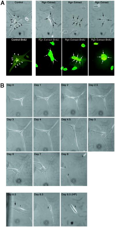
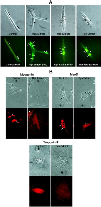
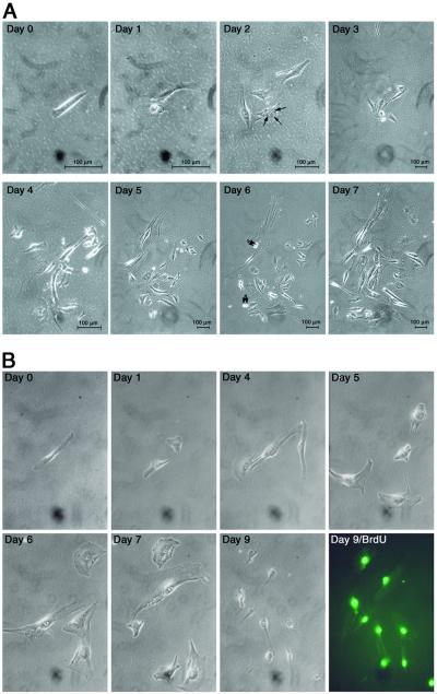
Newts are capable of regenerating several anatomical structures and organs, including their limbs. This remarkable regenerative capacity is thought to depend on cellular dedifferentiation. Terminally differentiated mammalian cells, by contrast, are normally incapable of reversing the differentiation process. Several factors could explain the absence of cellular dedifferentiation in mammals: (i) inadequate expression of genes that initiate dedifferentiation; (ii) insufficient intracellular signaling pathways; (iii) irreversible expression of differentiation factors; and (iv) structural characteristics that make dedifferentiation impossible. To investigate the causes underlying the lack of cellular plasticity in mammalian cells, we examined the effect of an extract derived from newt regenerating limbs on terminally differentiated mouse C2C12 myotubes. Approximately 18% of murine myotubes reentered the cell cycle when treated with regeneration extract, whereas 25% of newt myotubes exhibited cell cycle reentry. The muscle differentiation proteins MyoD, myogenin, and troponin T were reduced to undetectable levels in 15-30% of treated murine myotubes. We observed cellular cleavage in 11% of the treated murine myotubes and approximately 50% of these myotubes continued to cleave to produce proliferating mononucleated cells. These data indicate that mammalian myotubes can dedifferentiate when stimulated with the appropriate factors and suggest that one mechanism preventing dedifferentiation of mammalian cells is inadequate spatial or temporal expression of genes that initiate dedifferentiation.


Figures

Figure 1
Figure 1
Limb regeneration extract induces newt myotube dedifferentiation. (A) Newt myotubes reentered the cell cycle when stimulated with limb regeneration extract. Top row shows phase contrast photomicrographs of newt myotubes. Bottom row shows fluorescent photomicrographs of the corresponding newt myotubes. Left panel shows a control myotube treated with nonregeneration extract. No nuclei reentered the cell cycle as evidenced by the lack of BrdUrd incorporation (arrowheads point to nuclei). The three panels on the right show myotubes that were treated with limb regeneration extract. All nuclei reentered the cell cycle as evidenced by BrdUrd incorporation (arrows point to representative nuclei that have reentered the cell cycle). (B) Limb regeneration extract induced newt myotube cleavage. Newt A1 myotubes were plated at very low density (≈2 cells per mm2) in DM on day 0 and treated daily with DM containing regeneration extract for 8.5 days. Multinucleated myotube is shown before treatment with limb regeneration extract (day 0). On day 1, the myotube began to exhibit altered morphology by elongating. This elongation process continued through day 4.5. By day 5, elongating myotube began to pull apart. Many centrally located nuclei are now readily visible. The cleavage process continued on days 6 and 7. By day 8, the myotube completely cleaved at the center to produce three smaller cells numbered 1, 2, and 3. The three products of the myotube cleavage are shown in separate photomicrographs at day 8.5. Cleavage product 3 is shown at high power (32× objective lens). All other photomicrographs in this figure are shown at low power (10× objective lens). Arrow points to a small cellular fragment protruding from the largest cleavage product, which suggests that additional cleavage is occurring (1). Control newt myotubes treated with nonregeneration limb extract or kept in DM alone showed no evidence of cleavage.
Figure 2
Figure 2
Regeneration extract induces cell cycle reentry and reduction in myogenic protein levels in mouse myotubes. Mouse C2C12 myotubes were treated with regeneration or nonregeneration extract. (A) Myotubes were treated with BrdUrd for 12 h and then BrdUrd incorporation assays were performed. (Upper) Phase contrast photomicrographs; (Lower) corresponding fluorescent photomicrographs. Control-BrdU panel shows a myotube treated with nonregenerating limb extract. No nuclei reentered the cell cycle in this control myotube. Rgn Extract-BrdU panels show three mouse myotubes containing nuclei that reentered the cell cycle following treatment with regeneration extract. Arrowheads point to nuclei that did not incorporate BrdUrd. Arrows point to nuclei that incorporated BrdUrd in myotubes treated with regeneration extract. (B) Regeneration extract reduced myogenic protein levels in murine myotubes. (Upper) Phase contrast photomicrographs; (Lower) corresponding fluorescent photomicrographs. C2C12 myotubes cultured in DM containing regeneration extract (Rgn Extract) or nonregeneration limb extract (Control) are shown. The muscle-specific transcription factors myogenin and MyoD were reduced in myotubes treated with regeneration extract (arrowheads point to representative unstained nuclei), but present at normal levels in the controls (arrows point to representative fluorescent red nuclei). The muscle contractile protein troponin T was reduced in a treated myotube (unstained cytoplasm), but present in the control (fluorescent red cytoplasm).
Figure 3
Figure 3
Regeneration extract induces mouse myotube cleavage and cellular proliferation. (A) An isolated C2C12 myotube was treated daily for 7 days with DM containing regeneration extract. Day 0 shows the myotube cultured in DM before the addition of regeneration extract. Days 1–7 show the morphologic changes that occur as treatment continues for 7 days. On day 1, the myotube showed initial signs of fragmentation and cleavage. By day 2, the myotube cleaved to form four smaller fragments. Arrows point to three nuclei contained within a smaller myotube that resulted from the cleavage event. On days 3–5 the myotube continued to cleave to form numerous smaller cells. On day 6, there was evidence of cellular proliferation. Arrows point to cells progressing through cell division. On day 7, the cells continued to cleave and proliferate. (Scale bars, 100 μm.) (B) An example of a second isolated C2C12 myotube that was treated with newt regeneration extract for 9 days. Day 0, the myotube before treatment with the extract. Within 1 day of treatment, the myotube cleaved to form two smaller cellular products. Cleavage continued through day 9. On day 8 the cleaved cells were treated with BrdUrd, and on day 9 the mononucleated cells were assayed for BrdUrd incorporation (Day 9/BrdU).

References

    1. Morgan T H. Regeneration. New York: Columbia University; 1901.
    1. Becker R O, Chapin S, Sherry R. Nature (London) 1974;248:145–147. - PubMed
    1. Davis B M, Ayers J L, Koran L, Carlson J, Anderson M C, Simpson S B., Jr Exp Neurol. 1990;108:198–213. - PubMed
    1. Brockes J P. Science. 1997;276:81–87. - PubMed
    1. Zottoli S J, Bentley A P, Feiner D G, Hering J R, Prendergast B J, Rieff H I. Prog Brain Res. 1994;103:219–228. - PubMed

Publication types