. 2001 Nov 20;98(24):13734-8.
doi: 10.1073/pnas.241522698.

Conservation of methylation reprogramming in mammalian development: aberrant reprogramming in cloned embryos

Affiliations

Conservation of methylation reprogramming in mammalian development: aberrant reprogramming in cloned embryos

W Dean et al. Proc Natl Acad Sci U S A. .

Abstract

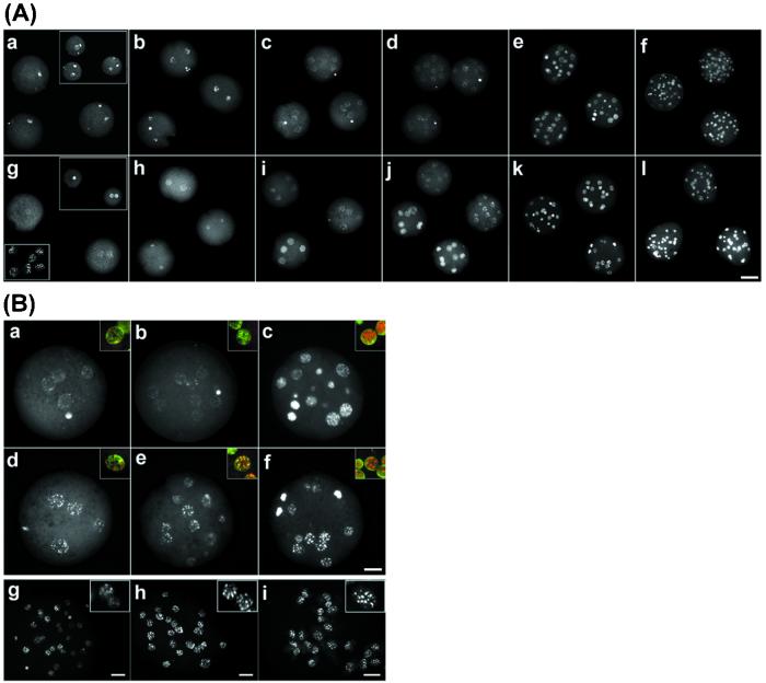
Mouse embryos undergo genome-wide methylation reprogramming by demethylation in early preimplantation development, followed by remethylation thereafter. Here we show that genome-wide reprogramming is conserved in several mammalian species and ask whether it also occurs in embryos cloned with the use of highly methylated somatic donor nuclei. Normal bovine, rat, and pig zygotes showed a demethylated paternal genome, suggesting active demethylation. In bovine embryos methylation was further reduced during cleavage up to the eight-cell stage, and this reduction in methylation was followed by de novo methylation by the 16-cell stage. In cloned one-cell embryos there was a reduction in methylation consistent with active demethylation, but no further demethylation occurred subsequently. Instead, de novo methylation and nuclear reorganization of methylation patterns resembling those of differentiated cells occurred precociously in many cloned embryos. Cloned, but not normal, morulae had highly methylated nuclei in all blastomeres that resembled those of the fibroblast donor cells. Our study shows that epigenetic reprogramming occurs aberrantly in most cloned embryos; incomplete reprogramming may contribute to the low efficiency of cloning.


Figures

Figure 1
Figure 1
Selective demethylation of the male pronucleus is conserved. Indirect immunofluorescence with the use of an antibody to 5-methyl cytosine (in red) shows that the male pronucleus is selectively demethylated immediately after fertilization, whereas the female pronucleus remains methylated, in mouse (a and b), rat (c and d), pig (e and f), and cow (g and h). DNA staining is in blue. The larger of the two pronuclei is the male, except in cows, where they are of the same size. In all cases embryos were collected and fixed before DNA replication. (Scale bar, 20 μm.)
Figure 2
Figure 2
Demethylation and remethylation are conserved during preimplantation development. (A) Normal mouse (af) and bovine (gl) embryos were stained for 5-methyl cytosine (red) from the zygote to the blastocyst stage. In the mouse (af), there is an initial loss of methylation specifically from the male pronucleus [(Inset) DNA stained to identify two pronuclei, green]. Thereafter the remaining decline in signal occurs in a stepwise fashion up to the morula stage (e). The ICM, but not the trophectoderm, has undergone de novo methylation by the blastocyst stage (f). Bovine zygotes also show loss of methylation from one pronucleus (g) followed by a further stepwise decline in methylation to the eight-cell stage (hj). De novo methylation by the 16-cell stage results in heterogeneity with highly and moderately methylated nuclei (k) such that at the blastocyst stage (l) the ICM contains highly methylated nuclei and the trophectoderm moderately methylated ones. (B) To better define the location of the methylated nuclei images are presented with the methylation signal (red) and the merged image of the DNA (blue) superimposed on the methylation signal (pink). This superimposition of images clearly shows that in the mouse the ICM has become remethylated, but in bovine nuclei both ICM and trophectoderm are methylated.
Figure 3
Figure 3
Aberrant methylation patterns in cloned bovine preimplantation embryos. (AaAf) Anti-5-methyl cytosine immunofluorescence of normal bovine embryos. (a) Zygotes 12 h after fertilization [(Inset) staining of the DNA; n = 25, number of embryos analyzed]. (b) Two-cell embryos stain intensely (n > 10). (c and d) Four-cell embryos (n > 15; c) and eight-cell embryos (d) show reduced staining (n >10). (e) Ten- to sixteen-cell embryos undergo a dramatic increase in methylation (n >10). ( f) Morulae (>24 cells) maintain the high-intensity signal (n > 10). (gl) Cloned embryos. (g) Cloned one-cell embryos stain faintly (n > 10). (Inset) DNA staining. (Lower Inset) Fibroblast donor nuclei. (h) Two-cell cloned embryos (n = 15). (i and j) In four- (i) and eight-cell (j) clones there are two populations of embryos with high and low staining, respectively (n = 7/14), in each group. (k) Ten to sixteen cells (n = 20). (l) Morula stage (n > 10). (Scale bar, 50 μm.) (B) Organization of methylation patterns in normal and cloned bovine embryos. (ac) Normal bovine embryos at four- to 16-cell stage. (df) Cloned bovine embryos at the 4- to 16-cell stage. Note the heterogeneous de novo methylation and fine granular staining in normal 16-cell embryos (c) and the precocious de novo methylation and organization into fewer and more intense foci in cloned 4- to 16-cell embryos (df). (Insets) Merged images of methylation staining (red) and DNA staining (green), with yellow showing overlapping patterns. (gi) Confocal projection of normal and cloned morula, and fibroblast donors. (g) Normal morula stains heterogeneously. (Inset) Nuclei indicate the two patterns of staining. (h) Cloned morula stains homogeneously. (i) Methylation organization in fetal fibroblast donor cells. (Scale bar, 20 μm.) All insets are enlarged ×3.

References

    1. Bird A P, Wolffe A P. Cell. 1999;99:451–454. - PubMed
    1. Bestor T H. Hum Mol Genet. 2000;9:2395–2402. - PubMed
    1. Stancheva I, Meehan R R. Genes Dev. 2000;14:313–327. - PMC - PubMed
    1. Li E, Bestor T H, Jaenisch R. Cell. 1992;69:915–926. - PubMed
    1. Okano M, Bell D W, Haber D A, Li E. Cell. 1999;99:247–257. - PubMed

Publication types