

High-level HIV-1 viremia suppresses viral antigen-specific CD4(+) T cell proliferation
- PMID: 11717444
- PMCID: PMC61135
- DOI: 10.1073/pnas.251539598
High-level HIV-1 viremia suppresses viral antigen-specific CD4(+) T cell proliferation
Abstract
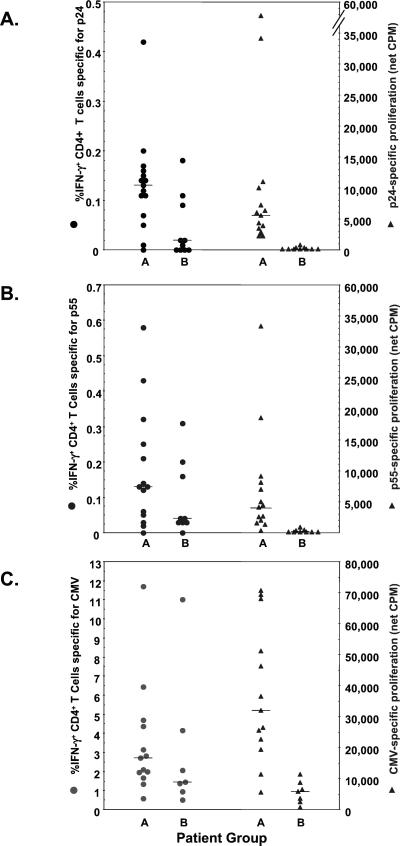
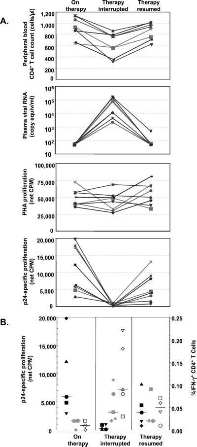
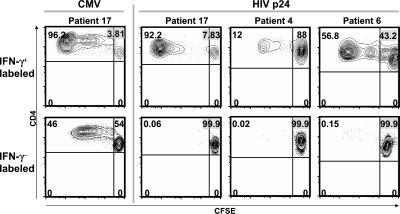
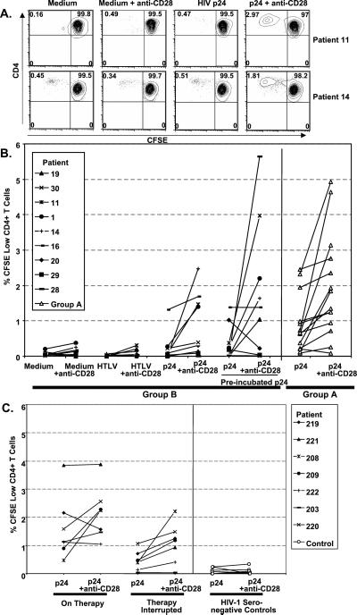
In chronic viral infections of humans and experimental animals, virus-specific CD4(+) T cell function is believed to be critical for induction and maintenance of host immunity that mediates effective restriction of viral replication. Because in vitro proliferation of HIV-specific memory CD4(+) T cells is only rarely demonstrable in HIV-infected individuals, it is presumed that HIV-specific CD4(+) T cells are killed upon encountering the virus, and maintenance of CD4(+) T cell responses in some patients causes the restriction of virus replication. In this study, proliferative responses were absent in patients with poorly restricted virus replication although HIV-specific CD4(+) T cells capable of producing IFN-gamma were detected. In a separate cohort, interruption of antiretroviral therapy resulted in the rapid and complete abrogation of virus-specific proliferation although HIV-1-specific CD4(+) T cells were present. HIV-specific proliferation returned when therapy was resumed and virus replication was controlled. Further, HIV-specific CD4(+) T cells of viremic patients could be induced to proliferate in response to HIV antigens when costimulation was provided by anti-CD28 antibody in vitro. Thus, HIV-1-specific CD4(+) T cells persist but remain poorly responsive (produce IFN-gamma but do not proliferate) in viremic patients. Unrestricted virus replication causes diminished proliferation of virus-specific CD4(+) T cells. Suppression of proliferation of HIV-specific CD4(+) T cells in the context of high levels of antigen may be a mechanism by which HIV or other persistently replicating viruses limit the precursor frequency of virus-specific CD4(+) T cells and disrupt the development of effective virus-specific immune responses.
Figures
References
-
- Leist T P, Cobbold S P, Waldmann H, Aguet M, Zinkernagel R M. J Immunol. 1987;138:2278–2281. - PubMed
-
- Rosenberg E S, Billingsley J M, Caliendo A M, Boswell S L, Sax P E, Kalams S A, Walker B D. Science. 1997;278:1447–1450. - PubMed
-
- Schwartz D, Sharma U, Busch M, Weinhold K, Matthews T, Lieberman J, Birx D, Farzedagen H, Margolick J, Quinn T, et al. AIDS Res Hum Retroviruses. 1994;10:1703–1711. - PubMed
Publication types
MeSH terms
Substances
LinkOut - more resources
Full Text Sources
Medical
Research Materials
