. 2001 Nov 20;98(24):13895-900.
doi: 10.1073/pnas.241516998.

Cubilin dysfunction causes abnormal metabolism of the steroid hormone 25(OH) vitamin D(3)

Affiliations

Cubilin dysfunction causes abnormal metabolism of the steroid hormone 25(OH) vitamin D(3)

A Nykjaer et al. Proc Natl Acad Sci U S A. .

Abstract

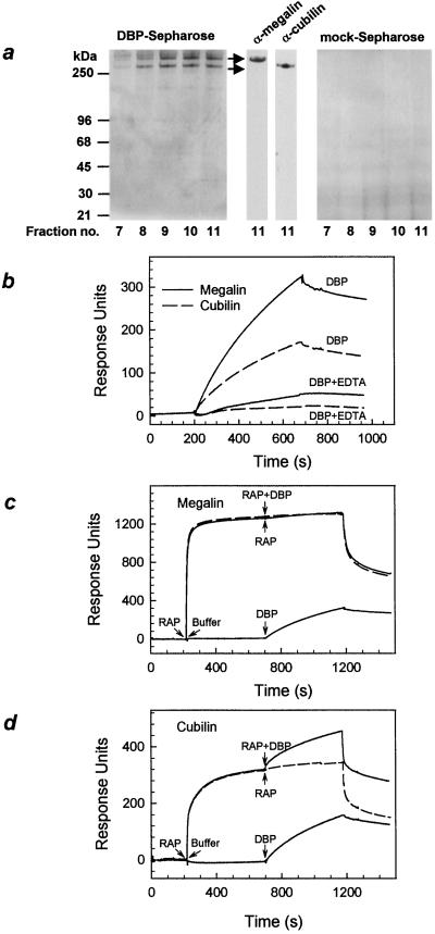
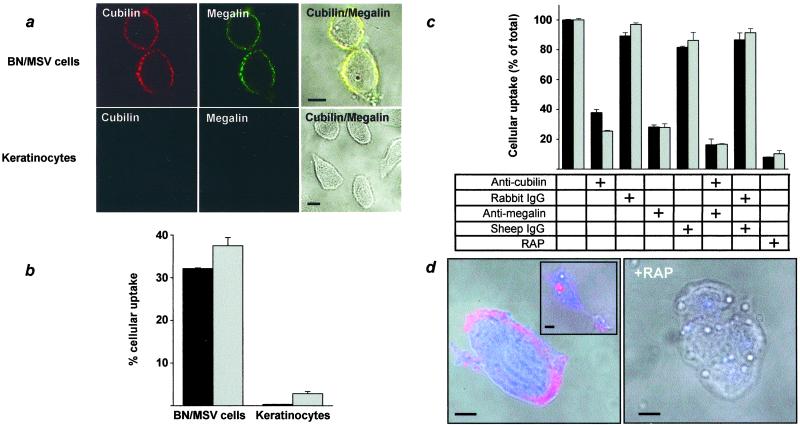
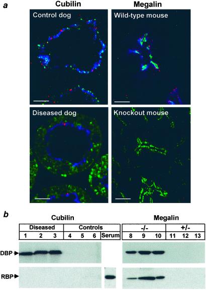
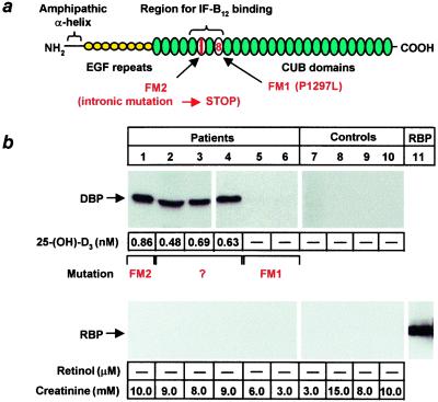
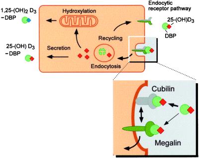
Steroid hormones are central regulators of a variety of biological processes. According to the free hormone hypothesis, steroids enter target cells by passive diffusion. However, recently we demonstrated that 25(OH) vitamin D(3) complexed to its plasma carrier, the vitamin D-binding protein, enters renal proximal tubules by receptor-mediated endocytosis. Knockout mice lacking the endocytic receptor megalin lose 25(OH) vitamin D(3) in the urine and develop bone disease. Here, we report that cubilin, a membrane-associated protein colocalizing with megalin, facilitates the endocytic process by sequestering steroid-carrier complexes on the cellular surface before megalin-mediated internalization of the cubilin-bound ligand. Dogs with an inherited disorder affecting cubilin biosynthesis exhibit abnormal vitamin D metabolism. Similarly, human patients with mutations causing cubilin dysfunction exhibit urinary excretion of 25(OH) vitamin D(3). This observation identifies spontaneous mutations in an endocytic receptor pathway affecting cellular uptake and metabolism of a steroid hormone.


Figures

Figure 1
Figure 1
Characterization of DBP binding to cubilin and megalin. (a) DBP affinity chromatography of rabbit kidney membranes. Fractions eluted at pH 4.0 in the presence of 5 mM EDTA from the DBP affinity column (Left) or mock column (Right) were subjected to SDS/PAGE and silver staining. The two proteins eluted were identified as megalin (≈600 kDa) and cubilin (≈450 kDa) using Western blot analysis of fraction 11 (Center). (b–d) SPR analysis of DBP (1 μM) binding to immobilized megalin and cubilin. (b) The on and off rates were recorded, and the Kd values were 133 nM and 129 nM for binding to megalin and cubilin in the displayed experiment, respectively. EDTA (10 mM) inhibits binding to both receptors. (c) RAP (10 μM) was prebound to immobilized megalin at 200–700 s followed by the addition of 0.5 μM DBP (full line) or continuous infusion with 10 μM RAP alone (dashed line). Binding of DBP after preincubation with buffer alone is shown for comparison. (d) RAP (10 μM) was prebound to cubilin at 200–700 s, and the experiment was carried out as described for megalin in c.
Figure 2
Figure 2
Cellular uptake of DBP and 25(OH)D3 in BN/MSV cells and keratinocytes. (a) Cellular expression of cubilin and megalin as determined by confocal immunofluorescence microscopy. The panels show cubilin (red) and megalin expression (green) and differential interference contrast images with the fluorescence images superimposed. (b) Cells were incubated for 2 h at 37°C with ≈50 pM 25(OH)D3-125I-DBP or 3H-25(OH)D3–DBP complex. As cell-associated 125I-DBP was negligible, the percent-degraded DBP was taken as a measure of cellular uptake during the incubation period (black bars). Uptake of steroid was determined as percentage of total 3H-25(OH)D3 present in the cell lysates (gray bars). Values are means of triplicates ± SD. (c) Inhibition of DBP and 25(OH)D3 uptake in BN/MSV cells. The experiment was carried out as described in b, except for the addition of IgG (200 μg/ml) or RAP (2 μM) as indicated. The results are expressed in percent of uptake without additions ± SD. (d) Cellular uptake of DBP and 25(OH)D3 determined by confocal immunofluorescence microscopy. BN/MSV cells were incubated with 200 nM biotin-25(OH)D3–DBP complex in the absence or presence of 5 μM RAP for 30 or 120 min (Inset). DBP staining is depicted in red, biotin-25(OH)D3 in blue. (Scale bars, 10 μm.)
Figure 3
Figure 3
Reduced endocytic uptake of DBP in cubilin-malexpressing dogs and megalin knockout mice. (a) Confocal immunofluorescence microscopy of kidney proximal tubules showing reduced endocytic uptake of DBP in cubilin-diseased dogs. Renal cortical cryosections were stained with sheep anti-megalin, rabbit anti-cubilin, and goat anti-DBP. Megalin is shown in blue, cubilin in green, and DBP in red. Similar stainings were conducted on wild-type and megalin knockout kidneys. (b) Urinary excretion of DBP in cubilin-affected dogs. Urine samples from cubilin-affected and control dogs as well as megalin knockouts and control mice were applied to SDS/PAGE followed by anti-DBP and anti-RBP Western blotting. One microliter of dog serum served as positive control for reactivity with canine RBP.
Figure 4
Figure 4
Urinary excretion of DBP and 25(OH)D3 in patients with Imerslund–Gräsbeck disease. (a) Schematic representation of the structural organization of cubilin. The region for IF–B12 binding is depicted together with the FM1 and FM2 mutations in CUB domains 8 and 6. (b) Urine samples from six patients and four healthy controls were applied to anti-DBP and anti-RBP Western blot analysis. Purified human RBP (10 ng) was used as positive control. Urinary excretion of 25(OH)D3 and retinol are shown. Limits of detection were 0.15 nM and 0.1 μM, respectively. The type of mutation present in the cubilin gene is shown. Creatinine is indicated for each patient.
Figure 5
Figure 5
Schematic model of the tandem function of megalin and cubilin in renal uptake and activation of 25(OH)D3. Filtered 25(OH)D3–DBP is endocytosed by the proximal tubular epithelium via the endocytic receptor pathway recognizing DBP. The complexes are delivered to lysosomes where DBP is degraded and 25(OH)D3 is released to the cytosol; 25(OH)D3 is either secreted or hydroxylated in the mitochondria to 1,25(OH)2D3 before release into the interstitial fluid and complex formation with DBP. (Inset) Molecular dissection of the endocytic pathway. Cubilin greatly facilitates the endocytic process by sequestering the steroid–carrier complex on the cell surface before association with megalin and internalization of the cubilin-bound 25(OH)D3–DBP. Some 25(OH)D3–DBP binds directly to megalin.

References

    1. Mendel C M. Endocr Rev. 1989;10:232–274. - PubMed
    1. Nykjaer A, Dragun D, Walther D, Vorum H, Jacobsen C, Christensen E I, Willnow T. Cell. 1999;96:507–515. - PubMed
    1. Safadi F F, Thornton P, Magiera H, Hollis B W, Gentile M, Haddad J G, Liebhaber S A, Cooke N E. J Clin Invest. 1999;103:239–251. - PMC - PubMed
    1. Gliemann J. Biol Chem. 1998;379:951–964. - PubMed
    1. Kristiansen M, Kozyraki R, Jacobsen C, Nexo E, Verroust P J, Moestrup S K. J Biol Chem. 1999;274:20540–20544. - PubMed

Publication types

Substances

LinkOut - more resources