. 2001 Nov 26;155(5):747-54.
doi: 10.1083/jcb.200104063. Epub 2001 Nov 26.

The nonhelical tail domain of keratin 14 promotes filament bundling and enhances the mechanical properties of keratin intermediate filaments in vitro

Affiliations

The nonhelical tail domain of keratin 14 promotes filament bundling and enhances the mechanical properties of keratin intermediate filaments in vitro

O Bousquet et al. J Cell Biol. .

Abstract

Keratin filaments arise from the copolymerization of type I and II sequences, and form a pancytoplasmic network that provides vital mechanical support to epithelial cells. Keratins 5 and 14 are expressed as a pair in basal cells of stratified epithelia, where they occur as bundled arrays of filaments. In vitro, bundles of K5-K14 filaments can be induced in the absence of cross-linkers, and exhibit enhanced resistance to mechanical strain. This property is not exhibited by copolymers of K5 and tailless K14, in which the nonhelical tail domain has been removed, or copolymers of K5 and K19, a type I keratin featuring a short tail domain. The purified K14 tail domain binds keratin filaments in vitro with specificity (kD approximately 2 microM). When transiently expressed in cultured cells, the K14 tail domain associates with endogenous keratin filaments. Utilization of the K14 tail domain as a bait in a yeast two-hybrid screen pulls out type I keratin sequences from a skin cDNA library. These data suggest that the tail domain of K14 contributes to the ability of K5-K14 filaments to self-organize into large bundles showing enhanced mechanical resilience in vitro.


Figures
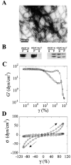
Figure 1.
Figure 1.
Rheological assessment of bulk properties of keratin polymers. All studies were conducted in low–ionic strength Tris-HCl buffer at pH 7.0. (A) Electron micrograph of K5–K14ΔT polymer subjected to negative staining. (B) Sedimentation assays for wt K5–K14 and K5–K14ΔT polymers spun at 150,000 × g (left) and 8,200 × g (right). P, pellet; S, supernatant. See Materials and methods for details. (C) Strain dependence of elastic modulus G' of the wt K5–K14 (○) and K5–K14ΔT (□) polymers. These findings are highly reproducible between experiments. γ, strain amplitude. (D) Time-resolved stress versus strain relationship experiment was conducted at a 100% strain amplitude. Wild-type K5–K14 (○); K5–K14ΔT (□). The arrows depict the direction of the oscillations of the strain-inducing plate. Data corresponding to the first cycle of shear are shown (Ma et al., 2001).
Figure 2.
Figure 2.
The purified recombinant K14 tail domain binds keratin filaments in vitro. (A) Electrophoretic analysis of purified recombinant His-T14. (Lane 1) Coomassie blue staining. (Lanes 2 and 3) Western immunoblots probed with antibodies directed against the His tag (2) and the K14 tail domain (3). (B) Cosedimentation of His-T14 with wt K5–K14 filaments in Tris-HCl buffer at pH 7.4 (standard conditions). His-T14 was mixed in a 1:1 molar ratio with preformed filaments (10 μM; left) or not (right). As controls, His-NPT or insulin do not cosediment with wt K5–K14 under the same conditions. (B') Binding curve defining the interaction between wt K5–K14 (X axis) and His-T14 (fixed concentration). The Y axis corresponds to the fraction of His-T14 bound. The inset shows some of the cosedimentation data (K, K5–K14; T14, His-T14). Nonlinear regression analysis indicates that His-T14 binds keratin IFs with an apparent kD of 2 μM. (C) Assessment of His-T14 binding to other IF polymers. His-T14 binds reconstituted K8–K18 filaments (10 μM) and K5–K14ΔT filaments (10 μM) but not vimentin filaments (10 μM). (D) Impact of adding purified His-T14 on the low pH-induced bundling of wt K5–K14 filaments (10 μM), as assessed by a low-speed centrifugation assay. P, pellet; S, supernatant. Under such conditions, the bulk of K5-K14 is retrieved in the supernatant when assembled at pH 7.4, and in the pellet when assembled at pH 7.0. Addition of His-T14 at a 1:4 stoichiometry partially inhibits the low pH induced sedimentation. Paradoxically, adding His-T14 at a higher stoichiometry (1:1.5) has no effect.
Figure 3.
Figure 3.
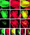
Transient expression of Myc-T14 or controls in various cell lines. (A–A'' and B–B'') Expression of myc-T14 in Ptk2 cells. Myc immunostaining (A and B) localizes to the nucleus and the cytoplasm, where it occurs in a fibrous pattern that coaligns to a significant extent with the endogenous keratin filament network (A' and B'), as shown in the merged images (A'' and B''). (C–C'') Expression of myc-PTE1 in Ptk2 cells. Myc immunostaining (C) shows a puncate pattern in the cytoplasm that is clearly distinct from the endogenous keratin filament network (C' and C''). (D–D'' and E–E'') Coexpression of K5, K14, along with either myc-T14 (D–D'') or myc-PTE1 (E–E'') in BHK-21 cells. Myc-T14 distributes to both the nucleus (N) and the cytoplasm; in this latter instance, it colocalizes with keratin polymers (D' and D''). In contrast, the localization of Myc-PTE1 (E) and keratin polymers (E') are clearly distinct (E'').
Figure 4.
Figure 4.
Outcome of yeast two-hybrid screen and model for tail domain-mediated bundling. (A) List of keratin sequences found to interact with the K14 tail domain in a yeast two-hybrid screen. All are type I keratins. The NH2-terminal boundary of the individual clones, and the domain to which it maps to (B), are indicated. Full-length human K14 exhibits 472 amino acid residues. (B) Schematic depicting the secondary structure of type I keratin proteins. The central rod domain is dominated by α-helical subsegments (1A, 1B, 2A, and 2B) separated by short linker regions (L1, L12, and L2). The rod is flanked by nonhelical head and tail domains at the NH2- and COOH-termini. (C) Modeling the interaction mediated by the K14 tail domain between two closely apposed keratin filaments. Coiled-coil heterodimers involving K5 and K14 are depicted near the filament-filament interface. For the sake of clarity, the nonhelical head domains of K5 and K14 and the tail domain of K5 are omitted, whereas the subdomain 1A of K5 is shown in black. An antiparallel tetramer (Herrmann and Aebi, 1998) is shown in filament 1. It should be noted that, on average, mature cytoplasmic IFs are comprised of 16 dimers in cross-section, that the coiled-coil central rod domain is 42–44 nm long, whereas the filaments themselves are 10–12 nm wide (Herrmann and Aebi, 1998). Individual elements are therefore not drawn to scale in this schematic. On average, 16 copies of the K14 tail domain should be present per 44–50-nm filament length. We postulate that a subset of these copies of the K14 tail domain are surface-exposed and engaged in a direct interaction with type I keratins in subunits from a closely apposed neighboring filament (*). These interactions would assist in the process of keratin filament bundling. The portion of the ∼50 residue–long K14 tail domain mediating those interactions is unknown, but steric considerations along with lack of sequence homology with K16 suggest that it should reside within the distal half of the sequence. Reversible changes in the balance of charges (i.e., through phosphorylation) and other factors (i.e., associated proteins) are postulated to influence the stability of filament–filament interactions mediated by the exposed tail domains, and hence modulate self-induced bundling.

Similar articles

Cited by

References

    1. Albers, K., and E. Fuchs. 1987. The expression of mutant epidermal keratin cDNAs transfected in simple epithelial and squamous cell carcinoma lines. J. Cell Biol. 105:791–806. - PMC - PubMed
    1. Bader, B.L., T.M. Magin, M. Freudenmann, S. Stumpp, and W.W. Franke. 1991. Intermediate filaments formed de novo from tailless cytokeratins in the cytoplasm and in the nucleus. J. Cell Biol. 115:1293–1307. - PMC - PubMed
    1. Boussif, O., Lezoualc'h, F., Zanta, M.A., Mergny, M.D., Scherman, D., Demeneix, B., Behr, J.P. 1995. A versatile vector for gene and oligonucleotide transfer into cells in culture and in vivo: polyethylenimine. Proc. Natl. Acad. Sci. USA. 92:7297–7301. - PMC - PubMed
    1. Coulombe, P.A., and E. Fuchs. 1990. Elucidating the early stages of keratin filament assembly. J. Cell Biol. 111:153–169. - PMC - PubMed
    1. Coulombe, P.A., Y.M. Chan, K. Albers, and E. Fuchs. 1990. Deletions in epidermal keratins leading to alterations in filament organization in vivo and in intermediate filament assembly in vitro. J. Cell Biol. 111:3049–3064. - PMC - PubMed

Publication types