. 2001 Nov 26;155(5):809-20.
doi: 10.1083/jcb.200106139. Epub 2001 Nov 26.

Direct interaction of insulin-like growth factor-1 receptor with leukemia-associated RhoGEF

Affiliations

Direct interaction of insulin-like growth factor-1 receptor with leukemia-associated RhoGEF

S Taya et al. J Cell Biol. .

Abstract

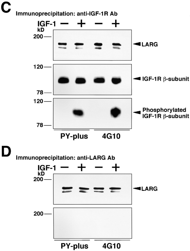
Insulin-like growth factor (IGF)-1 plays crucial roles in growth control and rearrangements of the cytoskeleton. IGF-1 binds to the IGF-1 receptor and thereby induces the autophosphorylation of this receptor at its tyrosine residues. The phosphorylation of the IGF-1 receptor is thought to initiate a cascade of events. Although various signaling molecules have been identified, they appear to interact with the tyrosine-phosphorylated IGF-1 receptor. Here, we identified leukemia-associated Rho guanine nucleotide exchange factor (GEF) (LARG), which contains the PSD-95/Dlg/ZO-1 (PDZ), regulator of G protein signaling (RGS), Dbl homology, and pleckstrin homology domains, as a nonphosphorylated IGF-1 receptor-interacting molecule. LARG formed a complex with the IGF-1 receptor in vivo, and the PDZ domain of LARG interacted directly with the COOH-terminal domain of IGF-1 receptor in vitro. LARG had an exchange activity for Rho in vitro and induced the formation of stress fibers in NIH 3T3 fibroblasts. When MDCKII epithelial cells were treated with IGF-1, Rho and its effector Rho-associated kinase (Rho-kinase) were activated and actin stress fibers were enhanced. Furthermore, the IGF-1-induced Rho-kinase activation and the enhancement of stress fibers were inhibited by ectopic expression of the PDZ and RGS domains of LARG. Taken together, these results indicate that IGF-1 activates the Rho/Rho-kinase pathway via a LARG/IGF-1 receptor complex and thereby regulates cytoskeletal rearrangements.


Figures

Figure 1.
Figure 1.
Identification of the IGF-1 receptor-interacting protein. (A) Amino acid sequences of the IGF-1 receptor-interacting peptide and human LARG. The numbers denote amino acid positions. Asterisks denote identical amino acids. The black bars show amino acid positions of PDZ domain. (B) Schematic representation of the IGF-1 receptor-interacting peptide and human LARG. The numbers indicate the amino acid sequence identity in the PDZ domains or amino acid numbers. (C) Interaction of MBP-LARG-PDZ domain with GST–IGF-1 receptor β-subunit in vitro. MBP, MBP-LARG-PDZ domain, or the AF-6–PDZ domain was mixed with GST- or GST–IGF-1 receptor-coated beads. The interacting proteins were coeluted with GST fusion proteins. The eluates were subjected to SDS-PAGE and subjected to immunoblot analysis with anti-MBP or anti–AF-6 antibody. The results shown are representative of three independent experiments.
Figure 2.
Figure 2.
Tissue and subcellular distributions of LARG. (A) Distribution of LARG in rat tissues. Immunoblot analysis of LARG was carried out using anti-LARG antibody. 1, E21 brain; 2, P3 brain; 3, P6 brain; 4, adult brain; 5, lung; 6, heart; 7, kidney; 8, liver; 9, spleen; 10, thymus; 11, testis; 12, ovary; 13, prostate; 14, small intestine; 15, colon. The arrowhead denotes the position of LARG. (B) Distribution of LARG in cultured cells. Immunoblot analysis of LARG was carried out using anti-LARG antibody. 1, C6; 2, MDCKII; 3, L; 4, NIH 3T3; 5, TMK1; 6, HT29 cells. The arrowhead denotes the position of LARG. (C) Subcellular distribution of endogenous LARG in MDCKII cells. Confluent MDCKII cells were stained with anti-LARG antibody. (D) Localization of LARG mutants. MDCKII cells were transfected with various LARG mutants and then stained with anti-HA antibody. The number of cells used for each calculation are >100, and the values shown are average in three independent experiments. (E) Colocalization of exogenous LARG and endogenous IGF-1 receptor. MDCKII cells were transfected with pEF-BOS-HA-LARG (top) or pEF-BOS-HA-LARG-ΔPDZ (middle and bottom) and then doubly stained with anti-HA and anti–IGF-1 receptor antibodies. The results are representative of three independent experiments. Bars, 10 μm.
Figure 2.
Figure 2.
Tissue and subcellular distributions of LARG. (A) Distribution of LARG in rat tissues. Immunoblot analysis of LARG was carried out using anti-LARG antibody. 1, E21 brain; 2, P3 brain; 3, P6 brain; 4, adult brain; 5, lung; 6, heart; 7, kidney; 8, liver; 9, spleen; 10, thymus; 11, testis; 12, ovary; 13, prostate; 14, small intestine; 15, colon. The arrowhead denotes the position of LARG. (B) Distribution of LARG in cultured cells. Immunoblot analysis of LARG was carried out using anti-LARG antibody. 1, C6; 2, MDCKII; 3, L; 4, NIH 3T3; 5, TMK1; 6, HT29 cells. The arrowhead denotes the position of LARG. (C) Subcellular distribution of endogenous LARG in MDCKII cells. Confluent MDCKII cells were stained with anti-LARG antibody. (D) Localization of LARG mutants. MDCKII cells were transfected with various LARG mutants and then stained with anti-HA antibody. The number of cells used for each calculation are >100, and the values shown are average in three independent experiments. (E) Colocalization of exogenous LARG and endogenous IGF-1 receptor. MDCKII cells were transfected with pEF-BOS-HA-LARG (top) or pEF-BOS-HA-LARG-ΔPDZ (middle and bottom) and then doubly stained with anti-HA and anti–IGF-1 receptor antibodies. The results are representative of three independent experiments. Bars, 10 μm.
Figure 3.
Figure 3.
Interaction of LARG with the IGF-1 receptor. (A) Interaction of endogenous LARG with the IGF-1 receptor in vivo. The lysates of MDCKII cells were incubated with rabbit IgG, anti-LARG, or the anti–IGF-1 receptor antibody. The immunocomplexes were then precipitated and subjected to immunoblot analysis with anti-LARG (top) or anti–IGF-1 receptor antibody (bottom). (B) Interaction of the recombinant LARG with the IGF-1 receptor β-subunit. L fibroblasts were cotransfected with pEF-BOS-Myc-LARG and pEF-BOS-HA–IGF-1 receptor β-subunit. These lysates were incubated with anti-HA (top) or with the anti-Myc antibody (bottom). The immunocomplexes were subjected to immunoblot analysis with anti-Myc (top) or anti-HA antibody (bottom). (C) Effect of IGF-1 on the binding state of LARG and the IGF-1 receptor. MDCKII cells were serum starved for 48 h and then stimulated with 10 nM IGF-1 for 20 min. The lysates of MDCKII cells were incubated with anti–IGF-1 receptor antibody. The immunoprecipitates were subjected to immunoblot analysis with anti-LARG antibody (top), anti–IGF-1 receptor antibody (middle), or antiphosphotyrosine antibodies (PY-plus and 4G10) (bottom). (D) Effect of IGF-1 on tyrosine phosphorylation of LARG. Under the same condition as C, the lysates of MDCKII cells were incubated with anti-LARG antibody, and then the precipitated LARG was subjected to immunoblot analysis with anti-LARG antibody (top) or antiphosphotyrosine antibodies (bottom). The results shown are representative of three independent experiments.
Figure 3.
Figure 3.
Interaction of LARG with the IGF-1 receptor. (A) Interaction of endogenous LARG with the IGF-1 receptor in vivo. The lysates of MDCKII cells were incubated with rabbit IgG, anti-LARG, or the anti–IGF-1 receptor antibody. The immunocomplexes were then precipitated and subjected to immunoblot analysis with anti-LARG (top) or anti–IGF-1 receptor antibody (bottom). (B) Interaction of the recombinant LARG with the IGF-1 receptor β-subunit. L fibroblasts were cotransfected with pEF-BOS-Myc-LARG and pEF-BOS-HA–IGF-1 receptor β-subunit. These lysates were incubated with anti-HA (top) or with the anti-Myc antibody (bottom). The immunocomplexes were subjected to immunoblot analysis with anti-Myc (top) or anti-HA antibody (bottom). (C) Effect of IGF-1 on the binding state of LARG and the IGF-1 receptor. MDCKII cells were serum starved for 48 h and then stimulated with 10 nM IGF-1 for 20 min. The lysates of MDCKII cells were incubated with anti–IGF-1 receptor antibody. The immunoprecipitates were subjected to immunoblot analysis with anti-LARG antibody (top), anti–IGF-1 receptor antibody (middle), or antiphosphotyrosine antibodies (PY-plus and 4G10) (bottom). (D) Effect of IGF-1 on tyrosine phosphorylation of LARG. Under the same condition as C, the lysates of MDCKII cells were incubated with anti-LARG antibody, and then the precipitated LARG was subjected to immunoblot analysis with anti-LARG antibody (top) or antiphosphotyrosine antibodies (bottom). The results shown are representative of three independent experiments.
Figure 4.
Figure 4.
GEF activity of LARG for the Rho family GTPases in vitro. (A) The effect of LARG on the dissociation of GDP from RhoA in vitro. The DH/PH domain of LARG and Dbl enhanced the dissociation of [3H]-labeled GDP from RhoA in a time-dependent manner. (B) The effect of LARG on other small GTPases in vitro. The DH/PH domain of LARG showed an exchange activity for RhoA but not one for Rac1, Cdc42, or Ras. The results shown are representative of three independent experiments.
Figure 5.
Figure 5.
Effect of LARG on the Rho activity in NIH 3T3 fibroblasts. Serum-deprived NIH 3T3 cells were transfected with the pEF-BOS-HA-LARG (A and C) or -Myc dominant negative form of RhoA/-HA-LARG (B and D). The transfected cells were doubly stained with anti-HA antibody and TRITC-labeled phalloidin. The results shown are representative of three independent experiments. Bar, 20 μm.
Figure 6.
Figure 6.
Effect of IGF-1 on the Rho/Rho-kinase signaling pathway. (A) Activation of exogenous RhoA by IGF-1. MDCKII cells stably expressing EGFP-RhoA were serum starved and then stimulated with 10 nM IGF-1 for the indicated minutes. The lysates were incubated with GST–Rho-binding domain of rhotekin. The bound EGFP-RhoA was subjected to immunoblot analysis with anti-RhoA antibody. Arrowheads indicate the positions of GTP-bound EGFP-RhoA and total EGFP-RhoA. (B) IGF-induced phosphorylation of MBS at Ser-854. Serum-deprived MDCKII cells were stimulated with 10 nM IGF-1 for the indicated minutes, and the whole cell lysates were resolved by SDS-PAGE followed by immunoblot analysis with anti-pS854 (top) or anti-MBS antibody (bottom). Arrowheads indicate the positions of the phosphorylated MBS and total MBS. (C) Inhibition of the IGF-1–induced MBS phosphorylation by Rho-kinase inhibitors. HA1077 (right)- or Y-32885 (left)-pretreated serum-deprived MDCKII cells were stimulated with 10 nM IGF-1 for 10 min, and then the lysates were resolved by SDS-PAGE followed by immunoblot analysis with anti-pS854 (top) or anti-MBS antibody (bottom). Arrowheads indicate the positions of phosphorylated MBS and total MBS. The results shown are representative of three independent experiments.
Figure 7.
Figure 7.
Effect of the PDZ domain of LARG. (A) Inhibition of the complex formation of IGF-1 receptor and LARG by the PDZ domain of LARG. Endogenous LARG from MDCKII lysate and MBP, MBP-LARG-PDZ domain, or the AF-6–PDZ domain (500 pmol) were mixed with GST or GST–IGF-1 receptor-coated beads (100 pmol). The bound LARG was subjected to immunoblot analysis with anti-LARG antibody. (B) Effect of the PDZ and RGS domains of LARG on the IGF-1–induced MBS phosphorylation. MDCKII cells were transfected with pEF-BOS-HA, or -LARG-PDZ, -LARG-RGS, or -AF-6–PDZ domain. These transfected cells were serum starved for 48 h and then stimulated or not with 10 nM IGF-1 for 10 min. The lysates were resolved by SDS-PAGE followed by immunoblot analysis with anti-pS854 (top), anti-MBS antibody (middle), or anti-HA antibody (bottom). Arrowheads indicate the positions of phosphorylated MBS, total MBS, or expressed proteins. Asterisks indicate nonspecific bands. The results shown are representative of five independent experiments.
Figure 8.
Figure 8.
Effect of LARG on the IGF-1–induced enhancement of stress fibers. (A) IGF-1–induced enhancement of stress fiber formation. MDCKII cells were serum starved for 48 h and then stimulated with 10 nM IGF-1 for the indicated minutes. The stimulated cells were stained with TRITC-labeled phalloidin. Arrowheads indicate the cells showing IGF-1–induced enhancement of stress fibers. (B and C) Effect of the PDZ and RGS domains of LARG on the IGF-1–induced enhancement of stress fibers. MDCKII cells were transfected with the pEF-BOS-HA-LARG-PDZ domain. The transfected cells were serum starved for 48 h and then stimulated with 10 nM IGF-1 for 60 min. The transfected cells were doubly stained with anti-HA antibody and TRITC-labeled phalloidin (B). The cells showing the IGF-1–induced enhancement of stress fibers, which were transfected with pEF-BOS-GST, -HA-LARG-PDZ, -HA-LARG-RGS, or -HA–AF-6–PDZ, were calculated (C). The number of cells used for each calculation is >300, and the values shown are means ± SE of triplicates. **p < 0.01, significance of difference from the cells of IGF-1–induced stress fibers analyzed by Student's t test.

Similar articles

Cited by

References

    1. Abe, K., K.L. Rossman, B. Liu, K.D. Ritola, D. Chiang, S.L. Campbell, K. Burridge, and C.J. Der. 2000. Vav2 is an activator of Cdc42, Rac1, and RhoA. J. Biol. Chem. 275:10141–10149. - PubMed
    1. Blangy, A., E. Vignal, S. Schmidt, A. Debant, C. Gauthier-Rouviere, and P. Fort. 2000. TrioGEF1 controls Rac- and Cdc42-dependent cell structures through the direct activation of rhoG. J. Cell Sci. 113:729–739. - PubMed
    1. Cerione, R.A., and Y. Zheng. 1996. The Dbl family of oncogenes. Curr. Opin. Cell Biol. 8:216–222. - PubMed
    1. Cheng, H.L., M.L. Steinway, J.W. Russell, and E.L. Feldman. 2000. GTPases and phosphatidylinositol 3-kinase are critical for insulin-like growth factor-I-mediated Schwann cell motility. J. Biol. Chem. 275:27197–27204. - PubMed
    1. Crespo, P., K.E. Schuebel, A.A. Ostrom, J.S. Gutkind, and X.R. Bustelo. 1997. Phosphotyrosine-dependent activation of Rac-1 GDP/GTP exchange by the vav proto-oncogene product. Nature. 385:169–172. - PubMed

Publication types

MeSH terms

Substances