. 2001 Nov 26;155(5):833-44.
doi: 10.1083/jcb.200103005. Epub 2001 Nov 26.

Synchronized activation and refolding of influenza hemagglutinin in multimeric fusion machines

Affiliations

Synchronized activation and refolding of influenza hemagglutinin in multimeric fusion machines

I Markovic et al. J Cell Biol. .

Abstract

At the time of fusion, membranes are packed with fusogenic proteins. Do adjacent individual proteins interact with each other in the plane of the membrane? Or does each of these proteins serve as an independent fusion machine? Here we report that the low pH-triggered transition between the initial and final conformations of a prototype fusogenic protein, influenza hemagglutinin (HA), involves a preserved interaction between individual HAs. Although the HAs of subtypes H3 and H2 show notably different degrees of activation, for both, the percentage of low pH-activated HA increased with higher surface density of HA, indicating positive cooperativity. We propose that a concerted activation of HAs, together with the resultant synchronized release of their conformational energy, is an example of a general strategy of coordination in biological design, crucial for the functioning of multiprotein fusion machines.


Figures
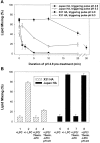
Figure 1.
Figure 1.
X31 and Japan HA differ in activation rather than inactivation rates. (A) The time course of Japan (squares) and X31 HA (circles) activation/inactivation at low pH in the absence of target membrane. HA-cells were treated with a pH 4.9 activating pulse for 1 to 30 min. Then, after RBC binding, cells were treated with a fusion-triggering pulse of pH 4.9 for 2 min (closed symbols) or pH 5.3 for 5 min (open symbols). A higher degree of HA activation during the activating pulse resulted in a lower extent of lipid mixing after the triggering pulse. (B) X31 (bars 1–4) or Japan HA-cells (bars 5–8) with bound RBCs were incubated for 5 min in pH 4.9 medium containing LPC. Although no lipid mixing was observed in the presence of LPC (bars 1 and 5), its removal completely restored fusion (bars 2 and 6). In the experiments represented in bars 3, 4, 7, and 8, cells at the LPC-arrested fusion stage were treated with thermolysin to cleave low pH–activated HA. Lipid mixing was assayed either directly after LPC removal (bars 3 and 7) or after application of an additional 5-min pulse of pH 4.9 (bars 4 and 8). Points are means ± SE, n > 3.
Figure 2.
Figure 2.
Activation of X31 and Japan HA assessed by Western blotting as exposure of HA1–HA2 S–S bond. (A) Japan or X31 HA viral particles (the two left and two right panels, respectively) were acidified (pH 4.9, 10 min, 22°C) or not and reduced with DTT. Minor loss of the HA1–HA2 band for Japan is in apparent contrast to X31 HA1–HA2 disappearance. (B) Time course of activation for Japan and X31 HA expressed in cells. Cells were incubated at 22°C in pH 4.9 medium for a given time interval in the presence of protease inhibitors.
Figure 3.
Figure 3.
NaBut-induced increase in the surface density of Japan HA. (A) Increase in the surface HA expression after preincubation with NaBut was detected by biotinylation (open circles) and trypsinization (closed circles) of surface HA and using flow cytometry (closed triangles). (B) The percentage of surface HA among total cellular HA for cells treated with 0–9 mM NaBut was determined by cell-surface biotinylation and trypsinization. Note that the majority of HA is expressed at the cell surface.
Figure 4.
Figure 4.
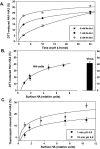
Cooperative activation of Japan HA. Japan HA activation was monitored by Western blotting as HA sensitivity to DTT (A and B) or thermolysin (C). (A) Time course of HA activation for different HA expression levels. HA expression was varied by preincubation of HA-cells with 0, 1, or 5 mM NaBut. The duration of the pH 4.9 pulse ranged from 1 to 30 min (B) Efficiency of Japan HA activation after a 10-min pulse of pH 4.9 as a function of the surface density of HA. Relative surface density of HA after cell preincubation with 0 to 9 mM NaBut was assayed by measuring the changes in total cellular HA and normalized by that in NaBut-untreated HAb2 cells. Points are means ± SE, n = 4. (C) The percentage of low pH–activated HA molecules that reached the early stage of fusion peptide exposure was assayed by means of thermolysin cleavage. HA expression in cells was altered by pretreatment with 0 to 9 mM NaBut. Cells were incubated at pH 4.9 for 1 or 10 min, reneutralized, and treated with thermolysin to cleave exposed fusion peptides. Cleavage of activated HA resulted in a decrease in the HA1–HA2 band, which was normalized by the pH 7.4 band taken as 100%.
Figure 5.
Figure 5.
Cooperative activation of X31 and Udorn HA. In all experiments, activation was monitored by Western blotting as HA sensitivity to DTT. (A) The surface density of activation-competent X31 HA molecules in HA-cells was varied by altering trypsin concentration (1–10 μg/ml). The increase in the percentage of the HA1–HA2 form, out of total HA, gave the increase in percentage of activation. (B) X31 HA reconstituted in virosomes at a higher HA to lipid ratio, and thus at a higher surface density, activates to a higher level. The percentage of HA activated after a 10-min pulse of pH 4.9 is shown for two independent experiments (open and closed bars). (C) The surface density of Udorn HA was altered by varying the multiplicity of infection of CV1 cells with SV-40 recombinant virus carrying the HA gene. Increases in the median cell surface fluorescence assessed by FACS® analysis (closed bars) confirmed an increase in the average amount of HA per cell in the specific range of the recombinant virus concentrations (0.01–1 mg/ml total viral protein). Open bars represent the percentage of activated HA molecules after a 10-min pulse of pH 4.9.
Figure 6.
Figure 6.
Functional assay confirms the acceleration of activation/inactivation upon increase in the level of HA expression. Japan HA-cells incubated with 0 (closed circles) or 2 mM NaBut (open circles) were pretreated with an activating pulse, pH 4.9. Next, RBCs were added, and fusion was triggered by a 10-min pulse of pH 5.2. As in biochemical experiments, the efficiency of HA activation is higher for the cells with an HA expression level increased by NaBut.
Figure 7.
Figure 7.
A target membrane enhances activation to the level observed at high HA density. (A) Closed circles: Japan HA-cells at different HA densities with saturating concentrations of liposomes. Japan HA-cells with levels of HA expression altered by pretreatment with 0 to 9 mM NaBut were plated as single cells on poly-l-lysine–treated flasks and incubated at 4°C with saturating concentrations of liposomes. HA expression was normalized by that in NaBut-untreated HAb2 cells. After removal of unbound liposomes, cells were treated with a 10-min pulse, pH 4.9 (22°C). Open circles: control, analogous cells in the absence of liposomes. Points are means ± SE, n= 4. (B) The cartoon illustrates HA enrichment in the contact zones between an HA-cell and liposomes. HA1-receptor interaction induces HA concentration in the contact zone. Therefore, as long as the contact area is less than the total area of HA-membrane, effective HA density and hence the level of HA activation in the presence of a target membrane exceed those in the absence of a target membrane. The graph represents a theoretical curve based on the estimate of HA enrichment and activation in the HA-cell–target membrane contact region for cells with low HA density. The expected level of activation, shown as a function of the ratio of the contact zone area to the total HA-membrane area, α, approaches the activation level observed for cells with high HA density in the absence of a target membrane.
Figure 8.
Figure 8.
Schematic diagram showing the hypothetical mechanism of cooperative activation of HA at low pH. Low pH application triggers restructuring of HA trimers from initial conformation (depicted as blue) to an early, transient activated form (yellow) followed by the final, lowest energy conformation (orange). (A) Membrane-anchored HA trimers before low pH application. (B–D) After acidification, inter-trimer interactions promote transition from a transient activated form of HA to the lowest energy protein conformation, and increases the probability of activation for native HA molecules proximal to activated ones. Activation consequently spreads among neighboring HAs (red arrows). At a high local density of HA, such concerted activation leads to the synchronized release of the conformational energy by multiple trimers assembled around the fusion site.

Similar articles

Cited by

References

    1. Armstrong, R.T., A.S. Kushnir, and J.M. White. 2000. The transmembrane domain of influenza hemagglutinin exhibits a stringent length requirement to support the hemifusion to fusion transition. J. Cell Biol. 151:425–438. - PMC - PubMed
    1. Blumenthal, R., D.P. Sarkar, S. Durell, D.E. Howard, and S.J. Morris. 1996. Dilation of the influenza hemagglutinin fusion pore revealed by the kinetics of individual cell–cell fusion events. J. Cell Biol. 135:63–71. - PMC - PubMed
    1. Bron, R., A. Ortiz, J. Dijkstra, T. Stegmann, and J. Wilschut. 1993. Preparation, properties, and applications of reconstituted influenza virus envelopes (virosomes). Methods Enzymol. 220:313–331. - PubMed
    1. Bullough, P.A., F.M. Hughson, J.J. Skehel, and D.C. Wiley. 1994. Structure of influenza haemagglutinin at the pH of membrane fusion. Nature. 371:37–43. - PubMed
    1. Carr, C.M., and P.S. Kim. 1993. A spring-loaded mechanism for the conformational change of influenza hemagglutinin. Cell. 73:823–832. - PubMed

MeSH terms