. 2001 Dec 4;98(25):14669-74.
doi: 10.1073/pnas.251341998. Epub 2001 Nov 27.

Minocycline prevents nigrostriatal dopaminergic neurodegeneration in the MPTP model of Parkinson's disease

Affiliations

Minocycline prevents nigrostriatal dopaminergic neurodegeneration in the MPTP model of Parkinson's disease

Y Du et al. Proc Natl Acad Sci U S A. .

Abstract

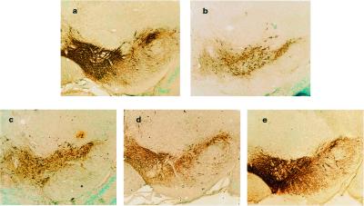
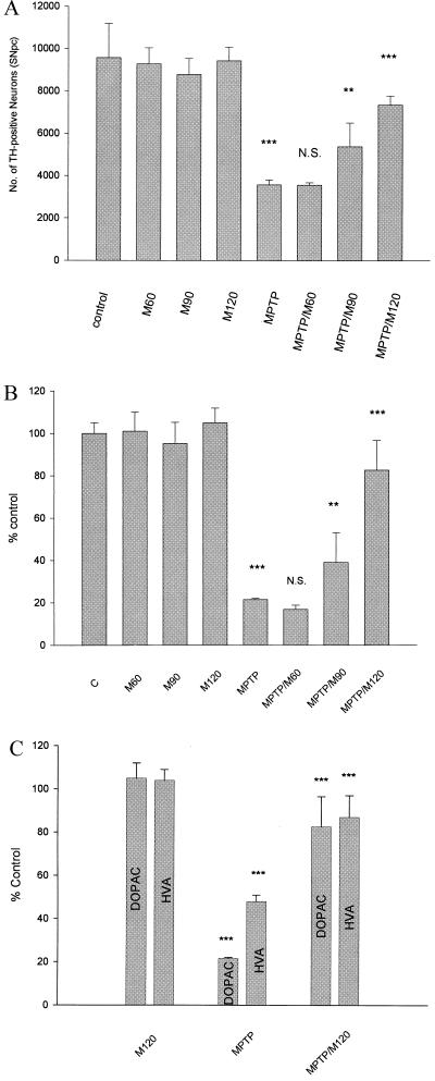
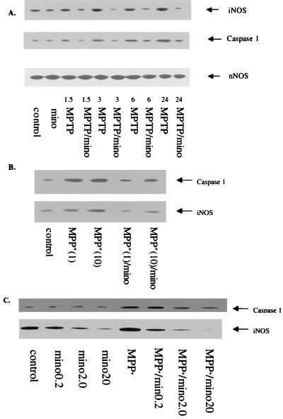
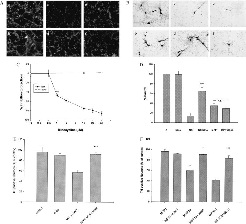
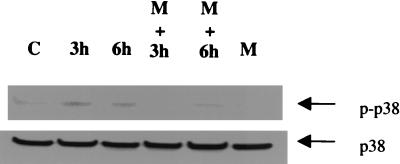
Parkinson's disease is a chronic neurodegenerative disorder characterized by the loss of dopamine neurons in the substantia nigra, decreased striatal dopamine levels, and consequent extrapyramidal motor dysfunction. We now report that minocycline, a semisynthetic tetracycline, recently shown to have neuroprotective effects in animal models of stroke/ischemic injury and Huntington's disease, prevents nigrostriatal dopaminergic neurodegeneration in the 1-methyl-4-phenyl-1,2,3,6-tetrahydropyridine (MPTP) mouse model of Parkinson's disease. Minocycline treatment also blocked dopamine depletion in the striatum as well as in the nucleus accumbens after MPTP administration. The neuroprotective effect of minocycline is associated with marked reductions in inducible NO synthase (iNOS) and caspase 1 expression. In vitro studies using primary cultures of mesencephalic and cerebellar granule neurons (CGN) and/or glia demonstrate that minocycline inhibits both 1-methyl-4-phenylpyridinium (MPP(+))-mediated iNOS expression and NO-induced neurotoxicity, but MPP(+)-induced neurotoxicity is inhibited only in the presence of glia. Further, minocycline also inhibits NO-induced phosphorylation of p38 mitogen-activated protein kinase (MAPK) in CGN and the p38 MAPK inhibitor, SB203580, blocks NO toxicity of CGN. Our results suggest that minocycline blocks MPTP neurotoxicity in vivo by indirectly inhibiting MPTP/MPP(+)-induced glial iNOS expression and/or directly inhibiting NO-induced neurotoxicity, most likely by inhibiting the phosphorylation of p38 MAPK. Thus, NO appears to play an important role in MPTP neurotoxicity. Neuroprotective tetracyclines may be effective in preventing or slowing the progression of Parkinson's and other neurodegenerative diseases.


Figures

Figure 1
Figure 1
Minocycline prevents loss of dopamine neurons after MPTP administration. Dopamine neurons and processes were identified by TH immunostaining of representative midbrain sections 7 days after MPTP treatment with or without treatment with minocycline (60, 90, and 120 mg/kg daily, see Materials and Methods for details). (a) dH2O. (b) MPTP treated. (c) MPTP treated after 2 days pretreatment with minocycline 60 mg/kg. (d) MPTP treated after 2 days pretreatment with minocycline 90 mg/kg. (e) MPTP treated after 2 days pretreatment with minocycline 120 mg/kg. Note the marked reduction in TH-positive cell bodies and processes after MPTP administration (compare a and b) and the protection by minocycline (b vs. e). Photomicrographs are from a representative experiment repeated three times with similar results.
Figure 2
Figure 2
Minocycline prevents loss of TH-positive neurons, striatal dopamine, and dopamine metabolites after MPTP administration. (A) Quantification of TH-positive neurons in the SNpc was carried out as described in the text (21). Minocycline at 90 and 120 mg/kg significantly protects TH-positive neurons from death induced by MPTP exposure (one-way ANOVA; **, P < 0.01; ***, P < 0.001; N.S., not significant) (see text for details). (B and C) Dopamine, HVA, and DOPAC were measured by HPLC (see text and ref. for details). Mice administered MPTP showed significant reductions in striatal dopamine, HVA, and DOPAC compared with controls. Minocycline treatment significantly protected animals from MPTP-induced reductions in dopamine, HVA, and DOPAC (one-way ANOVA; **, P < 0.01; ***, P < 0.001; N.S., not significant). See text for details. Each group consisted of 5–7 animals, and the data are from a representative experiment repeated at least twice with similar results.
Figure 3
Figure 3
(A) Minocycline blocks MPTP-induced expression of iNOS and caspase 1 in vivo and in vitro. Immunoblot analyses were performed with polyclonal antibodies against iNOS, nNOS, and caspase 1 (Santa Cruz Biotechnology). Minocycline doses and concentrations as well as the time course after MPTP or MPP+ administration exposure are indicated. MPTP treatment increases iNOS and caspase 1 expression by 3 h posttreatment. Minocycline treatment blocks the increase in both iNOS and caspase 1. Numbers (i.e., 1.5–24) represent the hours of treatment. Note that MPTP treatment fails to alter nNOS expression in these same samples. (B) Minocycline (20 μM) inhibits caspase 1 and iNOS expression induced by MPP+ (1 and 10 μM, 18 h) in primary cultures of mouse astrocytes. Astrocytes from neonatal mouse cerebral cortex were prepared as described (35). Lane 1 (left to right) = control; lane 2 = MPP+ (1 μM); lane 3 = MPP+ (10 μM); lane 4 = MPP+ (1 μM) minocycline (20 μM); and lane 5 = MPP+ (10 μM)/minocycline (20 μM). (C) Minocycline inhibits caspase 1 and iNOS expression induced by MPP+ in a mouse microglial cell line (BV2). BV2 cells (36) were cultured to near confluency and then treated with various concentrations of minocycline (0.2–20 μM) with and without MPP+ (10 μM, 18 h). Note that minocycline reduces basal iNOS expression in BV2 cells and completely blocks iNOS and caspase 1 expression induced by MPP+.
Figure 4
Figure 4
Effects of minocycline on NO and MPP+ toxicity of cultured CGN and RMN. (A) Minocycline blocks NO-induced neuronal death of CGN, but not MPP+-induced neurotoxicity. CGN were exposed to increasing concentrations of minocycline (0.5–50 μM) for 24 h in the presence of SNP (50 μM, 24 h) or MPP+ (70 μM, 72 h). Viable and dead CGN were quantified by using fluorescein diacetate (yellow-green) and propidium iodide (red) staining as described (22). (af) Representative fields of CGN were photographed (×100) after double staining in the absence (a, c, and e) or presence (b, d, and f) of minocycline (20 μM). (a and b) No treatment. (c and d) SNP treatment. (e and f) MPP+ treatment. (C) Quantification of the effects of minocycline on MPP+-treated CGN. Values are expressed as a % of control (untreated) cultures for each concentration of minocycline. Data represent the mean ± SE (bars) values of triplicate determinations from a single but representative experiment repeated three times with similar results (***, P < 0.001 by one-way ANOVA; N.S., not significant). (B) Minocycline blocks NO-induced neuronal death of cultured RMN but not MPP+-induced neurotoxicity. (af) Representative fields of fetal RMN (20) were photographed (×200) after TH staining (see text for details). Compare untreated control and minocycline-treated cultures (a and b) with those exposed to 10 μM SNP (NO) (c) or 10 μM MPP+ (e) plus minocycline (10 μM) (d and f). Note that minocycline markedly attenuates NO neurotoxicity (d), but not MPP+ neurotoxicity (f). (D) Quantification of the effects of minocycline on SNP (10 μM) and MPP+ (10 μM)-treated fetal rat RMN. TH-positive cells were counted from photomicrographs like those shown in B above. Data are from a representative experiment repeated twice with similar results (***, P < 0.001 compared with NO alone). (E) Minocycline blocks combined NO/MPP+ toxicity of cultured CGN. Quantification of the effects of minocycline on both SNP (5 μM) and MPP+ (0.1 μM)-treated CGN. Data are from a representative experiment repeated three times with similar results [***, P < 0.001 compared with SNP (5 μM) and MPP+ (0.1 μM) alone]. (F) Minocycline blocks MPP+ neurotoxicity in neuron/glia cocultures. Quantification of the effects of minocycline on MPP+ (1–50 μM)-treated fetal rat RMN/glia cocultures (24). TH-positive cells were quantified from photomicrographs like those shown in B above. Note that in the presence of glia higher concentrations of MPP+ are required to kill dopamine neurons. Nonetheless, in the presence of glia the neurotoxic effects of MPP+ are completely blocked by minocycline. Data are from a representative experiment repeated twice with similar results. (*, P < 0.05; ***, P < 0.001 compared with MPP+ alone).
Figure 5
Figure 5
The effect of NO and minocycline on p38 MAPK phosphorylation in CGN. CGN were exposed to SNP (50 μM) in the absence or presence of minocycline (20 μM) for the indicated times (see text for details). Cell lysates were immunoblotted with anti-phospho-p38 and anti-p38 antibody (New England Biolabs). Note that the increase in phospho-p38 MAPK observed after NO (SNP) treatment is blocked by minocycline (Upper). No changes in p38 MAPK itself was observed (Lower). Similar results were obtained in three independent experiments. C = control, M = minocycline, p-p38 = phosphorylated p38 MAPK; 3 h and 6 h represent the treatment times of SNP.

References

    1. Oertel W H, Quinn N P. In: Neurological Disorders: Course and Treatment. Brandt T, Diener H C, Caplan L R, Kennard C, Dichgans J, editors. San Diego: Academic; 1996. pp. 715–772.
    1. Quinn N P. Neurology. 1998;51:S25–S29. - PubMed
    1. Tolwani R J, Jakowec M W, Petzinger G M, Green S, Waggie K. Lab Anim Sci. 1999;49:363–371. - PubMed
    1. Langston J W, Ballard J W, Tetrud J W, Irwin I. Science. 1983;219:979–980. - PubMed
    1. Uhl G R, Javitch J A, Snyder S H. Lancet. 1985;1:956–957. - PubMed

MeSH terms