. 2001 Dec 4;98(25):14422-7.
doi: 10.1073/pnas.251401598. Epub 2001 Nov 27.

The tumor autocrine motility factor receptor, gp78, is a ubiquitin protein ligase implicated in degradation from the endoplasmic reticulum

Affiliations

The tumor autocrine motility factor receptor, gp78, is a ubiquitin protein ligase implicated in degradation from the endoplasmic reticulum

S Fang et al. Proc Natl Acad Sci U S A. .

Abstract

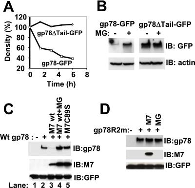
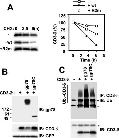
gp78, also known as the tumor autocrine motility factor receptor, is a transmembrane protein whose expression is correlated with tumor metastasis. We establish that gp78 is a RING finger-dependent ubiquitin protein ligase (E3) of the endoplasmic reticulum (ER). Consistent with this, gp78 specifically recruits MmUBC7, a ubiquitin-conjugating enzyme (E2) implicated in ER-associated degradation (ERAD), through a region distinct from the RING finger. gp78 can target itself for proteasomal degradation in a RING finger- and MmUBC7-dependent manner. Importantly, gp78 can also mediate degradation of CD3-delta, a well-characterized ERAD substrate. In contrast, gp78 lacking an intact RING finger or its multiple membrane-spanning domains stabilizes CD3-delta. gp78 has thus been found to be an example of a mammalian cellular E3 intrinsic to the ER, suggesting a potential link between ubiquitylation, ERAD, and metastasis.


Figures

Figure 1
Figure 1
Subcellular localization of gp78. (A) U2OS cells stably expressing gp78 with GFP at its C terminus (gp78-GFP) were assessed by fluorescence microscopy. (B) U2OS transiently expressing plasmids encoding gp78 and either HA-MmUBC6 (M6) or myc-MmUBC7 (M7) were assessed for colocalization by confocal microscopy after double staining with anti-gp78 and either anti-Myc or anti-HA. (C) U2OS cells transiently expressing gp78C and MmUBC7 (M7) were assessed for colocalization by confocal microscopy as in B.
Figure 2
Figure 2
gp78 and MmUBC7 physically interact in HEK 293T cells. (A) Cells transfected with plasmids encoding gp78 or gp78C in combination with HA-MmUBC6 or Myc-MmUBC7 were fractionated into cytosolic (C) and microsomal (M) fractions and after SDS/PAGE evaluated for expression of the indicated proteins by immunoblotting. MmUBC6 (M6) and MmUBC7 (M7) were detected by using Abs against HA or Myc. Numbers on the left indicate apparent molecular weight × 10−3. Cxn, calnexin. (B) Cells were transfected as indicated. In the left two lanes, cell lysates were subject to immunoprecipitation with anti-gp78; in the right two lanes lysates from the same cells were directly resolved on SDS/PAGE. Immunoblotting (IB) was with anti-gp78. Relative amounts of gp78 migrating at its predicted molecular weight, as compared to high molecular weight complexes, is variable with IP. With direct resolution of lysates, gp78 migrates almost exclusively as a high molecular weight form. (C) Lysates from cells transfected with plasmid encoding gp78 with a mutation in a critical RING finger Cys (gp78C356G) and the indicated E2s were subjected to IP with either anti-HA (MmUBC6 and E2–25K) or anti-Myc (MmUBC7) and IB with Abs against either the epitope tags (Top) or anti-gp78 (Middle); *, IgG light chain. The same lysates were subsequently subjected to re-immunoprecipitation with anti-gp78 followed by IB, also with anti-gp78 (Bottom); *, IgG heavy chain; M6, MmUBC6; M7, MmUBC7. (D) Cells were transfected as indicated. gp78R2m is a mutant of gp78 in which predicted coordinating residues for both putative zinc binding sites have been mutated. (Top) IP with anti-gp78 followed by IB with mixture of anti-HA and anti-Myc to detect E2s. (Middle and Bottom) Immunoblots with indicated Abs of whole-cell lysates representing one-fourth of the material used for the IP presented in Top.
Figure 3
Figure 3
In vitro analysis of gp78 interaction with MmUBC7. (A) Schematic representation of the cytoplasmic domain of gp78 (gp78C) and its truncated forms expressed as C-terminal fusions with GST and used for binding in B and C. The RING finger (amino acids 341–378) is boxed. The Cue domain (30) (amino acids 456–497) is shaded. (B and C) The indicated bead-bound GST fusions were evaluated for binding to 35S-labeled in vitro translated MmUBC7. Left lanes represent one-third of [35S]MmUBC7 used for each binding reaction.
Figure 4
Figure 4
In vitro ubiquitylation of gp78. (A) In vitro ubiquitylation in the presence of E1, UbcH5B (H5B), 32P-labeled Ub, and 20 pmol of the indicated GST fusion proteins performed as described. (B) In vitro ubiquitylation using the same GST fusion proteins as in A, but MmUBC7 was used instead of UbcH5B. (C) gp78C and its truncated mutants as presented in Fig. 3A were assayed for ubiquitylation in the presence of MmUBC7 (M7) (Upper) or UbcH5B (H5B) (Lower).
Figure 5
Figure 5
RING-, proteasome-, and MmUBC7-dependent degradation of gp78. (A) Cells stably expressing GFP-tagged wt gp78 (gp78-GFP) or GFP-tagged gp78 lacking the C-terminal tail (gp78ΔTail-GFP) were pulse labeled with [35S]methionine for 30 min followed by removal of unincorporated [35S]methionine. gp78 was then immunoprecipitated by using anti-GFP, and, after resolution on SDS/PAGE, gp78 was quantified and plotted as a function of the amount at the initiation of the chase. (B) Cells stably expressing gp78-GFP or gp78ΔTail-GFP were treated with or without MG132 (MG) for 6 h followed by immunoblotting with anti-GFP; β-actin was used as a gel loading control. (C and D) Lysates from cells transfected as indicated were processed for immunoblotting for gp78, MmUBC7 (M7), and cotransfected GFP (transfection efficiency control). Cells exposed to MG132 (MG) were treated with 20 μM for 6 h.
Figure 6
Figure 6
gp78-mediated ERAD of CD3-δ. (A) HEK 293T cells transfected with plasmid encoding HA-CD3-δ and either full-length wt gp78 or gp78R2m were treated with cycloheximide (CHX), 50 μg/ml, for the indicated times followed by evaluation of cell lysates by IB with anti-HA to detect HA-tagged CD3-δ. Bands were quantified by densitometry and plotted as a function of the amount of material when CHX was added (Right). (B) HEK 293T cells were transfected as indicated with plasmids encoding HA-CD3-δ, full-length wt gp78, and the C-terminal tail of gp78 (gp78C). After transfection (24 h), cell lysates were processed for IB with anti-gp78, anti-HA, and anti-GFP (transfection efficiency control). (C) U2OS cells transfected as in B were subject to IP with anti-HA followed by IB first with either anti-Ub (Upper) followed by reprobing of the membrane with anti-HA (Lower). *, IgG heavy chain (Upper and Lower) and light chain (Upper).

Similar articles

Cited by

References

    1. Hershko A, Ciechanover A. Annu Rev Biochem. 1998;67:425–479. - PubMed
    1. Joazeiro C A, Weissman A M. Cell. 2000;102:549–552. - PubMed
    1. Weissman A M. Nat Rev Mol Cell Biol. 2001;2:169–178. - PubMed
    1. Bonifacino J S, Weissman A M. Annu Rev Cell Dev Biol. 1998;14:19–57. - PMC - PubMed
    1. Plemper R K, Wolf D H. Trends Biochem Sci. 1999;24:266–270. - PubMed

Publication types