. 2001 Dec 4;98(25):14631-6.
doi: 10.1073/pnas.251460998. Epub 2001 Nov 27.

Activation of the Mason-Pfizer monkey virus protease within immature capsids in vitro

Affiliations

Activation of the Mason-Pfizer monkey virus protease within immature capsids in vitro

S D Parker et al. Proc Natl Acad Sci U S A. .

Abstract

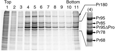
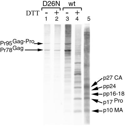
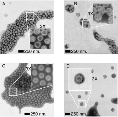
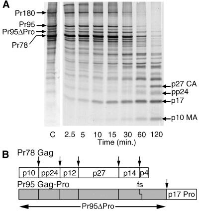
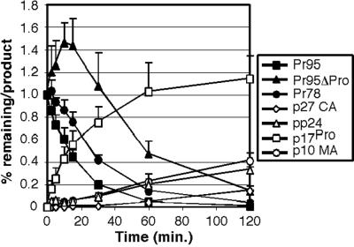
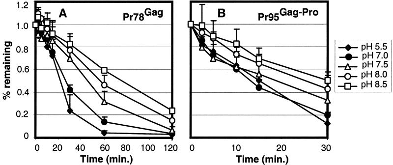
For all retroviruses, the completion of the viral budding process correlates with the activation of the viral protease by an unknown mechanism, and, as the structural (Gag) polyproteins are cleaved by the viral protease, maturation of the immature virus-like particle into an infectious virion. Unlike most retroviruses, the Mason-Pfizer monkey virus Gag polyproteins assemble into immature capsids within the cytoplasm of the cell before the viral budding event. The results reported here describe a unique experimental system in which Mason-Pfizer monkey virus immature capsids are removed from the cell, and the protease is activated in vitro by the addition of a reducing agent. The cleavage of the protease from the precursor form is a primary event, which proceeds with a half time of 14 min, and is followed by authentic processing of the Gag polyproteins. Activity of the viral protease in vitro depends on pH, with an increase in catalytic rates at acidic and neutral pH. The initiation of protease activity within immature capsids in vitro demonstrates that viral protease activity is sensitive to oxidation-reduction conditions, and that the viral protease can be activated in the absence of viral budding.


Figures

Figure 1
Figure 1
Separation of M-PMV immature capsids from cell lysates. As a final purification step, M-PMV immature capsids were centrifuged through a 5–20% (wt/vol) sucrose velocity gradient. The gradient fractions are labeled from Top (lane 1) to Bottom (lane 11, including the pellet). Of each gradient fraction, 1% is analyzed directly by SDS/PAGE. A region of the gel corresponding to lane 4, in which the Gag and Gag-Pro precursors are found, is expanded on the right of the figure for identification of the individual precursor polyproteins. Most of the M-PMV immature capsids sediment into fractions 4–6 and are composed of Pr78Gag, Pr95Gag-Pro, and Pr180Gag-Pro-Pol polyproteins, as well as the Pr95ΔPro cleavage product. Other Gag products include Pr85 and Pr68, which are a consequence of internal initiation during the translation of Pr95 and Pr78, respectively.
Figure 2
Figure 2
Activation of the M-PMV protease within immature capsids in vitro. The contents of M-PMV immature capsids with an inactive protease (D26N, lanes 1 and 2) were analyzed by SDS/PAGE with autofluorography and compared with immature capsids with an active protease (wt, lanes 3 and 4) before (lanes 1 and 3) and after (lanes 2 and 4) treatment with DTT. The cleavage products found in wt immature capsids are compared with the mature viral proteins found within M-PMV virions (lane 5) and are labeled as major capsid protein (p27 CA), uncleaved phosphoprotein (pp24), the cleaved phosphoprotein (pp16–18), the viral protease (p17Pro), and the matrix protein (p10 MA).
Figure 3
Figure 3
Electron microscopy of M-PMV immature capsids. M-PMV immature capsids with an inactive protease (C and D) were imaged by negative stain electron microscopy and compared with immature capsids with an active protease (A and B) before (A and C) and after (B and D) the addition of DTT. The particles within the high-magnification Insets are representative of a minimum of 50 particles observed in multiple images for each sample.
Figure 4
Figure 4
Processing of M-PMV Gag and Gag-Pro precursor polyproteins by the viral protease over time. (A) M-PMV immature capsids with an active viral protease were treated with DTT, and the precursor polyproteins, as well as the cleavage products, were analyzed by SDS/PAGE with autofluorography at measured intervals (2.5, 5, 10, 15, 30, 60, and 120 min) and compared with untreated immature capsids (control, labeled as “C”). The Gag, Gag-Pro, and Gag-Pro-Pol precursor polyproteins are identified as well as the final cleavage products—major capsid protein (p27 CA), phosphoprotein (pp24), protease (p17Pro), and matrix protein (p10 MA). (B) The relationship of the individual Gag and Gag-Pro cleavage products within the precursor polyproteins is diagramed, with arrows delineating the protease cleavage sites, and “fs” delineating the site of the frame shift.
Figure 5
Figure 5
Kinetics of M-PMV Gag and Gag-Pro cleavage into mature viral proteins. The amounts of the Gag (Pr78) and Gag-Pro (Pr95) precursor polyproteins, the Pr95ΔPro cleavage product, were quantitatively measured at precise intervals (2.5, 5, 10, 15, 30, 60, and 120 min) after the addition of DTT and plotted as “% remaining” relative to the amount of precursor measured in an equal quantity of an untreated control sample. The major capsid (p27 CA), phosphoprotein (pp24), protease (p17Pro), and matrix protein (p10 MA) cleavage products were also plotted (as “% product”) relative to the amount of the appropriate precursor(s) in the control sample.
Figure 6
Figure 6
Processing of M-PMV Gag and Gag-Pro precursor polyproteins by the viral protease as a function of reaction pH. The amounts of the Gag [Pr78 (A)] and Gag-Pro [Pr95 (B)] precursor polyproteins were quantitatively measured at precise intervals (2.5, 5, 10, 15, 30, 60, and 120 min) after the addition of DTT, at pH values ranging from 5.5 to 8.5, and plotted relative to the amount of precursor measured in an equal quantity of an untreated control sample. Values for pH 6.0 and 6.5 are omitted for clarity because they overlaid the values for pH 5.5.

Similar articles

Cited by

References

    1. Bryant M, Ratner L. Proc Natl Acad Sci USA. 1990;87:523–527. - PMC - PubMed
    1. Burstein H, Bizub D, Skalka A M. J Virol. 1991;65:6165–6172. - PMC - PubMed
    1. Bennett R P, Nelle T D, Wills J W. J Virol. 1993;67:6487–6498. - PMC - PubMed
    1. Kaplan A H, Manchester M, Everitt L, Swanstrom R. Methods Enzymol. 1994;241:58–69. - PubMed
    1. Kaplan A H, Swanstrom R. Proc Natl Acad Sci USA. 1991;88:4528–4532. - PMC - PubMed

Publication types

MeSH terms

LinkOut - more resources