. 2001 Dec 4;98(25):14428-33.
doi: 10.1073/pnas.261562698. Epub 2001 Nov 27.

Specific interference with gene expression induced by long, double-stranded RNA in mouse embryonal teratocarcinoma cell lines

Affiliations

Specific interference with gene expression induced by long, double-stranded RNA in mouse embryonal teratocarcinoma cell lines

E Billy et al. Proc Natl Acad Sci U S A. .

Abstract

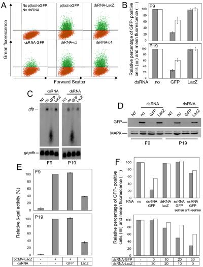
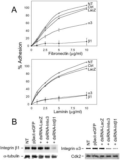
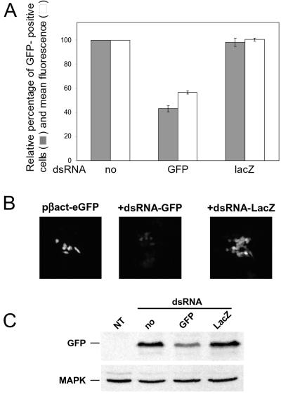
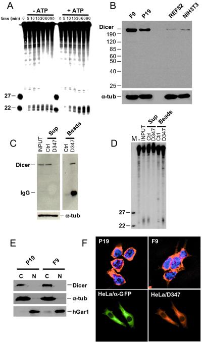
In eukaryotes, double-stranded (ds) RNA induces sequence-specific inhibition of gene expression, referred to as RNA interference (RNAi). In invertebrates, RNAi can be triggered effectively by either long dsRNAs or 21- to 23-nt-long short interfering (si) duplex RNAs, acting as effectors of RNAi. siRNAs recently have been shown to act as potent inducers of RNAi in cultured mammalian cells. However, studies of RNAi activated by long dsRNA are impeded by its nonspecific effects, mediated by dsRNA-dependent protein kinase PKR and RNase L. Here, we report that the RNAi response can be induced effectively by long dsRNA in nondifferentiated mouse cells grown in culture. Transfection of dsRNA into embryonal carcinoma (EC) P19 and F9 cells results in a sequence-specific decrease in the level of proteins expressed from either exogenous or endogenous genes. dsRNA-mediated inhibition of the reporter gene also occurs in mouse embryonic stem cells. The RNAi effect is mediated by siRNAs, which are generated by cleavage of dsRNA by the RNaseIII-like enzyme, Dicer. We demonstrate that extracts prepared from EC cells catalyze processing of dsRNA into approximately 23-nt fragments and that Dicer localizes to the cytoplasm of EC and HeLa cells.


Figures

Figure 1
Figure 1
Effect of dsRNA on activity of genes expressed in transfected EC cells. (A) FACS analysis of F9 cells transfected with pβact-eGFP in the absence or presence of different dsRNAs as indicated. Fluorescent cells are in green. (B) Effect of dsRNA on the number of GFP-positive EC cells and their fluorescence intensity. Cells were transfected with pβact-eGFP in the absence or presence of dsRNA-GFP or dsRNA-LacZ. Values (means ± SD; n = 4) for the number of fluorescent cells and mean fluorescence were calculated relative to the GFP-positive cells transfected with only pβact-eGFP. (C) Northern analysis of GFP mRNA levels in cells transfected with either pβact-eGFP alone or cotransfected with dsRNA-GFP or dsRNA-LacZ. NT, nontransfected cells. (Lower) Hybridization of the same blot with the glyceraldehyde-3-phosphate dehydrogenase mRNA-specific probe. (D) Western blot analysis of GFP levels in cells transfected with either pβact-eGFP alone or cotransfected with dsRNA-GFP or dsRNA-LacZ. (Lower) Reprobing with α-mitogen-associated protein kinase Ab. (E) Effect of dsRNA on β-gal activity in extracts of cells transfected with pCMV-LacZ and indicated dsRNA. Activity was measured by using the chemiluminescent β-gal assay (Roche). Values represent averages of two independent experiments, with the range indicated. (F) Effect of GFP ssRNAs (Upper) and increasing doses of cotransfected dsRNA-GFP (Lower) on the number and fluorescence intensity of GFP-positive P19 cells. Cells were transfected with pβact-eGFP alone or in the presence of dsRNA-GFP, or dsRNA-LacZ, or ssRNA-GFP of the sense or antisense orientation. (Lower) Cells were transfected with pβact-eGFP alone or in the presence of 10, 20, or 30 μg of dsRNA-GFP supplemented with dsRNA-LacZ to a total amount of 30 μg. Values are averages from two experiments.
Figure 2
Figure 2
dsRNA-mediated inhibition of integrin β1 and α3 expression in F9 cells. (A) Effect of dsRNA-β1 (β1) and dsRNA-α3 (α3) on adhesion to fibronectin (Upper) and laminin (Lower) of the FACS-sorted, GFP-positive cells. Aliquots of 103 cells were spotted in the plate wells precoated with indicated concentrations of either fibronectin or laminin. Values represent means of four determinations originating from two independent transfection and sorting experiments. SD values are shown only for cells treated with dsRNA-α3 and dsRNA-β1. Other curves represent nontransfected cells (NT) and cells transfected with pβact-eGFP alone (Ctrl) or in the presence of dsRNA-LacZ (LacZ). One hundred percent values correspond to the number of control (Ctrl) cells (60) attached at 10-μg/ml ligand concentration. (B) Western blot analysis of protein extracts (10 μg) from indicated transfected cells (described in A) with Abs specific for integrins β1 and α3. Blots were reprobed with anti-α-tubulin or α-Cdk2 Abs, as indicated.
Figure 3
Figure 3
Inhibition of GFP expression in mouse ES cells. (A) Effect of dsRNA on the number and fluorescence intensity of ES cells transfected with either pβact-eGFP alone or in the presence of dsRNA-GFP or dsRNA-LacZ. Values were calculated relative to the eGFP-positive cells transfected with only pβact-eGFP. They are averages of two independent experiments, with the range indicated. (B) A representative example of the in vivo imaging of ES cells transfected with pβact-eGFP alone or cotransfected with dsRNA-GFP or dsRNA-LacZ, as indicated. The fluorescence intensity scale obtained from pβact-eGFP-transfected cells was applied to cells cotransfected with pβact-eGFP and dsRNA-eGFP or dsRNA-LacZ. Cells were grown on coverslips and imaged 48 h posttransfection. (C) Western blot analysis of GFP levels in extracts of ES cells transfected with either pβact-eGFP alone or cotransfected with either dsRNA-GFP or dsRNA-LacZ. NT, nontransfected cells. GFP was detected with α-GFP Ab. (Lower) Reprobing with α-mitogen-associated protein kinase Ab.
Figure 4
Figure 4
In vitro cleavage of dsRNA and cellular localization of Dicer. (A) Processing of dsRNA to ≈22-nt RNAs in cytoplasmic extract of F9 cells. Reactions were incubated for the indicated time, in the presence or absence of ATP, and analyzed by PAGE-Urea. Extracts used for reactions without ATP were preincubated with hexokinase and glucose. Size markers are 22- and 27-nt oligoribonucleotides. (B) Western blot analysis of total cell extracts prepared from F9, P19, NIH 3T3, and REF52 cells. Proteins (≈75 μg) were separated in an 8% SDS/PAGE, and the blot was probed sequentially with α-Dicer D347 and anti-α-tubulin Abs. (C) Immunodepletion of Dicer from F9 cytoplasmic extract by incubation with D347 Ab-coated Protein A-Sepharose beads. As control (Ctrl), the extract was incubated with noncoated Protein A-Sepharose beads. Supernatant (Sup) and beads fractions were analyzed by Western blots, using D347 and anti-α-tubulin Abs. (D) Analysis of the dsRNA-processing activity present in the immunoprecipitation fractions described in C. dsRNA (123 bp) was used as a substrate. (E) Western blot analysis of cytoplasmic (C) and nuclear (N) fractions of EC cells using D347 Ab. Reprobing of the blot with Abs against the nuclear protein hGAR1 and the cytoplasmic α-tubulin indicated no major cross-contamination of fractions. (F Upper) Indirect immunolocalization of endogenous Dicer in F9 and P19 cells, using D347 Ab and Texas red-labeled α-rabbit secondary Ab. Nuclei are stained with 4′,6-diamidino-2-phenylindole. (Lower) Localization of the CFP-Dicer fusion protein in HeLa cells transfected with pCFP-Dicer, using α-GFP and α-Dicer D347 Abs.

Similar articles

Cited by

References

    1. Hammond S M, Caudy A A, Hannon G J. Nat Rev Genet. 2001;2:110–119. - PubMed
    1. Matzke M A, Matzke A J, Pruss G J, Vance V B. Curr Opin Genet Dev. 2001;11:221–227. - PubMed
    1. Sharp P A. Genes Dev. 2001;15:485–490. - PubMed
    1. Grishok A, Pasquinelli A E, Conte D, Li N, Parrish S, Ha I, Baillie D L, Fire A, Ruvkun G, Mello C C. Cell. 2001;106:23–34. - PubMed
    1. Hutvagner G, McLachlan J, Pasquinelli A E, Balint E, Tuschl T, Zamore P D. Science. 2001;293:834–838. - PubMed

Publication types