. 2001 Dec 4;98(25):14541-6.
doi: 10.1073/pnas.261562798. Epub 2001 Nov 27.

Flk-2 is a marker in hematopoietic stem cell differentiation: a simple method to isolate long-term stem cells

Affiliations

Flk-2 is a marker in hematopoietic stem cell differentiation: a simple method to isolate long-term stem cells

J L Christensen et al. Proc Natl Acad Sci U S A. .

Abstract

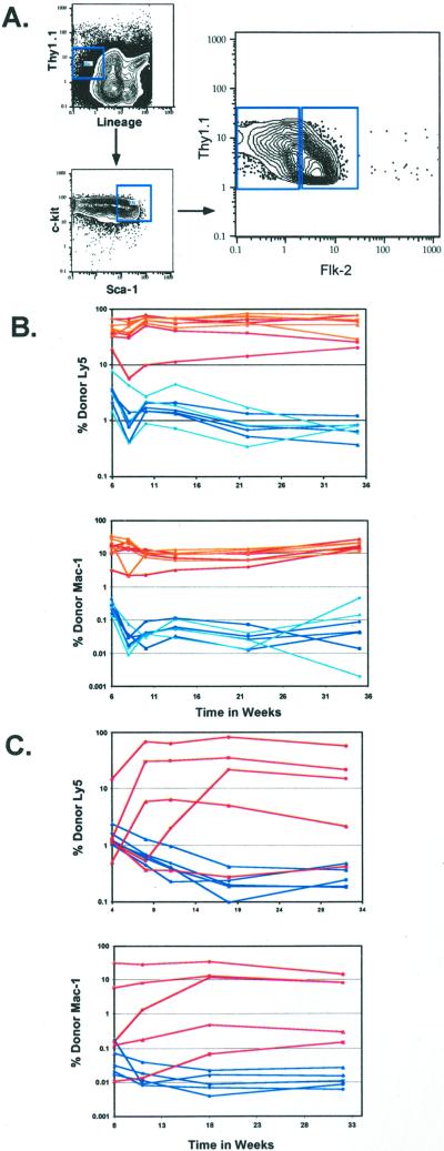
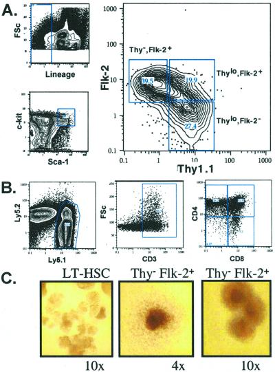
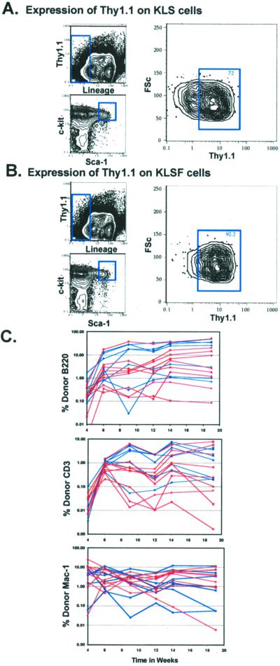
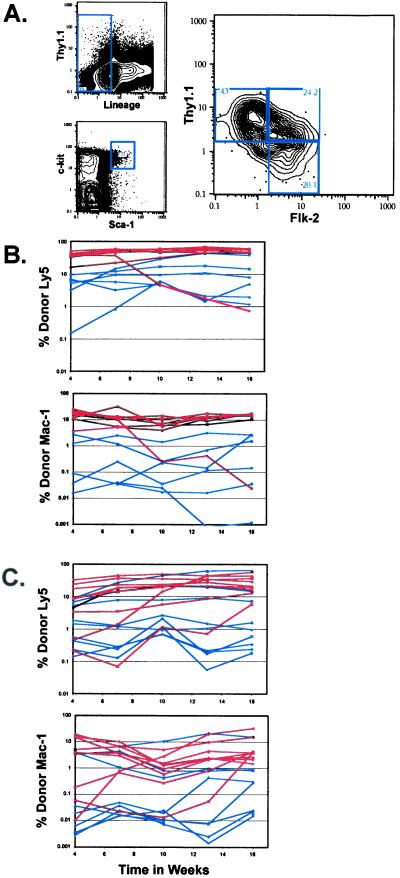
Clonogenic multipotent mouse hematopoietic stem cells (HSCs) and progenitor cells are contained within the c-kit(+) (K) lineage(-/lo) (L) Sca-1(+) (S) population of hematopoietic cells; long-term (LT) and short-term (ST) HSCs are Thy-1.1(lo). c-kit is a member of the receptor tyrosine kinase family, a class of receptors that are important in the proliferation and differentiation of hematopoietic cells. To establish whether the Flk-2/Flt3 receptor tyrosine kinase was expressed on the most primitive LT-HSCs, we sorted highly purified multipotent stem and progenitor cells on the basis of Flk-2 surface expression and used them in competitive reconstitution assays. Low numbers of Flk-2(-) HSCs gave rise to long-term multilineage reconstitution in the majority of recipients, whereas the transfer of Flk-2(+) multipotent cells resulted in mostly short-term multilineage reconstitution. The KLS subset of adult mouse bone marrow was analyzed for Flk-2 and Thy-1.1 expression. Three phenotypically and functionally distinct populations were isolated: Thy(lo) Flk-2(-) (LT-HSCs), Thy(lo) Flk-2(+) (ST-HSCs), and Thy(-) Flk-2(+) multipotent progenitors. The loss of Thy-1.1 and gain of Flk-2 expression marks the loss of self-renewal in HSC maturation. The addition of Flk-2 antibody to the lineage mix allows direct isolation of LT-HSC from adult bone marrow as c-kit(+) lin(-) Sca-1(+) Flk-2(-) from many strains of mice. Fetal liver HSCs are contained within Flk-2(-) and Flk-2(+) KTLS cells.


Figures

Figure 1
Figure 1
Heterogeneous expression of Flk-2 on the HSC population. (A) Flk-2 expression was analyzed on the HSC population; Lin−/lo Thy-1.1lo c-kit+ Sca-1+ of c-kit enriched BM. HSCs that were either Flk-2+ or Flk-2 were sorted. Lethally irradiated recipients were competitively reconstituted with 50–100 (B) or 20 (C) Flk-2+ or Flk-2 HSCs plus host-type radioprotective WBM. (B) Fifty Flk-2+ HSCs are shown in blue, 100 flk-2+ HSCs are in light blue, 50 Flk-2 HSCs are in red, and 100 Flk-2 HSCs are in orange. (C) Twenty Flk-2+ HSCs are shown in blue, and 20 Flk-2 HSCs are in red. Peripheral blood was analyzed for donor Ly5 B, T, and myeloid contribution. Each curve represents an individual animal. Because myeloid cells are present in the blood for a short period, myeloid activity is shown as a good indicator of stem cell activity. The background is 0.1%.
Figure 2
Figure 2
(A) Characterization of the Lin−/lo c-kit+ Sca-1+ population of c-kit-enriched murine WBM for Thy-1.1 and Flk-2 expression. (B) T cell read-out from 20 Thy-1.1 Flk-2+ cells injected intrathymically 3 weeks postinjection. (C) In vitro methylcellulose colonies derived from a single HSC or Thy-1.1 Flk-2+ progenitor. (Left) A single HSC forms many blast-like colonies at early time points (day 7). (Center and Right) Thy-1.1 Flk-2+ KLS cells do not form blast colonies. Although some form a single large colony of differentiated cell types, others form multifoci colonies.
Figure 3
Figure 3
HSC purification by the addition of Flk-2 to the lineage mix. Characterization of Thy-1.1 expression on the KLS (c-kit+ Lin Sca-1+) population, with the lineage mixture either including (B) or not including (A) anti-Flk-2. (C) Lethally irradiated recipients were reconstituted competitively with 10 Thy-1.1lo KTLS HSCs (c-kit+ Thy-1.1lo Lin Sca-1+) or 10 Flk-2 KLSF HSCs (c-kit+ Lin Sca-1+ Flk-2). Flk-2 HSCs (KLSF) were sorted by including Flk-2 in the lineage mixture while omitting Thy-1.1 from the sort conditions. Mice reconstituted with KTLS cells are shown in blue, and KLSF cells are shown in red.
Figure 4
Figure 4
(A) Analysis of Flk-2 and Thy-1.1 on the Lin−/lo c-kit+ Sca-1+ subset of fetal liver. Fetal liver HSCs (c-kit+ Thy-1.1lo Lin−/lo Sca-1+) that were either positive or negative for Flk-2 expression were sorted. Lethally irradiated recipients were reconstituted competitively with 50 (B) or 20 (C) Flk-2+ or Flk-2 fetal liver HSCs. Mice reconstituted with Flk-2+ fetal liver HSCs are shown in blue, and those reconstituted with Flk-2 fetal liver HSCs are in red. Peripheral blood was analyzed for donor B, T, or myeloid contribution.

Similar articles

Cited by

References

    1. Jones R J, Wagner J E, Celano P, Zicha M S, Sharkis S J. Nature (London) 1990;347:188–189. - PubMed
    1. Fleming W H, Alpern E J, Uchida N, Ikuta K, Spangrude G J, Weissman I L. J Cell Biol. 1993;122:897–902. - PMC - PubMed
    1. Spangrude G J, Johnson G R. Proc Natl Acad Sci USA. 1990;87:7433–7437. - PMC - PubMed
    1. Wiesmann A, Phillips R L, Mojica M, Pierce L J, Searles A E, Spangrude G J, Lemischka I. Immunity. 2000;12:193–199. - PubMed
    1. Morel F, Galy A, Chen B, Szilvassy S J. Exp Hematol (Charlottesville, Va) 1998;26:440–448. - PubMed

Publication types

MeSH terms