. 2001 Dec;25(6):676-84.
doi: 10.1165/ajrcmb.25.6.4321.

Ceramide-mediated apoptosis in lung epithelial cells is regulated by glutathione

Affiliations

Ceramide-mediated apoptosis in lung epithelial cells is regulated by glutathione

S N Lavrentiadou et al. Am J Respir Cell Mol Biol. 2001 Dec.

Abstract

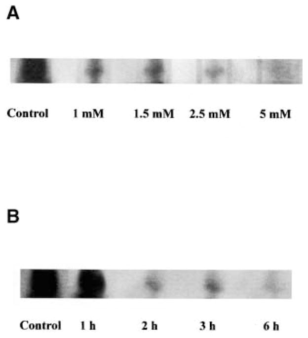
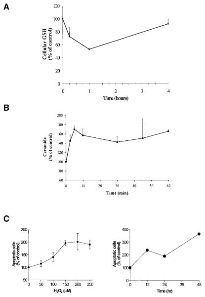
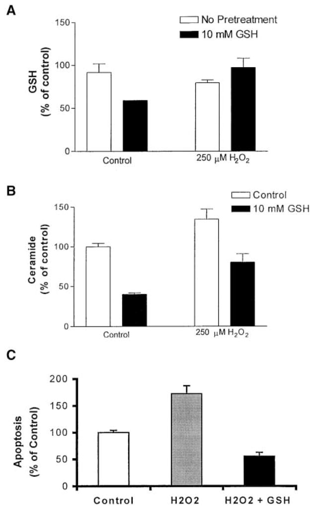
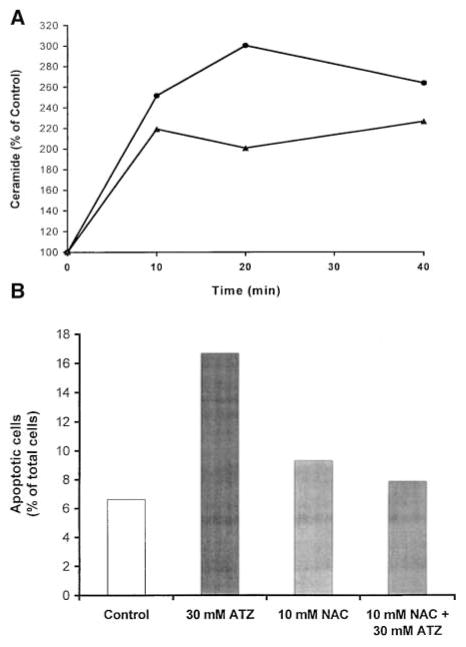
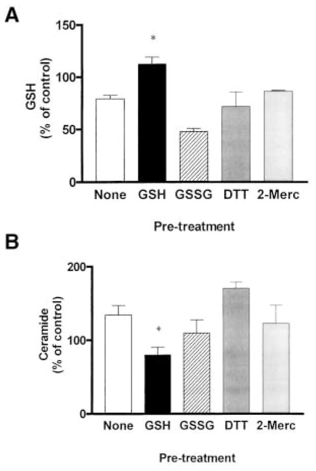
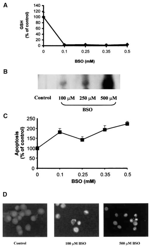
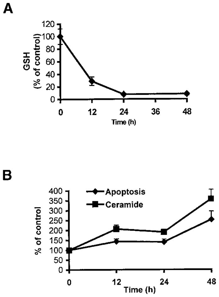
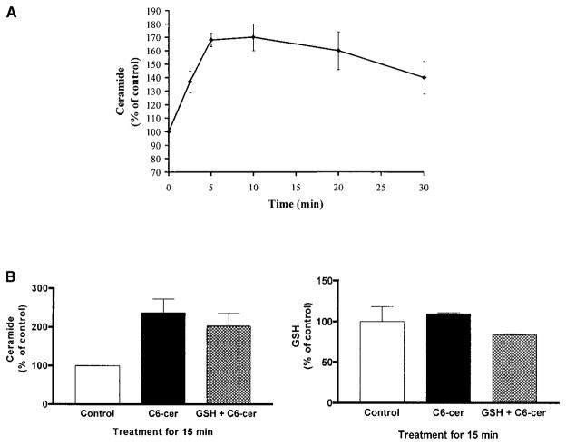
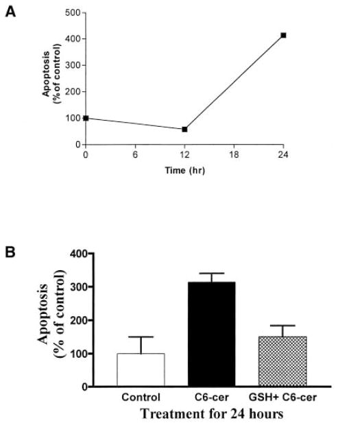
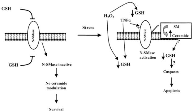
Reactive oxygen species (ROS) are mediators of lung injury, and glutathione (GSH) is the major nonprotein antioxidant that protects the cell from oxidative stress. We have recently shown that H(2)O(2) induces ceramide-mediated apoptosis in human lung epithelial cells. We hypothesized that ROS-mediated depletion of GSH plays a regulatory role in ceramide generation, and thus in the induction of apoptosis. Our present studies demonstrate that GSH at physiologic concentrations (1 to 10 mM) inhibits ceramide production in a time- and dose-dependent manner in A549 human alveolar epithelial cells. On the other hand, buthionine-sulfoximine-mediated depletion of intracellular GSH induces elevation of ceramide levels and apoptosis. In addition, GSH blocks H(2)O(2)-mediated induction of intracellular ceramide generation and apoptosis. These effects were not mimicked by oxidized GSH (GSSG) or other thiol antioxidants, such as dithiothreitol and 2-mercaptoethanol. Moreover, increase of intracellular H(2)O(2), mediated by inhibition of catalase by aminotriazole, also induces ceramide generation and apoptosis. These effects were blocked by N-acetylcysteine. Our results suggest that GSH depletion may be the link between oxidative stress and ceramide-mediated apoptosis in the lung.


Figures

Figure 1
Figure 1
Extracellularly supplemented GSH inhibits ceramide generation in A549 cells. Autoradiography of ceramide in lipid extracts of A549 cells treated in the presence of regular medium supplemented with 1% serum with (A) increasing concentrations of GSH (0 to 5 mM) for 3 h or (B) 5 mM GSH for different incubation times. Incubations were terminated by washes with ice-cold PBS, and the cells were harvested with 0.05% trypsin-EDTA. Cellular lipids were extracted and assayed for ceramide by the DAG kinase assay, as described previously (5, 6). The reaction products were analyzed by TLC and autoradiography.
Figure 2
Figure 2
H2O2 mediates induction of intracellular ceramide levels and apoptosis via depletion of intracellular GSH. A549 cells were incubated in medium supplemented with 1% serum with 250 μM H2O2 for the indicated times. (A). Cell lysates were analyzed for GSH as described in Materials and Methods. (B). Cellular lipids were extracted and assayed for ceramide by the DAG kinase assay (5, 6). The reaction products were analyzed by TLC and quantified using a phosphorimager. (C). To determine apoptosis, cells were stained with Annexin V-FITC and PI, and were evaluated by FACS analysis, as described in Materials and Methods. The values are represented as percent (%) of control, not treated, cells and represent mean ± SEM.
Figure 3
Figure 3
Extracellular supplementation of GSH prevents H2O2-induced GSH depletion, ceramide generation, and apoptosis in human lung epithelial cells. A549 cells were incubated in medium supplemented with 1% serum with 10 mM GSH for 30 min, followed by incubations with 250 μM H2O2 for an additional 30 min (A and B) or 24 h (C). (A). Cell lysates were analyzed for GSH as described in Materials and Methods. (B). Cellular lipids were extracted and assayed for ceramide by the DAG kinase assay (5, 6). The reaction products were analyzed by TLC and quantified using a phosphorimager. (C). Apoptotic cells were stained with Annexin V-PI and detected by FACS. The values are represented as percent (%) of control, not treated, cells.
Figure 4
Figure 4
ATZ induces ceramide and apoptosis, effects efficiently inhibited by NAC. (A). Primate tracheal epithelial cells were incubated with 30 mM ATZ for the indicated times with (solid triangles) or without (solid circles) preincubation with 10 mM NAC for 1 h. Cellular lipids were analyzed for ceramide by the DAG kinase assay, as described in Materials and Methods. (B). Cells were incubated with 30 mM ATZ for 18 h with or without preincubation with 10 mM NAC for 1 h. After treatments, cells were evaluated for apoptosis by TUNEL. The values represent apoptotic cells (percent of total cells counted) where at least 300 cells were counted.
Figure 5
Figure 5
GSH, but not GSSG or other antioxidants, inhibits ceramide generation induced by H2O2 in lung epithelial cells. A549 cells were preincubated for 1 h in medium supplemented with 1% serum in the presence or absence of 10 mM of GSH, GSSG, DTT, or 2-mercaptoethanol, followed by incubations with 250 μM H2O2 for an additional 30 min. The cells were rinsed with ice-cold PBS to terminate the treatments and harvested by trypsinization. GSH (A) and ceramide (B) levels were determined as described in Materials and Methods. Values represent mean ± SEM. *Mean of the group that was significantly different from the mean of the H2O2-treated group (P < 0.05). All other comparisons were statistically not significant (P > 0.05). Statistical analysis was performed using the Mann-Whitney, nonparametric, two-tailed test.
Figure 6
Figure 6
BSO depletes cellular GSH and induces ceramide production and apoptosis in a dose-dependent manner. After treatments with the indicated concentrations of BSO for 24 h, A549 cells were washed with ice-cold PBS and collected by trypsinization. (A). The cells were lysed and cellular GSH content was determined as previously described (21). (B). Treated cells were extracted with methanol:chloroform:1 N HCl, and cellular ceramide levels were determined by the DAG kinase assay (6). (C). To determine apoptosis, cells were stained with Annexin V-FITC and PI, and were evaluated by FACS analysis, as described in Materials and Methods. Values represent mean ± SEM. (D). Primary cells, after treatments with 250 or 500 μM BSO for 24 h, were evaluated for apoptosis by staining with Hoechst, as described in Materials and Methods.
Figure 7
Figure 7
BSO depletes cellular GSH and induces ceramide production and apoptosis in a time-dependent manner. After treatments with 250 μM BSO for the indicated times, A549 cells were washed with ice-cold PBS and collected by trypsinization. (A). Cells were lysed and cellular GSH content was determined as previously described (21). (B). Treated cells were extracted with methanol:chloroform:1 N HCl, and cellular ceramide levels were determined by the DAG kinase assay. Treated cells were also stained with Annexin V-FITC and PI, and were evaluated for apoptosis, as described in Materials and Methods. Values represent mean ± SEM.
Figure 8
Figure 8
C6-ceramide mediates ceramide generation without depletion of GSH. (A). A549 cells were treated with 25 μM C6-cer-amide for the indicated times. After treatment, cellular lipids were extracted and assayed for ceramide by the DAG kinase assay (5, 6). The reaction products were analyzed by TLC and quantified using a phosphorimager. (B). A549 cells were preincubated with (hatched bars) or without (solid bars) 10 mM GSH for 1 h before exposure to 25 μM C6-ceramide for 15 min. After treatment, cell lysates were analyzed for ceramide and GSH, as described in Materials and Methods. All values are represented as percent (%) of control.
Figure 9
Figure 9
C6-ceramide induces apoptosis, an effect efficiently inhibited by GSH. (A). A549 cells were treated with 25 μM C6-ceramide for the indicated times. (B). Cells were preincubated with (hatched bars) or without (solid bars) 10 mM GSH 1 h before exposure to 25 μM C6-ceramide for 24 h. After treatments, apoptosis was determined by Annexin V staining of apoptotic cells, as described in Materials and Methods. All values are represented as percent (%) of control.
Figure 10
Figure 10
Schematic representation of the role of oxidants (H2O2) and antioxidants (GSH) in ceramide generation and apoptosis. GSH inhibits N-SMase activity, thus maintaining low ceramide levels (19, 20). Stress factors (i.e., extracellularly administered H2O2) result in ROS-mediated depletion of GSH. This may be a critical event in the induction of apoptosis by activation of N-SMase and generation of ceramide. However, supplementation of antioxidants, such as GSH, counteract this effect by inhibiting N-SMase activity. Therefore, the redox state of the cell determines the activity of N-SMase and the levels of ceramide, thus modulating the apoptotic pathway in lung epithelial cells.

Comment in

References

    1. Cross CE, van der Vliet A, O’Neill CA, Louie S, Halliwell B. Oxidants, antioxidants, and respiratory tract lining fluids. Environ Health Perspect. 1994;102(Suppl 10):185–191. - PMC - PubMed
    1. Rahman I, Li XY, Donaldson K, Harrison DJ, MacNee W. Glutathione homeostasis in alveolar epithelial cells in vitro and lung in vivo under oxidative stress. Am J Physiol. 1995;269(3 Pt 1):L285–L292. - PubMed
    1. Mulier B, Rahman I, Watchorn T, Donaldson K, MacNee W, Jeffery PK. Hydrogen peroxide-induced epithelial injury: the protective role of intracellular nonprotein thiols (NPSH) Eur Respir J. 1998;11:384–391. - PubMed
    1. Rahman I, MacNee W. Oxidative stress and regulation of glutathione in lung inflammation. Eur Respir J. 2000;16:534–554. - PubMed
    1. Goldkorn T, Balaban N, Shannon M, Chea V, Matsukuma K, Gilchrist D, Wang H, Chan C. H2O2 acts on cellular membranes to generate ceramide signaling and initiate apoptosis in tracheobronchial epithelial cells. J Cell Sci. 1998;111(Pt. 21):3209–3220. - PubMed

MeSH terms