. 2001 Dec 3;20(23):6712-23.
doi: 10.1093/emboj/20.23.6712.

Nuclear export of phosphorylated C/EBPbeta mediates the inhibition of albumin expression by TNF-alpha

Affiliations

Nuclear export of phosphorylated C/EBPbeta mediates the inhibition of albumin expression by TNF-alpha

M Buck et al. EMBO J. .

Abstract

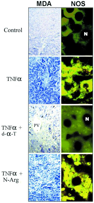
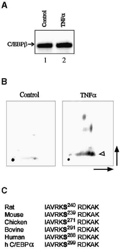
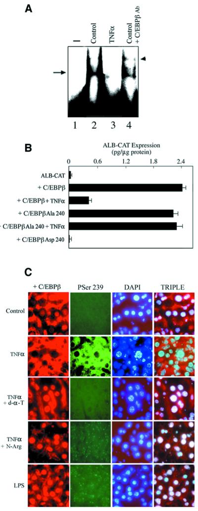
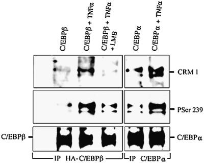
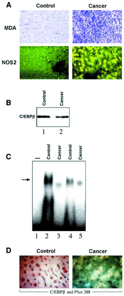
Decreased albumin expression is a frequent feature of cachexia patients afflicted with chronic diseases, including cancer, and a major contributor to their morbidity. Here we show that tumor necrosis-alpha (TNF-alpha) treatment of primary mouse hepatocytes or TNF-alpha overexpression in a mouse model of cachexia induces oxidative stress, nitric oxide synthase (NOS) expression and phosphorylation of C/EBPbeta on Ser239, within the nuclear localization signal, thus inducing its nuclear export, which inhibits transcription from the albumin gene. SIN-1, a NO donor, duplicated the TNF-alpha effects on hepatocytes. We found similar molecular abnormalities in the liver of patients with cancer-cachexia. The cytoplasmic localization and association of C/EBPbeta-PSer239 with CRM1 (exportin-1) in TNF-alpha-treated hepatocytes was inhibited by leptomycin B, a blocker of CRM1 activity. Hepatic cells expressing the non-phosphorylatable C/EBPbeta alanine mutant were refractory to the inhibitory effects of TNF-alpha on albumin transcription since the mutant remained localized to the nucleus. Treatment of TNF-alpha mice with antioxidants or NOS inhibitors prevented phosphorylation of C/EBPbeta on Ser239 and its nuclear export, and rescued the abnormal albumin gene expression.


Figures

None
Fig. 1. Increased oxidative stress and NOS2 in the liver of cachectic mice. The experimental groups are as described in Materials and methods. Representative examples (n = 8 in each group) of the immunohistochemistry for malondialdehyde (MDA)–protein adducts and NOS2, using antibodies specific for MDA–lysine adducts and NOS2. PV indicates portal venules. Negligible staining was observed in all immunohistochemistries when the first antibody was omitted.
None
Fig. 2. Oxidative stress and NO inhibit albumin expression in the liver of cachectic mice. (A) The experimental groups are as described in Materials and methods. Representative examples (n = 6 in each group) of albumin mRNA detected by an RNase protection assay with a specific riboprobe, using equal amounts of total RNA. The 18S RNA was utilized as a correction factor for loading. Albumin mRNA in livers from CHO (lane 1), TNF-α (lane 2), TNF-α/d-α-tocopherol (lane 3) and TNF-α/nitro-l-arginine (lane 4). P <0.05 for TNF-α. (B) Mice received TNF-α alone from 8 to 92 h (closed circles) or with d-α-tocopherol from 24 to 72 h (open circles) as described in Materials and methods. Animals were sacrificed as indicated and liver albumin mRNA was determined as in (A). The results are averages of at least triplicate samples. P <0.05 for d-α-tocopherol at 72 and 96 h. (C) Mobility shift analysis of liver nuclear extracts was performed using equal amounts of nuclear protein (5 µg) and following incubation with a 32P-labeled oligonucleotide (1 ng) spanning the D-site or the B-site of the albumin enhancer/promoter or the control Sp1 site. The position of the bound DNA is indicated by arrows. Representative samples (n = 5 in each group) are shown: CHO (2); TNF-α (3); TNF-α/d-α-tocopherol (4); TNF-α/BW755c (5); and TNF-α/nitro-l-arginine (6). On lane 1, the probe was processed without nuclear extracts. The D-site binding activities were CHO (100%), TNF-α (22%), TNF-α/d-α-tocopherol (84%), TNF-α/BW755c (67%) and TNF-α/nitro-l-arginine (76%). (D) HepG2 human hepatoma cells were transfected with ALB-CAT (1 µg) alone or with CMV-C/EBPβ (1 µg) and treated every 24 h with TNF-α (10 ng/ml); TNF-α + d-α-tocopherol (50 µM); TNF-α + nitro-l-arginine (500 µM); and SIN-1 (0.6 µM) as indicated. After 48 h, the cells were collected and CAT expression was determined. The results are averages (± SEM) of triplicate samples, and representative of four independent experiments. P <0.05 for C/EBPβ + TNF-α and C/EBPβ + SIN-1 compared with C/EBPβ.
None
Fig. 3. TNF-α stimulates phosphorylation of C/EBPβ in its DNA-binding domain. (A) Representative examples (n = 5 in each group) of C/EBPβ protein immunoblots in liver lysates from CHO (lane 1) and TNF-α (lane 2) groups. The antigen–antibody complexes were visualized using the Renaissance detection system (DuPont). (B) Tryptic phosphopeptide maps of C/EBPβ immunopurified from day-5 primary mouse hepatocytes cultured on a collagen type I matrix. Cells were labeled with [32P]orthophosphate (2 mCi/ml; total 30 mCi) for 18 h and either treated with TNF-α (10 ng/ml) for 30 min before harvest or not treated (control). C/EBPβ tryptic peptides were separated by high-voltage electrophoresis (horizontal dimension) followed by ascending thin-layer chromatography (vertical dimension). The level of the phosphopeptide containing Ser239 (arrowhead), identified as described previously (Trautwein et al., 1994), was increased after treatment with TNF-α. (C) The basic domain serine phosphoacceptor is conserved through evolution. Rat (S240), mouse (S239), chicken (S271), bovine (S291) and human (S288) phosphoacceptors, within the basic domain of C/EBPβ, are shown. An identical phosphoacceptor is present in human (h) C/EBPα (S299).
None
Fig. 4. Phosphorylation of C/EBPβ in its basic domain induced by TNF-α inhibits albumin expression. (A) Mobility shift analysis of nuclear extracts from day 5 primary mouse hepatocytes was performed using equal amounts of nuclear protein (5 µg) and following incubation with a 32P-labeled oligonucleotide (1 ng) spanning the D-site of the albumin enhancer/promoter. The position of the bound DNA is indicated by the arrow and the supershifted DNA is indicated by the arrowhead. Representative samples are shown: control (2); TNF-α (10 ng/ml for 30 min) (3); and control + C/EBPβ antibody (4). On lane 1, the probe was processed without nuclear extracts. (B) HepG2 cells were transfected with ALB-CAT (1 µg) alone or with 1 µg of CMV-C/EBPβ, CMV-C/EBPβ-Ala240 or CMV-C/EBPβ-Asp240, and treated with TNF-α (10 ng/ml) every 24 h as indicated. After 48 h, the cells were collected and CAT expression was determined. The results are averages (± SEM) of triplicate samples, and representative of five independent experiments. P <0.05 for C/EBPβ + TNF-α and C/EBPβ-Asp240 compared with C/EBPβ. (C) Representative examples (n = 8 in each group) of the triple channel immunofluorescence microscopy using antibodies for C/EBPβ and C/EBPβ-PSer239 simultaneously. Hepatocytes from control, TNFα/d-α-tocopherol-, TNF-α/nitro-l-arginine- and LPS-treated mice displayed nuclear C/EBPβ (in red) but not C/EBPβ-PSer-239 (in green). In hepatocytes from TNF-α livers, the cytoplasmic co-localization of antibodies against C/EBPβ (red) and C/EBPβ-PSer-239 (green) is shown in yellow. Nuclei are stained with DAPI (blue); co-localization of C/EBPβ (red) and DAPI (blue) is shown in white.
None
Fig. 5. Nuclear export of C/EBPβ-PSer-239 is mediated by CRM1 in hepatocytes treated with TNFα. Day 5 primary hepatocytes were isolated form C/EBPβ–/– mice. Representative confocal microscopy (n = 5 in each group). (A) Mouse hepatocytes were transfected with C/EBPβ, treated as described in Materials and methods and stained with antibodies against HA and CRM1. In C/EBPβ and TNF-α + leptomycin B cells, C/EBPβ (red) is nuclear and CRM1 (green) is nuclear and perinuclear. In TNF-α cells, C/EBPβ and CRM1 are co-localized in the cytoplasm (in yellow). Nuclei are stained with TOTO-3 (blue). (B) Mouse hepatocytes were transfected with CMV vectors expressing C/EBPβ-Ala239 or C/EBPβ-Asp239 for 24 h, treated as described in Materials and methods and stained with antibodies against HA and CRM1. C/EBPβ-Ala239 localized to the nucleus (in red) in untreated and TNF-α-treated hepatocytes. C/EBPβ-Asp239 co-localized with CRM1 in the perinuclear region (in yellow). Nuclei are stained with TOTO-3 (blue). (C) Cells were stained with antibodies against C/EBPα and CRM1. Control hepatocytes expressed nuclear C/EBPα. TNF-α treatment induced the nuclear export of C/EBPα, which co-localized with CRM1 in the perinuclear region (in yellow). Nuclei are stained with TOTO-3 (blue). (D) Mouse hepatocytes were treated with TNF-α or SIN-1 as described in Materials and methods, and stained with antibodies against HA and PSer239. TNFα and SIN-1 treatments induced the phosphorylation of C/EBPβ on Ser239 and its nuclear export. The cytoplasmic co-localization of C/EBPβ-PSer239 (green) and C/EBPβ (red) antibodies is shown in yellow. Control hepatocytes displayed mainly nuclear C/EBPβ (red) but not C/EBPβ-PSer239. Nuclei are stained with TOTO-3 (blue).
None
Fig. 6. C/EBPβ-PSer239 is associated with CRM1 in hepatocytes treated with TNF-α. Day 5 primary mouse C/EBPβ–/– hepatocytes were transfected with C/EBPβ-HA or not. After 24 h, cells were treated for 30 min as indicated. Cell lysates (500 µg) were precipitated with either HA or C/EBPα antibodies. Protein blotting of immunoprecipitates was performed as described in Materials and methods, using specific antibodies against CRM1, C/EBPβ-PSer239, C/EBPβ or C/EBPα.
None
Fig. 7. Nuclear export of C/EBPβ-Ser288 in the liver of patients with cancer-cachexia. (A) Representative immunohistochemistry for MDA–protein adducts, NOS2, C/EBPβ and C/EBPβ-PSer288 of control individuals (control) and cancer-cachexia patients (cachexia) was performed as described in Figures 1 and 4. (B) A representative C/EBPβ protein immunoblot in C/EBPβ immunoprecipitates from liver protein lysates (250 µg) of control (lane 1) and cancer-cachexia (lane 2) subjects was performed as described in Materials and methods. (C) Mobility shift analysis of liver nuclear extracts (5 µg of protein) and the 32P-labeled D-site of the albumin enhancer/promoter (1 ng) was performed as described in Figure 2. The position of the bound DNA is indicated by an arrow. Samples shown are: control (lanes 2 and 4); cancer-cachexia (lanes 3 and 5). On lane 1, the probe was processed without nuclear extracts. (D) Representative immunohistochemistry using antibodies for C/EBPβ (in red) and C/EBPβ-PSer288 (in green), simultaneously. In control liver, C/EBPβ was localized in the nucleus and C/EBPβ-PSer288 was undetectable. In cancer-cachexia liver, C/EBPβ-PSer288 was detected in the cytoplasm in yellow due to the superimposition of C/EBPβ (red) and C/EBPβ-PSer288 (green).

Similar articles

Cited by

References

    1. Akira S., Isshiki,H., Sugita,T., Tanabe,O., Kinoshita,S., Nishio,Y., Nakajima,T., Hirano,T. and Kishimoto,T. (1990a) A nuclear factor for IL-6 expression (NF-IL6) is a member of a C/EBP family. EMBO J., 9, 1897–1906. - PMC - PubMed
    1. Akira S., Hirano,T., Taga,T. and Kishimoto,T. (1990b) Biology of multifunctional cytokines; IL 6 and related molecules (IL and TNF). FASEB J., 4, 2860–2867. - PubMed
    1. Alonzi T., Maritano,D., Gorgoni,B., Rizzuto,G., Libert,C. and Poli,V. (2001) Essential role of STAT3 in the control of the acute-phase response as revealed by inducible gene activation in the liver. Mol. Cell. Biol., 21, 1621–1632. - PMC - PubMed
    1. Beutler B. (1992) Tumor Necrosis Factors: The Molecules and Their Emerging Role in Medicine. Raven Press, New York.
    1. Boyle W.J., van der Geer,P. and Hunter,T. (1991) Phosphopeptide mapping and phosphoamino acid analysis by two-dimensional separation on thin-layer cellulose plates. Methods Enzymol., 201, 110–149. - PubMed

Publication types

MeSH terms