. 2001 Dec 3;20(23):6772-82.
doi: 10.1093/emboj/20.23.6772.

Calreticulin and calnexin in the endoplasmic reticulum are important for phagocytosis

Affiliations

Calreticulin and calnexin in the endoplasmic reticulum are important for phagocytosis

A Müller-Taubenberger et al. EMBO J. .

Abstract

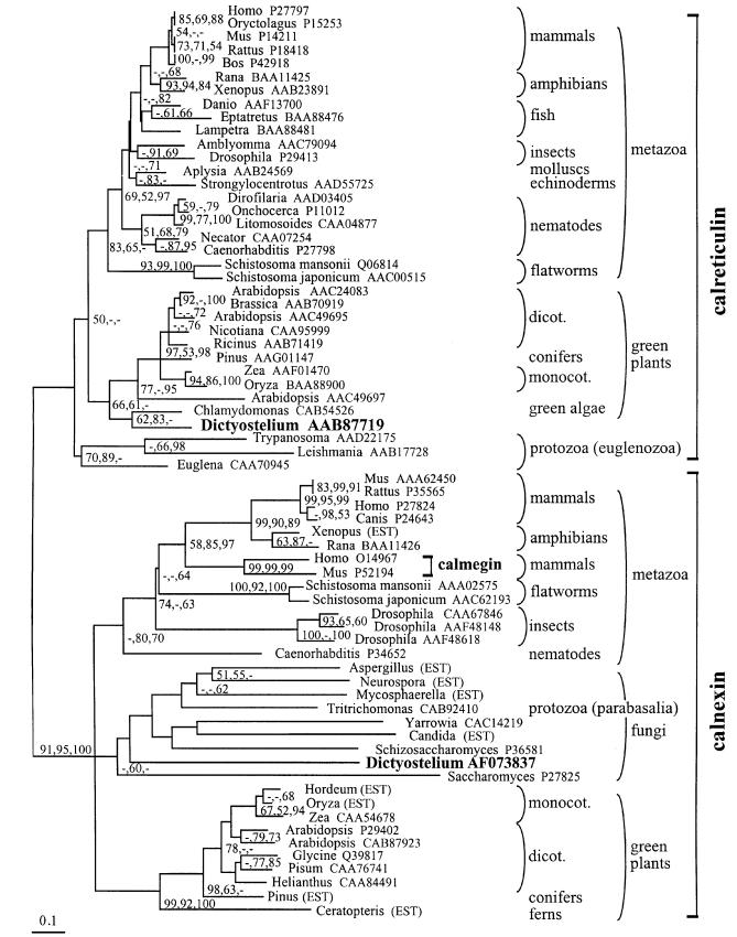
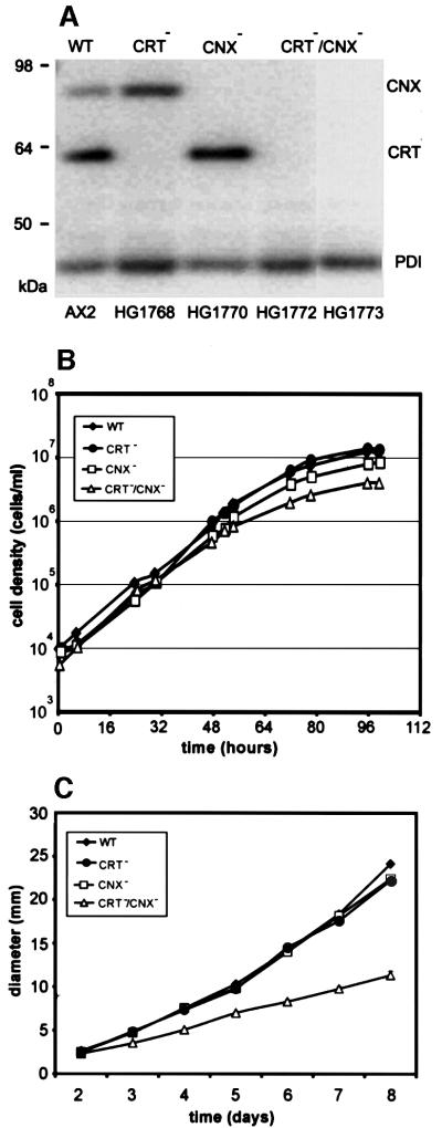
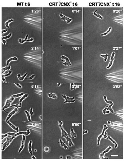
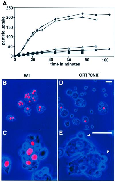
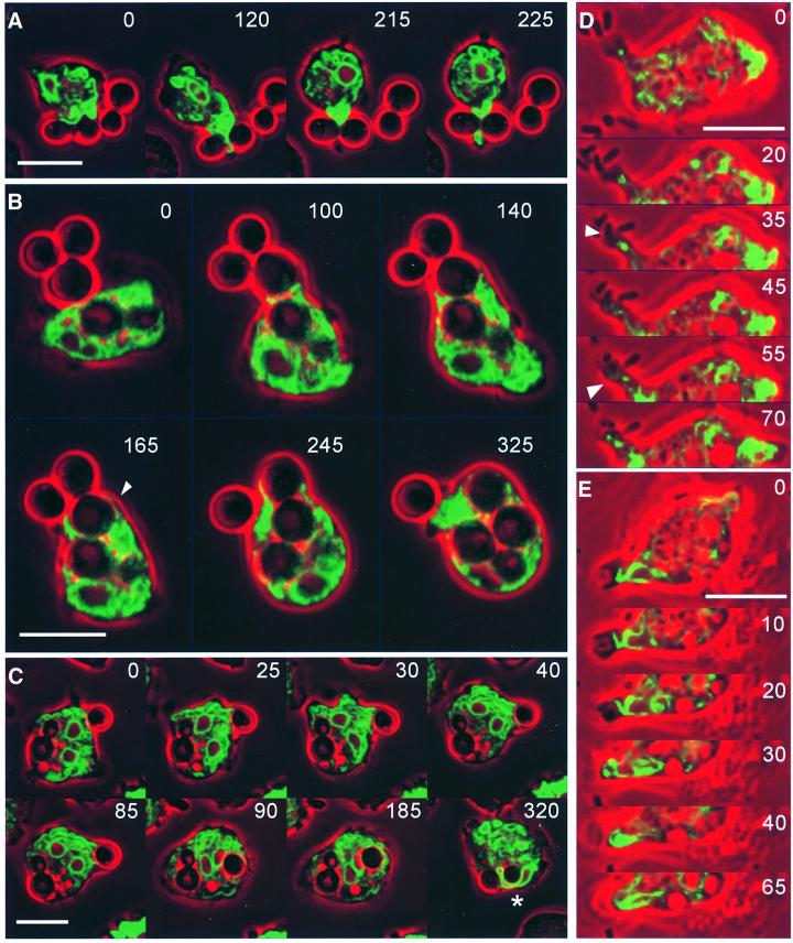
Calreticulin and calnexin are Ca2+-binding proteins with chaperone activity in the endoplasmic reticulum. These proteins have been eliminated by gene replacement in Dictyostelium, the only microorganism known to harbor both proteins; family members in Dictyostelium are located at the base of phylogenetic trees. A dramatic decline in the rate of phagocytosis was observed in double mutants lacking calreticulin and calnexin, whereas only mild changes occurred in single mutants. Dictyostelium cells are professional phagocytes, capable of internalizing particles by a sequence of activities: adhesion of the particle to the cell surface, actin-dependent outgrowth of a phagocytic cup, and separation of the phagosome from the plasma membrane. In the double-null mutants, particles still adhered to the cell surface, but the outgrowth of phagocytic cups was compromised. Green fluorescent protein-tagged calreticulin and calnexin, expressed in wild-type cells, revealed a direct link of the endoplasmic reticulum to the phagocytic cup enclosing a particle, such that the Ca2+ storage capacity of calreticulin and calnexin might directly modulate activities of the actin system during particle uptake.


Figures

None
Fig. 1. Distance-based phylogenetic tree of calnexins and calreticulins, with highlighted positions of the Dictyostelium proteins. The tree was computed with the Protdist, Seqboot and Neighbor modules of Phylip, and the alignment underlying the phylogenetic calculations was obtained as described in Materials and methods. Bootstrap support exceeding 50% of replicates by distance (Clustal; 1000 replicates), maximum likelihood (Puzzle; 1000 replicates) and maximum parsimony (PAUP; 100 replicates) methods is shown in that order, next to the corresponding node. The tree shown here was selected from 100 Seqboot replicates for the largest number of nodes supported by bootstrap analysis in Clustal, Puzzle and PAUP. Database accession codes for the non-redundant protein database at NCBI (http://www.ncbi.nlm.nih.gov/Entrez/) are shown next to each sequence, except where the sequence was reconstructed from ESTs.
None
Fig. 2. Immunolabeling of endogenous calreticulin and calnexin in Dictyostelium cells. (A) Specificity of mAbs used as ER markers, assayed in western blots of total cellular proteins. CNX, calnexin labeled with mAb 270-390-2; CRT, calreticulin labeled with mAb 252-234-2; PDI, protein disulfide isomerase labeled with mAb 221-135-1. The last lane (P) shows a parallel lane stained for proteins with Ponceau S. (B) Immunofluorescence labeling of fixed cells using the antibodies shown in (A). Immunofluorescence in red is superimposed on phase-contrast images in dark blue. All three antibodies label the reticulate structure of the ER. The perinuclear layer in the center of the cell is most prominently labeled with the calnexin antibody. Bar in (B), 10 µm.
None
Fig. 3. GFP fusion constructs of calreticulin and calnexin. (ADictyostelium calreticulin (top) shows the typical tripartite structure comprising an N-domain, a P-domain consisting of A and B repeats, and a C-domain ending with a HDEL retention signal. The GFP moiety has been placed behind the signal sequence of calreticulin. In calnexin (bottom), a signal sequence is followed by the luminal domain including a cluster of A and B repeats, which is separated by a transmembrane (TM) domain from the cytoplasmic domain. GFP is joined to the C-terminal end of this domain. (B) Cells expressing calnexin–GFP labeled with calreticulin-specific antibody or with phalloidin for F-actin. Upper panels show nuclei stained with 4′,6-diamidino-2-phenylindole in blue, superimposed on phase-contrast images in red. Lower panels depict the same cells with the fluorescence of calnexin–GFP in green, and antibody or phalloidin label in red. Areas of merging labels appear in yellow to brownish color. (C) Cells expressing GFP–calreticulin labeled with calnexin-specific antibody or with phalloidin. Top panels show phase-contrast images; lower panels show calnexin–GFP fluorescence in green, and the antibody or phalloidin label in red. Bars in (B) and (C), 10 µm.
None
Fig. 4. Retention of calnexin–GFP and GFP–calreticulin in the ER, shown in live Dictyostelium cells labeled with the endosome marker TRITC–dextran, which is taken up by macropinocytosis. (A) Cells expressing calnexin–GFP. (B) Cells expressing GFP–calreticulin. Upper panels show the fluorescence of TRITC–dextran in red. In the confocal images, cell bodies appear black with the exception of endosomes loaded with the marker. In the middle panels, GFP fluorescences are shown in green. In the lower panels, both images are superimposed on each other. Cells were incubated for the times indicated with TRITC–dextran. Until 30 min after internalization, endosomes are in the acidic phase (Aubry et al., 1997). After ≥90 min, endosomes of all stages until exocytosis should be loaded with the marker. Cells were incubated either in 10 mM TRICIN–HCl buffer pH 7.0 without Ca2+ or with 10 mM Ca2+. Bars, 10 µm.
None
Fig. 5. Wild-type AX2, single and double mutants, and their growth in liquid medium or on agar plates with bacteria. (A) Western blot of wild-type (WT), calreticulin-null (CRT), calnexin-null (CNX) and two independent CRT/CNX double-null mutants (CRT/CNX). Strain designations are given at the bottom. Equal amounts of protein were applied per lane, and the blot was probed for the presence of calreticulin, calnexin and PDI using a combination of calreticulin-, calnexin- and PDI-specific mAbs. (B) Semi-logarithmic plot of axenic growth with liquid nutrient medium in shaken suspension. Data are from one experiment. (C) Growth on bacterial lawns on agar plates measured as the increase in plaque diameter over time.
None
Fig. 6. Chemotaxis of CRT/CNX double-null mutant cells in comparison to wild type (WT). Cells were starved either for 6 h (t6) or 16 h (t16) and subsequently stimulated with cAMP through a micropipette. Orientation of cells in gradients of the chemoattractant is seen in all examples of wild-type and mutant cells. In the mutants, the typical elongation of aggregating cells needed a longer period of starvation than 6 h, which is sufficient for wild type. Numbers indicate times after positioning of the micropipette. Bar, 10 µm.
None
Fig. 7. Phagocytosis of yeast particles in wild-type and CRT/CNX double-null mutants. (A) Internalization of particles over time in two experiments (open and closed symbols) by wild-type AX2 cells (diamonds) and cells of two independent double mutants HG1772 (squares) and HG1773 (triangles). Particle uptake is plotted in arbitrary fluorescence units reflecting the number of internalized yeast particles. (BE) Internalized particles distinguished by red fluorescence from adherent particles whose fluorescence is quenched. Phase-contrast images are colored in blue. Wild-type cells (B and C) show numerous ingested particles. In double-null cells (D and E), only few internalized particles are found. Arrowheads in (E) point to adherent particles not enveloped by a phagocytic cup. Bars for the upper and lower panels, 10 µm.
None
Fig. 8. ER dynamics in phagocytosis. (A–C) Uptake of yeast particles by cells labeled with either calnexin–GFP (A and C) or GFP–calreticulin (B). The sequence in (A) shows the first engagement of a cell with a bipartite yeast particle. Initially, the ER forms a layer adjacent to the particle surface (0 s), and subsequently a hook around the cleft between the two halves of the particle (120 s). Strong accumulation of ER membranes at this cleft (215 s) is followed by the appearance in cross-section of an ER tube at the distal side of the particle (225 s). (B) Uptake of two yeast particles in tandem showing in optical section the extension of the ER along the particle surfaces. The arrowhead at 165 s points to a thin ER layer surrounding the particle that is taken up first. GFP fluorescence shown in green is superimposed on phase-contrast images in red. (C) Accumulation of the ER at a site of ongoing phagocytosis at the right edge of the cell. A previously internalized yeast cell with a bud located in the left half of the phagocyte shows no consistent association with ER membranes. The two nuclei of this cell are recognizable by their calnexin-enriched outer membrane; one of them is seen to be strongly deformed when squeezed between the yeast particles (asterisk in the last frame). (D and E) Phagocytosis of bacteria by cells labeled with either calnexin–GFP (D) or GFP–calreticulin (E). Both cells engulf bacteria at the tip of extensions that contain an ER core. In (D), a strand of the ER connecting the site of bacteria attachment with the cell body (arrowhead at 35 s) widens into a delicate bowl-shaped layer at the time of uptake (arrowhead at 55 s). In (E), the ER forms a layer on the bottom of the phagocytic cup (10 s) and turns into loops flanking the bacterium during its internalization (20–40 s). GFP fluorescence is shown in green, phase-contrast images are in red. Bars, 10 µm.

References

    1. Aizawa H., Sutoh,K., Tsubuki,S., Kawashima,S., Ishii,A. and Yahara,I. (1995) Identification, characterization and intracellular distribution of cofilin in Dictyostelium discoideum. J. Biol. Chem., 270, 10923–10932. - PubMed
    1. Arima H., Kinoshita,T., Ibrahim,H.R., Azakami,H. and Kato,A. (1998) Enhanced secretion of hydrophobic peptide fused lysozyme by the introduction of N-glycosylation signal and the disruption of calnexin gene in Saccharomyces cerevisiae. FEBS Lett., 440, 89–92. - PubMed
    1. Aubry L., Klein,G., Martiel,J.-L. and Satre,M. (1997) Fluid-phase endocytosis in the amoebae of the cellular slime mould Dictyostelium discoideum: mathematical modelling of kinetics and pH evolution. J. Theor. Biol., 184, 89–98.
    1. Cardelli J. (2001) Phagocytosis and macropinocytosis in Dictyostelium: phosphoinositide-based processes, biochemically distinct. Traffic, 2, 311–320. - PubMed
    1. Coppolino M.G., Woodside,M.J., Demaurex,N., Grinstein,S., St-Arnaud,R. and Dedhar,S. (1997) Calreticulin is essential for integrin-mediated calcium signalling and cell adhesion. Nature, 386, 843–847. - PubMed

Publication types

MeSH terms