. 2001 Dec 3;20(23):6793-804.
doi: 10.1093/emboj/20.23.6793.

Tyrosine phosphorylation of Grb2 by Bcr/Abl and epidermal growth factor receptor: a novel regulatory mechanism for tyrosine kinase signaling

Affiliations

Tyrosine phosphorylation of Grb2 by Bcr/Abl and epidermal growth factor receptor: a novel regulatory mechanism for tyrosine kinase signaling

S Li et al. EMBO J. .

Abstract

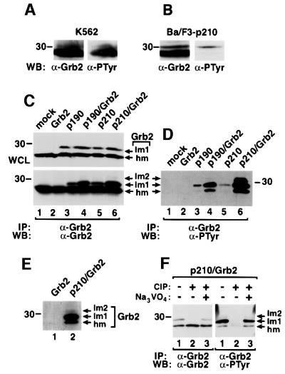
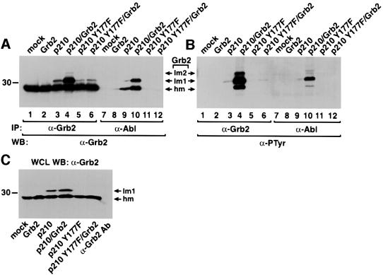
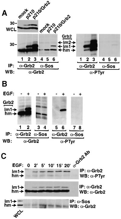
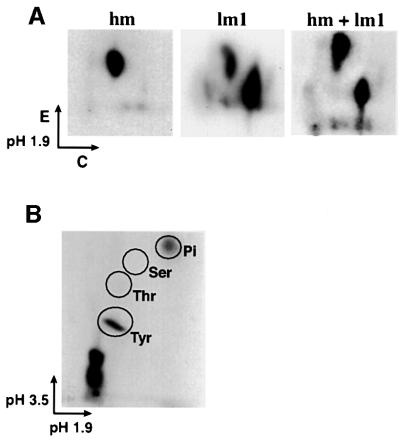
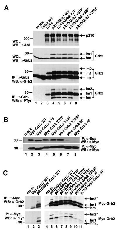
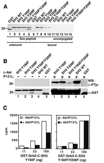
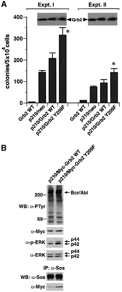
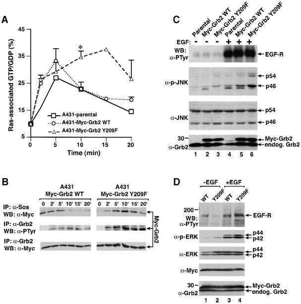
Growth factor receptor-binding protein-2 (Grb2) plays a key role in signal transduction initiated by Bcr/Abl oncoproteins and growth factors, functioning as an adaptor protein through its Src homology 2 and 3 (SH2 and SH3) domains. We found that Grb2 was tyrosine-phosphorylated in cells expressing BCR/ABL and in A431 cells stimulated with epidermal growth factor (EGF). Phosphorylation of Grb2 by Bcr/Abl or EGF receptor reduced its SH3-dependent binding to Sos in vivo, but not its SH2-dependent binding to Bcr/Abl. Tyr209 within the C-terminal SH3 domain of Grb2 was identified as one of the tyrosine phosphorylation sites, and phosphorylation of Tyr209 abolished the binding of the SH3 domain to a proline-rich Sos peptide in vitro. In vivo expression of a Grb2 mutant where Tyr209 was changed to phenylalanine enhanced BCR/ABL-induced ERK activation and fibroblast transformation, and potentiated and prolonged Grb2-mediated activation of Ras, mitogen-activated protein kinase and c-Jun N-terminal kinase in response to EGF stimulation. These results suggest that tyrosine phosphorylation of Grb2 is a novel mechanism of down-regulation of tyrosine kinase signaling.


Figures

None
Fig. 1. Phosphorylation of Grb2 in Bcr/Abl-expressing cells. Lysates from K562 leukemia cells (A) or Ba/F3 cells transformed by p210 BCR/ABL (B) were immunoprecipitated with anti-Grb2 antibodies followed by blotting with anti-Grb2 or anti-PTyr antibodies. (C and D) Lysates of 293T cells mock transfected (lane 1) or transfected with Grb2 (lane 2), p190 BCR/ABL (lane 3), p190 BCR/ABL/Grb2 (lane 4), p210 BCR/ABL (lane 5) and p210 BCR/ABL/Grb2 (lane 6) in pMINV were analyzed directly (whole-cell lysates, WCL) by western blot with anti-Grb2 antibodies (C, top panel) or immunoprecipitated with anti-Grb2 antibodies, followed by blotting with anti-Grb2 (C, bottom panel) and anti-PTyr (D) antibodies. Three Grb2 species were observed: the highest mobility species (hm), lower mobility species (lm1) and a minor species of the lowest mobility (lm2). (E) 293T cells transfected with Grb2 (lane 1) or with p210 BCR/ABL/Grb2 (lane 2) were labeled with [32P]orthophosphate, and immunoprecipitated Grb2 proteins analyzed by autoradiography. (F) 293T cells were transfected with p210 BCR/ABL/Grb2, Grb2 proteins were immunoprecipitated with anti-Grb2 antibody, and treated without (lane 1) or with (lane 2) calf intestinal phosphatase (CIP) or with CIP and the phosphatase inhibitor sodium vanadate (1 µM Na3VO4, lane 3), then analyzed by western blot using anti-Grb2 (left panel) and anti-PTyr (right panel) antibodies.
None
Fig. 2. Direct binding of Grb2 to Bcr/Abl facilitates Grb2 phosphorylation. (A and B) Lysates of 293T cells mock transfected (lanes 1 and 7), or transfected with Grb2 (lanes 2 and 8), p210 BCR/ABL (lanes 3 and 9), p210 BCR/ABL/Grb2 (lanes 4 and 10), p210 BCR/ABL Y177F (lanes 5 and 11) and p210 BCR/ABL Y177F/Grb2 (lanes 6 and 12) were immunoprecipitated with anti-Grb2 (lanes 1–6) and anti-Abl (lanes 7–12) antibodies, and analyzed by western blot using anti-Grb2 (A) and anti-PTyr (B) antibodies. (C) Whole-cell lysates (WCL) from the samples in (A) were analyzed directly by western blotting with anti-Grb2 antibodies, demonstrating an ∼50% increase in total Grb2 levels upon Grb2 overexpression. To exclude the possibility that some or most of the Grb2 signal in the immunoprecipitation/western blots in (A) resulted from detection of light chains of the precipitating antibodies by the secondary antibody, the same amount of anti-Grb2 antibody was added to lysis buffer alone (C, right lane) and processed as in (A), yielding very low signal in the region of Grb2.
None
Fig. 3. Phosphorylation of Grb2 reduces its binding to Sos in vivo. (A) Lysates of 293T cells mock transfected (lanes 1 and 4), or transfected with p210 BCR/ABL (lanes 2 and 5) or p210 BCR/ABL/Grb2 (lanes 3 and 6) were analyzed directly (WCL) by anti-Grb2 western blot (upper left panel) or immunoprecipitated with anti-Grb2 (lanes 1–3) or anti-Sos (lanes 4–6) antibodies, and analyzed by western blot using anti-Grb2 (left lower panel) and anti-PTyr (right lower panel) antibodies. Densitometric quantitation of the relative amount of hm and lm1 Grb2 species in the left panel, lanes 2 and 3, versus those in lanes 5 and 6 demonstrated that 40–50% of the total Grb2 but only 25% of the Sos-associated Grb2 is found as the lm1 species (data not shown). (B) A431 cells were serum starved (lanes 1, 3, 5 and 7) then stimulated with EGF (200 ng/ml) (lanes 2, 4, 6 and 8) for 10 min. Cell lysates were immunoprecipitated with anti-Grb2 (lanes 1, 2, 5 and 6) or anti-Sos (lanes 3, 4, 7 and 8) antibodies, and analyzed by western blot using anti-Grb2 (left two panels) and anti-PTyr (right two panels) antibodies. (C) A431 cells were starved then stimulated with EGF for the indicated times. Cell lysates were immunoprecipitated with anti-Grb2 (top and middle panels) or anti-Sos (bottom panel) antibodies and blotted with anti-PTyr (top panel) or anti-Grb2 (middle and bottom panels) antibodies. A sample of lysis buffer with anti-Grb2 antibodies was processed (right lane) to demonstrate that very little of the Grb2 signal is attributable to immunoglobulin light chains. A lane containing whole-cell lysate (WCL) from cells stimulated for 10 min was run adjacent to the anti-Sos IPs (left lane, lower panel) to confirm specific co-precipitation of the Grb2 hm species with Sos.
None
Fig. 4. Characterization of Grb2 phosphorylation. (A) Tryptic phosphopeptide mapping of phosphorylated Grb2. 293T cells were transfected with p210 BCR/ABL/Grb2, and 48 h later were incubated with 1 mCi of [32P]orthophosphate in 2 ml of phosphate-free DMEM for 3 h. Cell lysate was immunoprecipitated with anti-Grb2 antibody, separated by SDS–PAGE and blotted onto a nitrocellulose membrane. The highest mobility (hm) and lower mobility (lm1) species of Grb2 proteins containing phosphorylated Grb2 labeled with [32P]ortho phosphate were cut out of the membrane, digested with TPCK-treated trypsin and subjected to two-dimensional peptide mapping by electrophoresis in pH 1.9 buffer for the first dimension, followed by thin-layer chromotography for the second dimension, as described (Boyle et al., 1991). (B) Phosphoamino acid analysis of phosphorylated Grb2. The lower mobility species (lm1) of Grb2 proteins labeled with [32P]orthophosphate (A) was treated with 6 M HCl, and subjected to electrophoresis in pH 1.9 buffer for the first dimension, and in pH 3.5 buffer for the second dimension. Tyr, tyrosine; Thr, threonine; Ser, serine; Pi, free phosphate.
None
Fig. 5. Multiple tyrosine phosphorylation sites on Grb2. (A) Lysates of 293T cells mock transfected (lane 1) or transfected with wild-type Grb2 (lane 2), p210 BCR/ABL (lane 3), p210 BCR/ABL/Grb2 WT (lane 4), p210 BCR/ABL/Grb2 Y7F (lane 5), p210 BCR/ABL/Grb2 Y37F (lane 6), p210 BCR/ABL/Grb2 Y52F (lane 7) and p210 BCR/ABL/Grb2 Y209F (lane 8) were loaded directly (top panel) or immunoprecipitated with anti-Grb2 antibody (middle and bottom panels), and analyzed by SDS–PAGE and western blotting using anti-Abl (top panel), anti-Grb2 (middle panel) and anti-PTyr (bottom panel) antibodies. (B) Effect of tyrosine to phenylalanine mutations of Grb2 on binding to Sos in vivo. Lysates of 293T cells mock transfected (lane 1) or transfected with wild-type Myc-Grb2 (lane 2), Myc-Grb2 Y7F (lane 3), Myc-Grb2 Y37F (lane 4), Myc-Grb2 Y52F (lane 5), Myc-Grb2 Y209F (lane 6), Myc-Grb2 Y7F/Y52F/Y209F (3F) (lane 7) and Myc-Grb2 Y7F/Y37F/Y52F/Y209F (4F) were immunoprecipitated with anti-Sos (top panel) or anti-Myc (bottom panel) antibodies, and analyzed by western blot using anti-Myc antibody. (C) Lysates of 293T cells mock transfected (lanes 1 and 4), transfected with wild-type Myc-Grb2 alone (lane 2) or co-transfected with p210 BCR/ABL/Myc-Grb2 WT (lane 3 and 5), p210 BCR/ABL/Myc-Grb2 Y7F (lane 6), p210 BCR/ABL/Myc-Grb2 Y37F (lane 7), p210 BCR/ABL/Myc-Grb2 Y52F (lane 8), p210 BCR/ABL/Myc-Grb2 Y209F (lane 9), p210 BCR/ABL/Myc-Grb2 Y7F/Y52F/Y209F (3F) (lane 10) and p210 BCR/ABL/Myc-Grb2 Y7F/Y37F/Y52F/Y209F (4F) (lane 11) in pcDNA were immunoprecipitated with anti-Myc (9E10) antibody, and analyzed by western blot using anti-Grb2 (top panel) and anti-PTyr (bottom panel) antibodies.
None
Fig. 6. Grb2 phosphorylation at Tyr209 abolishes its binding to Sos. (A) Unphosphorylated GST–Grb2-C-SH3 WT or mutant fusion proteins bind Sos peptide beads. GST proteins were mixed with Affigel agarose beads coupled with Sos peptide (lanes 1–10) or unconjugated (lanes 11–15). Unbound (lanes 1–5) and bound (lanes 6–15) GST proteins were subjected to SDS–PAGE, and visualized by Coomassie Blue staining. (B) GST fusion proteins containing wild-type Grb2 C-terminal SH3 domain (Grb2-C-SH3) (lane 4), Y160F (lane 1), Y209F (lane 2), Y160F/Y209F (lane 3) or GST alone (lane 5) either untreated (left panels) or after phosphorylation by purified c-Abl P131L kinase (right panels) were analyzed by western blot using anti-PTyr (top panels) and anti-GST (bottom panels) antibodies. (C) GST–Grb2-C-SH3 Y160F (left panel) and Y160F/Y209F (right panel) were labeled with 32P using heart muscle kinase, then treated with or without purified c-Abl P131L kinase in the presence of non-radioactive ATP. Increasing amounts of each GST–Grb2 fusion protein were mixed with the same amount of Sos peptide beads, and total radioactivity (c.p.m.) associated with the beads in each binding reaction was measured. The binding of the phosphorylated GST–Grb2 Y160F single mutant to Sos beads was not tested because this residue is outside the SH3 domain, does not participate in ligand binding and is not phosphorylated significantly in vivo (Figure 5C).
None
Fig. 7. Mutation of Grb2 Tyr209 potentiates Bcr/Abl-induced transformation and ERK activation. (A) NIH-3T3 fibroblasts transduced with retrovirus expressing wild-type Grb2 alone or co-expressing p210 and either a neomycin resistance gene (neo), wild-type Grb2 or Grb2 Y209F were plated in soft agar and transformed colonies counted 3 weeks later (mean ± SE from triplicate plates). Two independent experiments with different retroviral stocks were performed. The increase in soft agar colonies with co-transduction of Grb2 Y209F relative to wild-type Grb2 in both experiments was statistically significant (asterisks, P <0.01, unpaired t-test). Lysates from the p210-transduced populations were blotted with anti-Grb2 antibodies (inserts) to demonstrate equivalent levels of Grb2 overexpression. (B) Cells co-transduced with p210 Bcr/Abl and either Myc-tagged wild-type Grb2 or the Y209F mutant were analyzed by direct western blotting of lysates with anti-PTyr, anti-Myc, anti-phospho-ERK and anti-pan-ERK antibodies (top four panels), or by immunoprecipitation with anti-Sos antibodies and blotting with anti-Sos and anti-Myc antibodies (bottom panels).
None
Fig. 8. Mutation of Grb2 Tyr209 potentiates and prolongs EGF signaling pathways. (A) Ras activation. A431 parental cells and populations stably expressing wild-type Myc-Grb2 and Myc-Grb2 Y209F were starved, labeled with [32P]orthophosphate and stimulated with EGF (200 ng/ml) for 0, 2, 5, 10, 15 or 20 min, and Ras-associated guanine nucleotides analyzed as described in Materials and methods. Bars indicate positive SE for the A431-Myc-Grb2 WT and A431-Myc-Grb2 Y209F cells; the 15 min time point was only done once. The difference in Ras GTP levels at 10 min between wild-type Grb2 and Grb2 Y209F cells (asterisk) is statistically significant (P ≤0.05, unpaired t-test). (B) Kinetic analysis of Grb2 tyrosine phosphorylation and Sos association from the A431-Myc-Grb2 WT- and A431-Myc-Grb2 Y209F-expressing cells in (A). Cells were starved and stimulated with EGF for the indicated times and lysates analyzed by immunoprecipitation with anti-Sos (top panel) or anti-Grb2 (middle and bottom panels) antibodies and blotting with anti-Myc (top and bottom panels) or anti-PTyr (middle panel) antibodies. (C) JNK activation. Parental (lanes 1 and 4), Myc-Grb2 WT- (lane 2 and 5) and Myc-Grb2 Y209F-expressing (lanes 3 and 6) A431 cells were starved (lanes 1–3) and stimulated with EGF (200 ng/ml, lanes 4–6), and lysates analyzed by western blot using anti-PTyr antibody for detecting EGF-stimulated phosphorylation of EGF receptor (top panel), anti-phospho-JNK antibody (second panel from the top), anti-JNK antibody (third panel from the top) or anti-Grb2 antibody (bottom panel). (D) MAPK/ERK activation. 293T cells transfected with EGF receptor and either Myc-tagged wild-type Grb2 (lanes 1 and 3) or Myc-tagged Grb2 Y209F (lanes 2 and 4) were starved (lanes 1 and 2) or stimulated with EGF for 10 min (200 ng/ml, lanes 3 and 4), and lysates were analyzed by western blot using anti-PTyr antibody for detection of phosphorylated EGF receptor (top panel), anti-phospho-ERK antibody (second panel from the top), anti-ERK antibody (third panel from the top), anti-Myc antibody (fourth panel from the top) and anti-Grb2 antibody (bottom panel).

References

    1. Baltensperger K., Kozma,L.M., Cherniack,A.D., Klarlund,J., Chawla,A., Banerjee,U. and Czech,M.P. (1993) Binding of the Ras activator son of sevenless to insulin receptor substrate-1 signaling complexes. Science, 260, 1950–1952. - PubMed
    1. Bedford M.T., Chan,D.C. and Leder,P. (1997) FBP WW domains and the Abl SH3 domain bind to a specific class of proline-rich ligands. EMBO J., 16, 2376–2383. - PMC - PubMed
    1. Benjamin C.W., Linseman,D.A. and Jones,D.A. (1994) Platelet-derived growth factor stimulates phosphorylation of growth factor receptor-binding protein-2 in vascular smooth muscle cells. J. Biol. Chem., 269, 31346–31349. - PubMed
    1. Boyle W.J., van Der Greer,P. and Hunter,T. (1991) Phosphopeptide mapping and phosphoamino acid analysis by two-dimensional separation on thin-layer cellulose plates. Methods Enzymol., 201, 110–149. - PubMed
    1. Brasher B.B. and Van Etten,R.A. (2000) c-Abl has high intrinsic tyrosine kinase activity that is stimulated by mutation of the Src homology 3 domain and by autophosphorylation at two distinct regulatory tyrosines. J. Biol. Chem., 275, 35631–35637. - PubMed

Publication types

MeSH terms

Associated data