. 2001 Dec 3;20(23):6816-25.
doi: 10.1093/emboj/20.23.6816.

CBP/p300 and muscle differentiation: no HAT, no muscle

Affiliations

CBP/p300 and muscle differentiation: no HAT, no muscle

A Polesskaya et al. EMBO J. .

Abstract

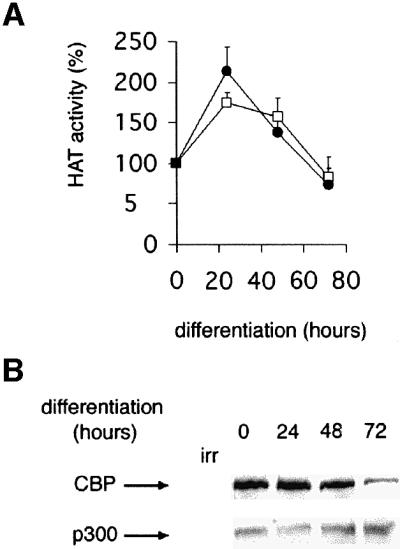
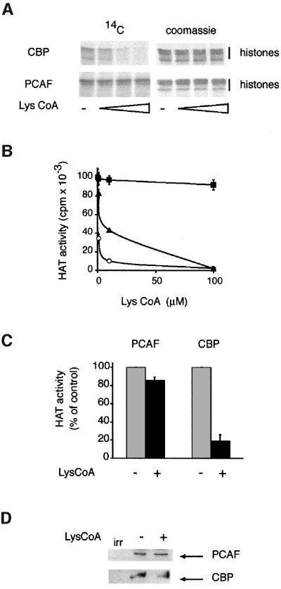
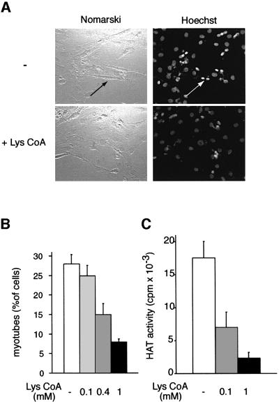
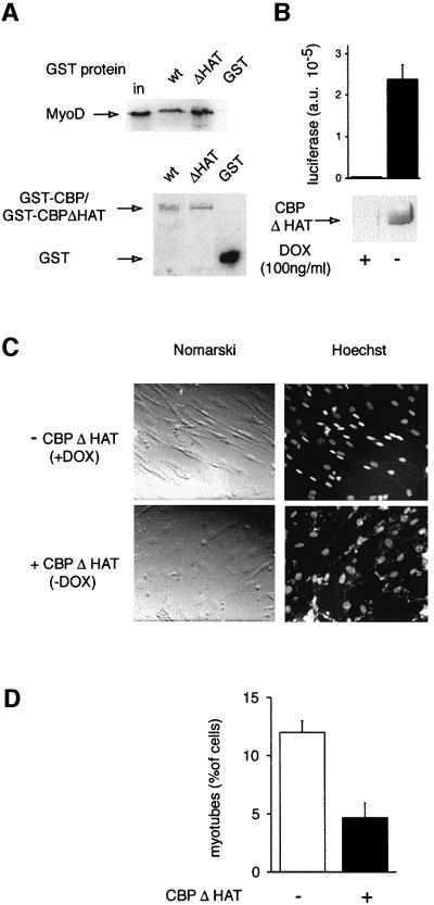
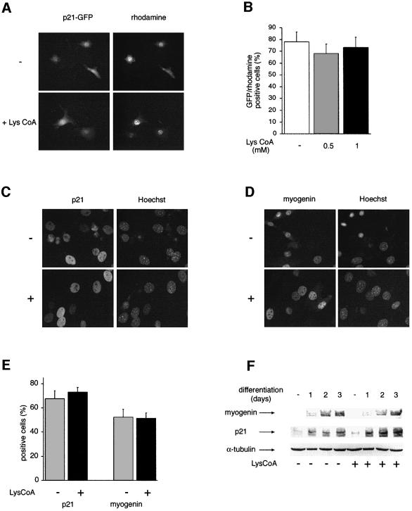
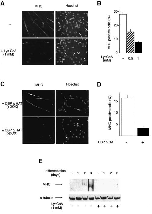
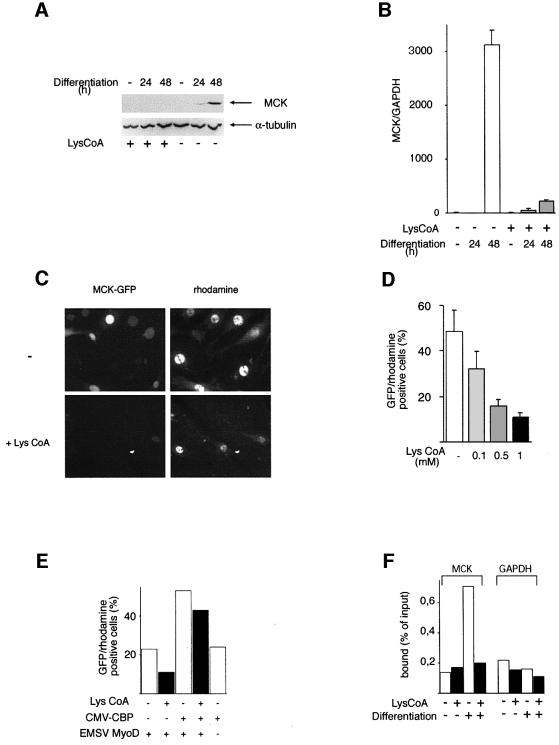
Terminal differentiation of muscle cells follows a precisely orchestrated program of transcriptional regulatory events at the promoters of both muscle-specific and ubiquitous genes. Two distinct families of transcriptional co-activators, GCN5/PCAF and CREB-binding protein (CBP)/p300, are crucial to this process. While both possess histone acetyl-transferase (HAT) activity, previous studies have failed to identify a requirement for CBP/p300 HAT function in myogenic differentiation. We have addressed this issue directly using a chemical inhibitor of CBP/p300 in addition to a negative transdominant mutant. Our results clearly demonstrate that CBP/p300 HAT activity is critical for myogenic terminal differentiation. Furthermore, this requirement is restricted to a subset of events in the differentiation program: cell fusion and specific gene expression. These data help to define the requirements for enzymatic function of distinct coactivators at different stages of the muscle cell differentiation program.


Figures

None
Fig. 1. Time course of CBP and p300 HAT activities during myogenic terminal differentiation. C2C12 cells were placed in differentiation medium, and CBP or p300 was immunoprecipitated at the times indicated and assayed for HAT activity (A) or analyzed by western blotting (B). Closed circle, CBP; open square, p300. The mean of three independent experiments is shown in (A). A value of 100% corresponds to 7470 c.p.m. for CBP and 8180 c.p.m. for p300.
None
Fig. 2. Lys-CoA specifically inhibits CBP/p300. (A) Bacterially produced recombinant CBP or PCAF (as indicated) was incubated with nucleosomes purified from HeLa cells and 14C-labeled acetyl-CoA; histones were analyzed by SDS–PAGE followed by autoradiography. (B) Recombinant CBP (closed triangles), p300 (open circles) or PCAF (closed squares) was incubated with a synthetic peptide corresponding to the first 24 amino acids of histone H3, together with [14C]AcCoA and the doses of Lys-CoA indicated. The radioactivity incorporated in the peptide was measured after 1 h; the mean of three independent experiments is shown. (C and D) C2C12 cells were permeabilized in the presence (+) or the absence (–) of 1 mM Lys-CoA. Extracts were prepared after 1 h. CBP/p300 and PCAF were immunoprecipitated and assayed for HAT activity (C) or analyzed by western blotting (D). The mean of three independent experiments is shown. A value of 100% corresponds to 15 053 c.p.m. for CBP and 2109 c.p.m. for PCAF.
None
Fig. 3. Lys-CoA inhibits myotube formation. C2C12 cells were permeabilized in the presence or absence of Lys-CoA, and placed in differentiation medium; after 3 days, cells were fixed and labeled with Hoechst 33258. (A) Typical fields: the arrows point to a myotube and its condensed nuclei. (B) Statistical analysis of the results: shown is the mean ± SE of three independent experiments; a minimum of 300 cells were counted in each experiment. (C) CBP/p300 HAT activity was measured as in Figure 2C. Shown is the mean ± SE of three independent experiments.
None
Fig. 4. CBPΔHAT, a negative transdominant mutant of CBP, inhibits myotube formation. (A) CBPΔHAT interacts with MyoD: GST pull-down analysis of CBP and CBPΔHAT. Upper panel: MyoD was incubated with beads coated with the GST proteins indicated, and bound MyoD was analyzed by western blotting; in, 10% of the input. Lower panel: the protein content of the beads used above was monitored by western blotting using an anti-GST antibody. (B) Induction of the mutant by doxycycline (DOX) removal: extracts of C2C12-CBPΔHAT cells were assayed for luciferase activity or immunoprecipitated with anti-CBP antibodies followed by western blot analysis with anti-HA. (C and D) C2C12-CBPΔHAT cells cultured in the presence of doxycycline or in its absence (as indicated) were fixed and labeled with Hoechst after 72 h in differentiation medium. (C) Typical fields. (D) Statistical analysis (mean of three independent experiments with 300 cells analyzed in each).
None
Fig. 5. Myogenin and p21 expression is not affected by Lys-CoA. (A and B) Lys-CoA has no effect on the activity of the p21 promoter. C3H 10T1/2 cells were microinjected with a p21–GFP construct, together with Lys-CoA where indicated (4 mM in the injection mix), recombinant MyoD and an injection marker coupled to rhodamine. Cells were analyzed 24 h later. (A) Typical fields; note that each field contains few microinjected cells, but significant numbers of cells were analyzed by cumulating numerous fields for each measure. (B) Statistical analysis: shown is the mean ± SE of three independent experiments, with 300 cells counted per experiment. (C–F) Endogenous p21 and myogenin proteins are not affected by Lys-CoA. C2C12 cells were permeabilized in the presence or absence of Lys-CoA (1 mM) as indicated, placed in differentiation medium for 24 h (C–E) for the periods of time indicated (F), fixed and labeled with Hoechst and either anti-p21 (C) or anti-myogenin (D) antibodies. (E) Statistical analysis of the results (mean ± SE of three independent experiments, with 300 cells analyzed per experiment). (F) Extracts of permeabilized cells were prepared at the times indicated and analyzed by western blotting using anti-myogenin, anti-p21 or anti-α-tubulin antibodies as indicated.
None
Fig. 6. Lys-CoA and CBPΔHAT both inhibit MHC protein expression. (A and B) C2C12 cells were permeabilized in the presence of Lys-CoA or in its absence (as indicated) and placed in differentiation medium for 72 h. Cells were fixed and analyzed by immunofluorescence using anti-MHC antibodies. (A) Typical fields. (B) Statistical analysis of the results (mean ± SE of three independent experiments, with 300 cells counted per experiment). (C and D) CBPΔHAT inhibits MHC expression. C2C12-CBPΔHAT were maintained in doxycycline (–) or cultured in the absence of doxycycline for 3 days (CBPΔHAT), then placed in differentiation medium for 72 h, fixed and analyzed by immunofluorescence using anti-MHC antibodies. (C) Typical fields; (D) statistical analysis of the results (mean ± SE of three independent experiments with 300 cells counted per experiment). (E) Western blot analysis; cells were treated with Lys-CoA as in (A) and placed in differentiation medium for the periods of time indicated; extracts were analyzed by western blotting using anti MHC or anti-α-tubulin antibodies.
None
Fig. 7. Lys-CoA inhibits MCK expression. (A and B) C2C12 cells were permeabilized in the presence of Lys-CoA (1 mM) or in its absence (as indicated) and placed in differentiation medium for the periods of time indicated. (A) MCK protein expression was analyzed by western blotting, using α-tubulin as an internal standard. (B) The MCK RNA steady-state level was analyzed by RT–PCR, using GAPDH as an internal standard; the ratio between MCK and GAPDH RNA is shown. (C–F) C3H 10T1/2 cells were microinjected with a mixture containing an MCK–GFP reporter construct, an expression vector for MyoD, an injection marker coupled to rhodamine, and Lys-CoA (as indicated). (C) Typical fields. (D) Statistical analysis of the results; shown is the mean ± SE of three independent experiments. (E) Cells were microinjected as in (A), but an expression vector for CBP was added where indicated (results of a typical experiment performed twice). (F) Lys-CoA inhibits histone H4 acetylation on the MCK promoter. Chromatin was prepared from C2C12 cells treated with Lys-CoA and induced to differentiate as indicated, immunoprecipitated using anti-acetylated H4 antibodies and analyzed by real-time PCR (on a Light Cycler) with primers amplifying the MCK promoter, or a GAPDH sequence used as a control. Shown are the numbers of copies as a percentage of the inputs (results of a typical experiment performed twice).

Similar articles

Cited by

References

    1. Ait-Si-Ali S., Ramirez,S., Robin,P., Trouche,D. and Harel-Bellan,A. (1998) A rapid and sensitive assay for histone acetyl-transferase activity. Nucleic Acids Res., 26, 3869–3870. - PMC - PubMed
    1. Ait-Si-Ali S. et al. (2000) CBP/p300 histone acetyl-transferase activity is important for the G1/S transition. Oncogene, 19, 2430–2437. - PubMed
    1. Arany Z., Sellers,W.R., Livingston,D.M. and Eckner,R. (1994) E1A-associated p300 and CREB-associated CBP belong to a conserved family of coactivators. Cell, 77, 799–800. - PubMed
    1. Bannister A.J. and Kouzarides,T. (1996) The CBP co-activator is a histone acetyltransferase. Nature, 384, 641–643. - PubMed
    1. Brownell J.E. and Allis,C.D. (1996) Special HATs for special occasions: linking histone acetylation to chromatin assembly and gene activation. Curr. Opin. Genet. Dev., 6, 176–184. - PubMed

Publication types

MeSH terms