. 2001 Dec 3;20(23):6856-65.
doi: 10.1093/emboj/20.23.6856.

Release of U18 snoRNA from its host intron requires interaction of Nop1p with the Rnt1p endonuclease

Affiliations

Release of U18 snoRNA from its host intron requires interaction of Nop1p with the Rnt1p endonuclease

C Giorgi et al. EMBO J. .

Abstract

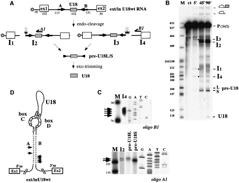
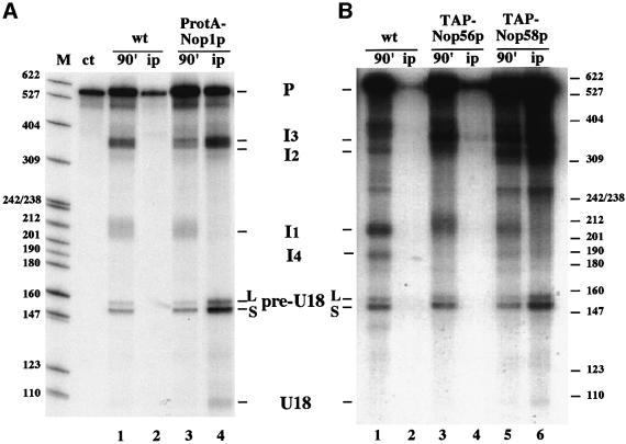
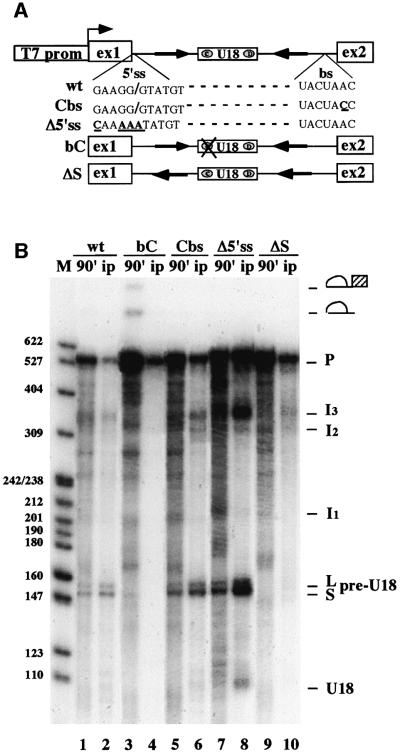
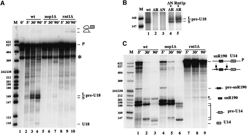
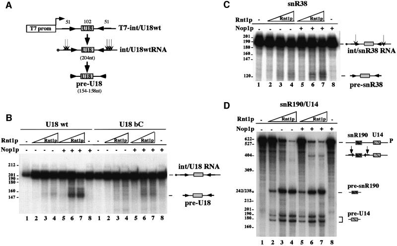
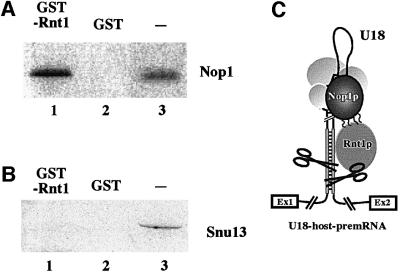
An external stem, essential for the release of small nucleolar RNAs (snoRNAs) from their pre-mRNAs, flanks the majority of yeast intron-encoded snoRNAs. Even if this stem is not a canonical Rnt1p substrate, several experiments have indicated that the Rnt1p endonuclease is required for snoRNA processing. To identify the factors necessary for processing of intron-encoded snoRNAs, we have raised in vitro extracts able to reproduce such activity. We found that snoRNP factors are associated with the snoRNA- coding region throughout all the processing steps, and that mutants unable to assemble snoRNPs have a processing-deficient phenotype. Specific depletion of Nop1p completely prevents U18 snoRNA synthesis, but does not affect processing of a dicistronic snoRNA-coding unit that has a canonical Rnt1p site. Correct cleavage of intron-encoded U18 and snR38 snoRNAs can be reproduced in vitro by incubating together purified Nop1p and Rnt1p. Pull-down experiments showed that the two proteins interact physically. These data indicate that cleavage of U18, snR38 and possibly other intron-encoded snoRNAs is a regulated process, since the stem is cleaved by the Rnt1p endonuclease only when snoRNP assembly has occurred.


Figures

None
Fig. 1. U18 snoRNA is faithfully processed in vitro from its host pre-mRNA. (A) Schematic representation of the ex/intU18 RNA containing the U18 host intron and part of the flanking EFB1 exons. In vitro transcription from this construct produces a 546 nucleotide transcript. Open boxes represent the first and second exons, while the U18 snoRNA coding region is represented by a gray box. The cap structure is represented by a filled circle. Arrows indicate the 14-nucleotide external stem [A and B sequences as indicated in (D)]. The different processing intermediates are indicated together with the formation of the mature U18 molecule. Vertical arrows indicate the position of the cleavage sites. Numbers refer to nucleotide lengths. (B32P-labeled ex/intU18 RNA was incubated, for the times indicated above each lane, in a yeast extract raised from strain CH1462. Input RNA was loaded on lane ct. Processing products were resolved on a 6% polyacrylamide–urea gel and visualized by autoradiography. Splicing and endonucleolytic processing intermediates are indicated on the right-hand side of the panel. Some of the intermediates (indicated by dots) are better visualized in the 5′ lane. Lane M: molecular weight marker (pBR322 plasmid DNA, MspI digested). (C) Primer extension analysis performed, with oligos A1 and B1 [see (A)], on gel-purified I2 and I4 molecules as well as on the short (S) and long (L) forms of the pre-U18 species. Extended products (indicated by horizontal arrows) are run in parallel with sequencing reactions and with molecular weight markers [as in (B)]. (D) Secondary structure for ex/intU18 RNA; the terminal conserved core structure is according to Watkins et al. (2000). The cleavage sites are indicated by arrows on the A/B external stem.
None
Fig. 2. snoRNP factors associate during processing. 32P-labeled ex/intU18 RNA was incubated for 90 min with extracts from strains CH1462 (lanes wt), ProtA-NOP1 (lanes ProtA-Nop1p, A), YAF2 (lanes TAP-Nop56p, B) and YAF3 (lanes TAP-Nop58p, B). One-third of each processing reaction was directly extracted (lanes 90′: 1, 3 and 5) and the remaining samples were immunoprecipitated with IgG–Sepharose (lanes ip: 2, 4 and 6). RNA was extracted and run on a 6% polyacrylamide–urea gel and visualized by autoradiography. Schematic representation of the different processing intermediates are shown at the sides of the panels.
None
Fig. 3. Effect of cis-acting mutations on U18 processing. (A) Schematic representation of the T7-ex/intU18 construct and of the mutant derivatives used in this study. Δ5′ss and Cbs contain site-specific mutations known to affect splicing, bC has nucleotide substitutions in the conserved box C (UGAUGA-CAUUGA) that affect snoRNP assembly and ΔS disrupts A–B pairing by inverting sequence A. (B32P-labeled ex/intU18 RNA, or its mutant derivatives, were incubated in a ProtA–Nop1p yeast extract and incubation was allowed to proceed for 90 min. One-third of each processing reaction was extracted directly (lanes 1, 3, 5, 7 and 9) and the remaining samples were immunoprecipitated with IgG–Sepharose (lanes 2, 4, 6, 8 and 10). RNA from total and pellet samples was resolved on a 6% polyacrylamide–urea gel and visualized by autoradiography. Splicing and processing intermediates are represented schematically at the side of the panel.
None
Fig. 4. Nop1p and Rnt1p proteins are required for U18 snoRNA processing. (A32P-labeled ex/intU18 RNA was incubated with extracts from strains CH1462 (lanes wt), D255 (lanes nop1Δ) and rnt1Δ (lanes rnt1Δ) for the time indicated above the lanes. The band indicated by an asterisk corresponds to a breakdown product already present in the input RNA (lane 1) and overlaps with molecules I2 and I3. (B32P-labeled ex/intU18 RNA was incubated with extracts from strains CH1462 (lane wt), rnt1Δ (lane ΔR), D255 + rnt1Δ (ΔN + ΔR) and rnt1Δ + recombinant GST–Rnt1p (5 ng/20 µl, lane ΔR + Rnt1). After 90 min of incubation, RNA was extracted and run a 6% polyacrylamide–urea gel and visualized by autoradiography. The panel shows only the region of the gel containing the pre-U18 molecule, which is diagnostic of cleavage. (C) The experiment is the same as in (A), with the difference that the input RNA is the dicistronic precursor containing the snR190 and U14 snoRNAs. The asterisk indicates an unspecific cleavage product. The products of the reactions are schematically represented at the sides of the panels.
None
Fig. 5. Purified Nop1p and Rnt1p are able to process pre-U18 molecules. (A) Schematic representation of the int/U18wt construct utilized: the U18 coding region plus the flanking intronic regions containing the external stem were cloned under the T7 promoter. The transcribed RNA is 204 nucleotides long. (BIn vitro cleavage of the int/U18wt RNA (lanes U18wt) and of its mutant derivative in the box C (lanes U18bC) (see construct bC in Figure 3). 32P-labeled RNAs were incubated in a 10 µl reaction with increasing concentrations of GST–Rnt1p protein (0.05 ng, lanes 2 and 5; 0.5 ng, lanes 3 and 6; 1 ng, lanes 4 and 7). In lanes 5–7, 10 ng of GST–Nop1p protein were added. Lanes 1: input RNA; lanes 8: RNAs were incubated only with GST–Nop1p (10 ng in 10 µl reaction). Incubations were allowed to proceed for 60 min. The RNA was extracted and run on a 6% polyacrylamide–urea gel and visualized by autoradiography. The products of the reaction are represented schematically at the side of the panel. (CIn vitro cleavage of a model snR38 precursor RNA. The 32P-labeled int/38 RNA containing the snR38 snoRNA coding sequences plus the external stem and flanking intron sequences was treated as the RNA in (B). The int/snR38 RNA is 190 nucleotides long and the pre-snR38 is ∼120 nucleotides long. (DIn vitro cleavage of 32P-labeled snR190/U14dicistronic precursor RNA. The RNA transcript is the same as that used in Figure 4C. The reactions were performed as in (B). Reactions and lanes in (C) and (D) are numbered as in (B).
None
Fig. 6. Nop1p and Rnt1p proteins interact physically. In vitro-translated 35S-labeled Nop1p (A) and Snu13p (B) were loaded on a GST–Rnt1p column (lanes 1) and, as control, on a GST only column (lanes 2). Proteins recovered from each column were analyzed on a SDS–polyacrylamide gel in parallel with the in vitro translated input proteins (lanes 3). (C) Proposed model of how cleavage of the non-canonical Rnt1p-substrate is stimulated by the snoRNP-specific factor Nop1p.

Similar articles

Cited by

References

    1. AbouElela S.A., Igel,H. and Ares,M.,Jr (1996) RNase III cleaves eukaryotic preribosomal RNA at a U3 snoRNP-dependent site. Cell, 85, 115–124. - PubMed
    1. Allmang C., Kufel,J., Chanfreau,G., Mitchell,P., Petfalski,E. and Tollervey,D. (1999) Functions of the exosome in rRNA, snoRNA and snRNA synthesis. EMBO J., 18, 5399–5410. - PMC - PubMed
    1. Ansari A. and Schwer,B. (1995) SLU7 and a novel activity, SSF1, act during the PRP16-dependent step of yeast pre-mRNA splicing. EMBO J., 14, 4001–4009. - PMC - PubMed
    1. Bachellerie J.-P., Michot,B., Nicoloso,M., Balakin,A., Ni,J. and Fournier,M.J. (1995) Antisense snoRNAs: a family of nucleolar RNAs with long complementarities to rRNA. Trends Biochem. Sci., 20, 261–264. - PubMed
    1. Caffarelli E., Fatica,A., Prislei,S., De Gregorio,E., Fragapane,P. and Bozzoni,I. (1996) Processing of the intron-encoded U16 and U18 snoRNAs: the conserved C and D boxes control both the processing reaction and the stability of the mature snoRNA. EMBO J., 15, 1121–1131. - PMC - PubMed

Publication types