. 2001 Dec 3;20(23):6866-76.
doi: 10.1093/emboj/20.23.6866.

Control of branch-site choice by a group II intron

Affiliations

Control of branch-site choice by a group II intron

V T Chu et al. EMBO J. .

Abstract

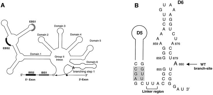
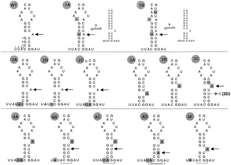
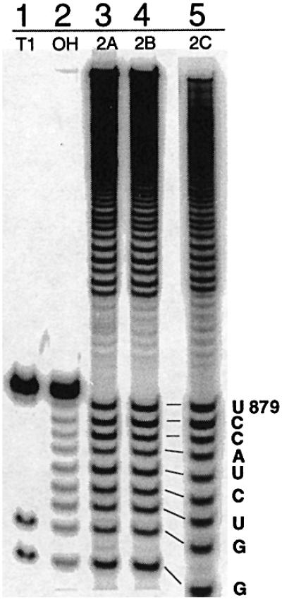
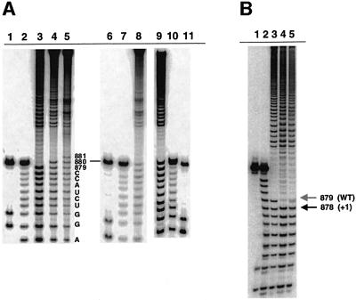
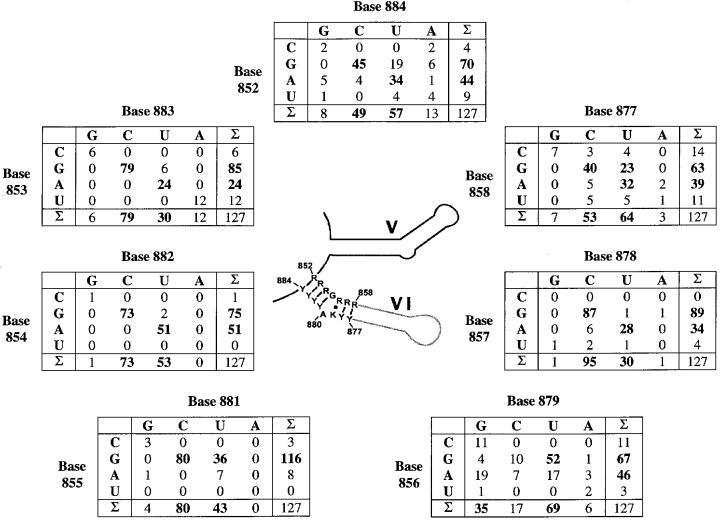
The branch site of group II introns is typically a bulged adenosine near the 3'-end of intron domain 6. The branch site is chosen with extraordinarily high fidelity, even when the adenosine is mutated to other bases or if the typically bulged adenosine is paired. Given these facts, it has been difficult to discern the mechanism by which the proper branch site is chosen. In order to dissect the determinants for branch-point recognition, new mutations were introduced in the vicinity of the branch site and surrounding domains. Single mutations did not alter the high fidelity for proper branch-site selection. However, several combinations of mutations moved the branch site systematically to new positions along the domain 6 stem. Analysis of those mutants, together with a new alignment of domain 5 and domain 6 sequences, reveals a set of structural determinants that appear to govern branch-site selection by group II introns.


Figures

None
Fig. 1. Secondary structural elements from group II intron ai5γ. (A) Schematic secondary structure of the ai5γ group II intron. The 5′ and 3′ splice sites are indicated as open and closed circles, respectively. The exon/intron pairing interactions (EBS1 and EBS2 pairing with IBS1 and IBS2) are shown as bold shaded boxes. Branching occurs during the first step of splicing, when the 2′-hydroxyl of A880 in D6 (bold) attacks the 5′-splice site (curved arrow). (B) The secondary structure of D56 is shown, with the branch site indicated by an arrow and the linker region by a bracket. Catalytically important residues in D5 are shaded.
None
Fig. 2. Schematic secondary structure of D6 mutants. Mutations to the abbreviated D6 sequence are shown in bold and highlighted in gray. The name of each variant is indicated to the upper left. The branch site of each mutant is indicated with an arrow. For the WT variant, the 7 nt downstream from D5 are shown. For mutants 1A and 1B, the two possible equilibrium conformations of D6 are indicated. For mutants 4B–E, brackets indicate additional base pairs that may serve to extend the D6 stem. RNA 3D is shown as a mutant of 3C, from which it is derived, with an arrow indicating the site of uridine to cytosine mutation.
None
Fig. 3. Schematic of the DNAzyme method for mapping group II intron branch points from both the 5′ and 3′ ends.
None
Fig. 4. Analysis of branch-site choice for intron revertants. (A) The branch points for mutants 1A (lane 4) and 1B (lane 1) were mapped by limited alkaline hydrolysis of branched fragments labeled at their 5′-termini. These were subjected to electrophoresis next to alkaline hydrolysis ladders of an oligonucleotide marker corresponding to the 17 nt immediately upstream of the WT branch-point adenosine (lanes 2 and 3). For the mapping procedure employed (Figure 3), the last band before the gap indicates the nucleotide 5′ of the selected branch point (A880, in each of these cases). (B) Partial alkaline hydrolysis of 3′-labeled branched fragments. For the mapping procedure employed (Figure 3), the band directly beneath the gap will correspond to a position 2 nts downstream from the branch point. Thus, the gap beginning at position C882 (lanes 6 and 7) corresponds to a WT branch-site choice at position A880 for mutants 1A (lane 6) and the prA–G (lane 7; Chu et al., 1998). Partial alkaline hydrolysis of these fragments is shown next to corresponding T1 (lane 4) and hydroxide (lane 5) ladders on an oligonucleotide marker corresponding to the terminal 13 nt of the intron. Partial alkaline hydrolysis of a cryptic branching mutant (RNA 4B, lane 1) is shown next to T1 (lane 3) and hydroxide (lane 2) ladders, and corresponds to branching at position U881 (arrow).
None
Fig. 5. Mapping the branch points of linker mutants 2A–C. Partial alkaline hydrolysis of branched fragments labeled at the 5′-end indicates that, like the WT intron, these mutants all branch at position A880 (lanes 3–5). The T1 digest (T1) and alkaline hydrolysis (OH) ladders on the 13 nt oligonucleotide serve as markers.
None
Fig. 6. Mapping the branch point of cryptic branching mutants. (A) Partial alkaline hydrolysis of branched fragments labeled at the 5′-end indicates that, unlike the WT intron (lane 3), mutants 4E (lane 4), 4C (lane 8) and 4B (lane 9) all branch at the nucleotide 3′ of the normal branch site (U881). Mutant 4D (lane 5) branches at a position 2 nt 3′ of the normal branch-site (U882). T1 nuclease (lanes 1, 6 and 11) and alkaline hydroxide (lanes 2, 7 and 10) ladders are shown as markers. (B) The 3C mutant (lane 4) and the related 3D mutant both react at a bulged adenosine that has been shifted by one nt upstream in contrast to WT (lane 3), and relative to the T1 (lane 1) and alkaline hydrolysis (lane 2) ladders. The solid and gray arrows indicate sites of upstream and WT branch-point selection, respectively. For mapping of mutants 3C and 3D, DNAzyme 1 (which cleaves 3′ of A862) was replaced by DNAzyme 3 (which cleaves 3′ of G866). Accordingly, the 17 nt marker oligonucleotide (lanes 1 and 2) spans intronic sequences U867 to G883.
None
Fig. 7. Covariation of D6 nucleotides. The secondary structure consensus of D5 and D6 are shown in the center. The numbering of nucleotides is derived from the ai5γ nomenclature. Each table displays the co-variation of 2 nt putatively involved in base pairing around the branching adenosine. The D56 alignment from which these matrices were calculated contains 127 sequences belonging to fungal, plant and algae organelles, and bacteria. All were downloaded from DDBJ/EMBL/GenBank and aligned combining the comparative approach with thermodynamic methods (Mfold server, Zuker, 1995–2000, Rensselaer Polytechnic Institute). Introns were classified by gene family and parsed into groups IIA and IIB according to standardized parameters (Michel et al., 1989). This resulted in the inclusion of 68 group IIB introns, 45 IIA introns and 14 unclassified bacterial introns that adopt features from both subgroups. Seven introns belonging to group IIA were excluded due to the lack of a bulged adenosine. Five of these are inserted into chloroplastid tRNAVal genes (trnV, J.Vogel, personal communication) and two into chloroplastic protease genes [clpP/2 or ORF203/2, sequence numbers 24 and 25 in Michel et al. (1989)]. Alignment of all sequences is provided as Supplementary data available at The EMBO Journal Online.

Similar articles

Cited by

References

    1. Bonen L. and Vogel,J. (2001) The ins and outs of group II introns. Trends Genet., 17, 322–331. - PubMed
    1. Boudvillain M. and Pyle,A.M. (1998) Defining functional groups, core structural features and inter-domain tertiary contacts essential for group II intron self-splicing: a NAIM analysis. EMBO J., 17, 7091–7104. - PMC - PubMed
    1. Boulanger S.C., Faix,P.H., Yang,H., Zhou,J., Franzen,J.S., Peebles,C.L. and Perlman,P.S. (1996) Length changes in the joining segment between domain 5 and 6 of a group II intron inhibit self-splicing and alter 3′ splice site selection. Mol. Cell. Biol., 16, 5896–5904. - PMC - PubMed
    1. Burkard M.E., Kierzek,R. and Turner,D.H. (1999) Thermodynamics of unpaired terminal nucleotides on short RNA helices correlates with stacking at helix termini in larger RNAs. J. Mol. Biol., 290, 967–982. - PubMed
    1. Cech T.R. (1986) The generality of self-splicing RNA: relationship to nuclear mRNA splicing. Cell, 44, 207–210. - PubMed

Publication types