Review
. 2001 Dec 1;29(23):4767-82.
doi: 10.1093/nar/29.23.4767.

Misreading of termination codons in eukaryotes by natural nonsense suppressor tRNAs

Affiliations
Review

Misreading of termination codons in eukaryotes by natural nonsense suppressor tRNAs

H Beier et al. Nucleic Acids Res. .

Abstract

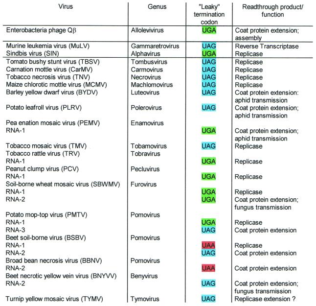
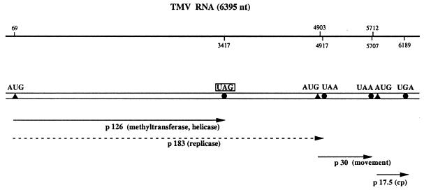
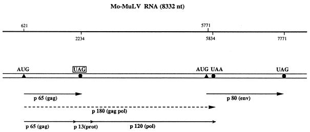
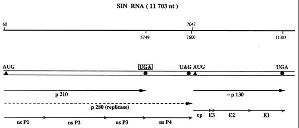
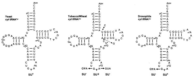
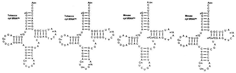
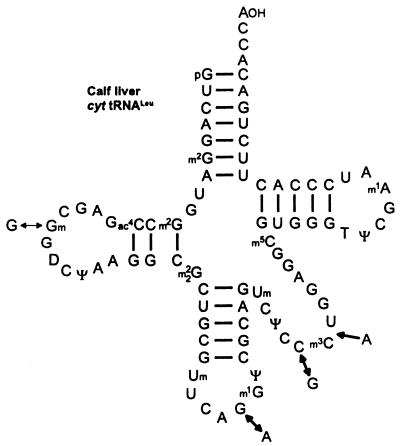
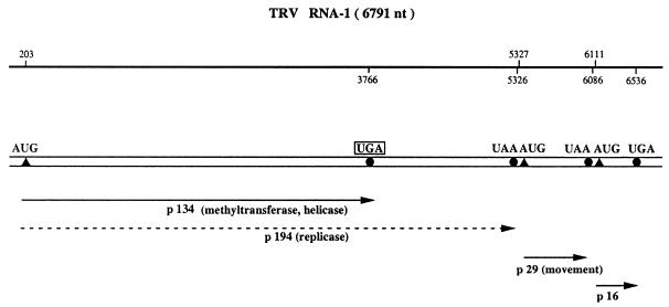
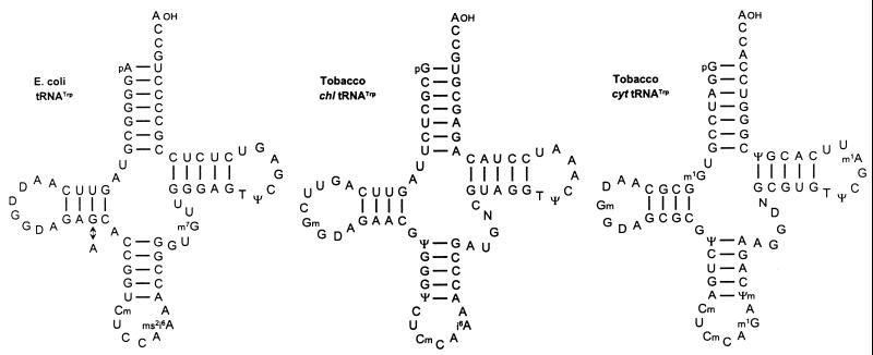
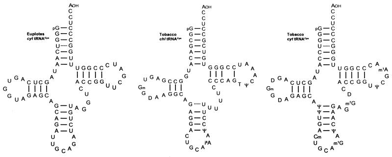
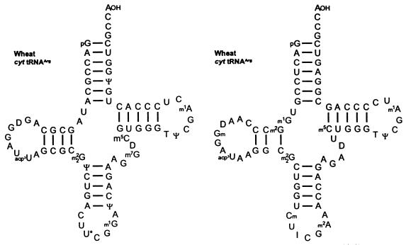
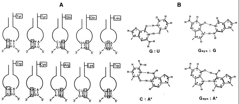
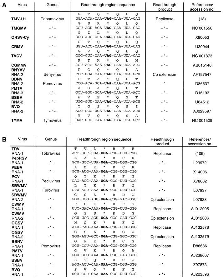
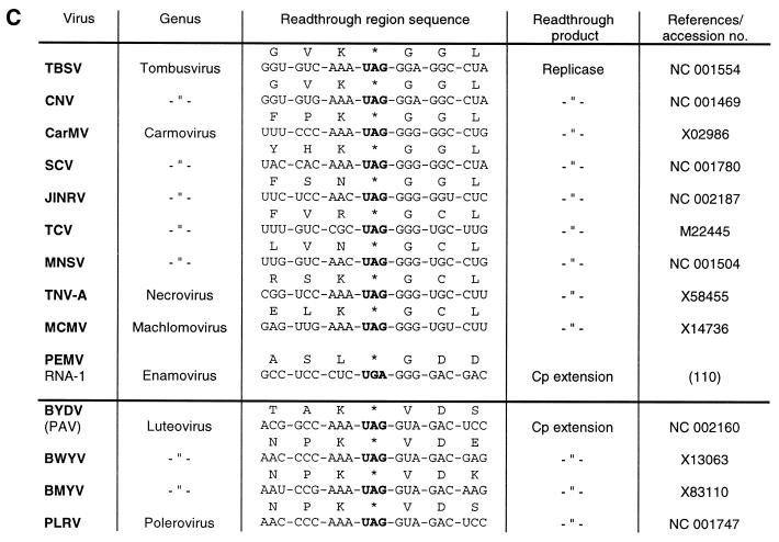
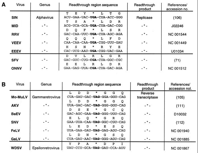
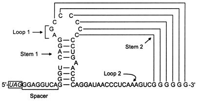
Translational stop codon readthrough provides a regulatory mechanism of gene expression that is extensively utilised by positive-sense ssRNA viruses. The misreading of termination codons is achieved by a variety of naturally occurring suppressor tRNAs whose structure and function is the subject of this survey. All of the nonsense suppressors characterised to date (with the exception of selenocysteine tRNA) are normal cellular tRNAs that are primarily needed for reading their cognate sense codons. As a consequence, recognition of stop codons by natural suppressor tRNAs necessitates unconventional base pairings in anticodon-codon interactions. A number of intrinsic features of the suppressor tRNA contributes to the ability to read non-cognate codons. Apart from anticodon-codon affinity, the extent of base modifications within or 3' of the anticodon may up- or down-regulate the efficiency of suppression. In order to out-compete the polypeptide chain release factor an absolute prerequisite for the action of natural suppressor tRNAs is a suitable nucleotide context, preferentially at the 3' side of the suppressed stop codon. Three major types of viral readthrough sites, based on similar sequences neighbouring the leaky stop codon, can be defined. It is discussed that not only RNA viruses, but also the eukaryotic host organism might gain some profit from cellular suppressor tRNAs.


Figures

Figure 1
Figure 1
Suppressible termination codons in positive sense ssRNA viruses, representing mostly type species from the corresponding genus. The classification of viruses is according to Pringle (104). Detailed information about sequences and function of readthrough products is listed in the legends to Figures 13 and 14.
Figure 2
Figure 2
Schematic structure of TMV RNA. The 6.4 kb genomic ssRNA of TMV (18) is designated by a double horizontal line. The upper line indicates the locations of ORFs as determined from the sequence. The positions of initiation and termination codons are specified by closed triangles and circles, respectively. The proteins synthesised in vivo are shown as single lines with arrowheads, the 183 kDa readthrough product is indicated by a broken line. The ‘leaky’ UAG stop codon at the end of the 126 kDa cistron is boxed.
Figure 3
Figure 3
Schematic structure of Mo-MuLV RNA. The 8.3 kb genomic ssRNA of Mo-MuLV (105) is represented by a double line. The positions of initiation and termination codons are indicated by closed triangles and circles, respectively. The ‘leaky’ UAG codon is boxed. The two primary polypeptides of 65 and 80 kDa are indicated by single lines. The gag/pol readthrough product of 180 kDa is shown as a broken line. This polypeptide is subsequently cleaved into the gag protein, a protease of 13 kDa and the reverse transcriptase of 120 kDa.
Figure 4
Figure 4
Schematic structure of sindbis virus (SIN) RNA. The 11.7 kb genomic ssRNA of SIN (106) is represented by a double line. The positions of initiation and termination codons are specified by closed triangles and circles, respectively. The translation of SIN non-structural (ns) proteins starts from the AUG codon at the beginning of nsP1 and proceeds to the UGA at the end of the nsP3 cistron. Partial readthrough of this ‘leaky’ stop codon yields the polypeptide of 280 kDa which is subsequently processed by proteolytic cleavage to produce the non-structural proteins nsP1 to nsP4.
Figure 5
Figure 5
Nucleotide sequences of cytoplasmic (cyt) tRNAsTyr from S.cerevisiae (34), Nicotiana rustica (19), wheat germ (29) and Drosophila melanogaster (107). The tRNATyr isoacceptors with GΨA anticodon have been shown to be active UAG suppressors in vitro, whereas the corresponding species with a GUA or QΨA anticodon are unable to misread this stop codon (19,27,29,39). U* (in position 20:A of Drosophila and 47 of plant tRNATyr), acp3U; m1G, 1-methylguanosine; m2G, N2-methylguanosine; m22G, N2,N2-dimethylguanosine; m7G, 7-methylguanosine; m1A, 1-methyladenosine; m5C, 5-methylcytidine; Ψ, pseudouridine; T, ribosylthymine; Q, queuosine; D, dihydrouridine; Gm, 2′-O-methylguanosine; Um, 2′-O-methyluridine.
Figure 6
Figure 6
Nucleotide sequences of cytoplasmic (cyt) tRNAsGln from N.rustica (48) and mouse liver (46). All of these tRNAGln isoacceptors suppress either the UAG, UAA or both stop codons in a wheat germ (41,48) and in a reticulocyte lysate, respectively (46). A major tRNAGln with UmUG anticodon from T.thermophila is also a strong UAG/UAA suppressor (41,108). The modified nucleosides not listed in theFigure 5 legend are: m2A, 2-methyladenosine; Cm, 2′-O-methylcytidine.
Figure 7
Figure 7
Nucleotide sequences of cytoplasmic (cyt) tRNAsLeu from calf liver (52). The two tRNALeu isoacceptors have been shown to misread the TMV-specific ‘leaky’ UAG codon in a reticulocyte lysate. The modified nucleoside not listed in the Figure 5 legend is m3C, 3-methylcytidine.
Figure 8
Figure 8
Schematic structure of TRV RNA-1. The 6.8 kb genomic ssRNA-1 of TRV (109) is designated by a double horizontal line. The positions of initiation and termination codons are indicated by closed triangles and circles, respectively. The first ORF codes for a non-structural protein of 134 kDa that is terminated by a UGA codon. Partial readthrough of this ‘leaky’ UGA stop codon yields the 194 kDa polypeptide (broken line).
Figure 9
Figure 9
Nucleotide sequences of chloroplast (chl) and cytoplasmic (cyt) tRNAsTrp from N.rustica.. The two tRNATrp isoacceptors promote UGA readthrough preferentially in the TRV-specific codon context in wheat germ extract (54,88). For comparison, the sequences of wild-type tRNATrp from E.coli and a mutated derivative are also shown. The mutated tRNATrp contains a single nucleotide exchange (G→A) at position 24, and exhibits increased UGA suppressor activity as compared to the wild type (14). The modified nucleosides not listed in the Figure 5 legend are: i6A, N6-isopentenyladenosine; ms2i6A, 2-methylthio-N6-isopentenyladenosine; Ψm, 2′-O-methylpseudouridine.
Figure 10
Figure 10
Nucleotide sequences of chloroplast (chl) and cytoplasmic (cyt) tRNAsCys from N.rustica. The two tRNACys isoacceptors promote UGA readthrough in wheat germ extract (63). The sequence of tRNACys from E.octocarinatus has been deduced from the known gene sequence contained within a cloned macronuclear DNA molecule. This tRNACys isoacceptor presumably reads the in-frame UGA codons present in numerous gene-derived mRNAs in Euplotes cells (64).
Figure 11
Figure 11
Nucleotide sequences of cytoplasmic (cyt) tRNAsArg from wheat germ. The tRNAArg with U*CG and to a minor extent tRNAArg with the ICG anticodon promote UGA readthrough preferentially in the PEMV-specific codon context in a wheat germ extract (70). U* (in position 34 of tRNAArg), mcm5U (5-methoxy-carbonylmethyluridine) and/or mcm5s2U (5-methoxy-carbonylmethyl-2-thiouridine); I, inosine.
Figure 12
Figure 12
Unconventional base interactions involved in the misreading of termination codons by natural suppressor tRNAs. (A) Schematic presentation of putative anticodon–codon interactions by eukaryotic suppressor tRNAs. With the exception of tRNALys(CUU) and tRNATrp(CmCA) misreading the UAG codon, all types of non-canonical codon reading by means of an appropriate suppressor tRNA have been demonstrated directly in vitro (19,39,48,52,54,63,70,89). In the case of the two former examples, incorporation of lysine and tryptophan (in addition to tyrosine) has been elucidated by amino acid sequence analysis of the translation protein produced by readthrough of an in-frame UAG codon within the yeast Ste6 gene (78). The tRNAGln isoacceptors with a UmUG anticodon from Tetrahymena, Nicotiana and mouse liver are able to also read UAG besides UAA (41,46,48). (B) Hypothetical interactions between non-complementary bases. In purine–purine mismatches, the nucleoside of the anticodon is presented in the syn configuration (isomerisation about the glycosyl bond of the nucleotide). The space-filling cyclopentene-diol side chain of the queuine base is attached to guanine at the indicated position (*) via a C–C bond, thus impairing the proposed G–G interaction (39). The adenosine in G:A and C:A mismatches is shown in the protonated form.
Figure 13
Figure 13
Plant viral readthrough sites. (A) Type I: TMV, tobacco mosaic virus; TMGMV, tobacco mild green mosaic virus; ORSV-Cy, odontoglossum ringspot tobamovirus; CRMV, chinese rape mosaic virus; TVCV, turnip vein clearing virus; CGMMV, cucumber green mottle mosaic virus; BNYVV, beet necrotic yellow vein virus; BBNV, broad bean necrosis virus; PMTV, potato mop-top furovirus; BSBV, beet soil-borne virus; BVQ, beet virus Q; TYMV, turnip yellow mosaic virus. (B) Type II: TRV, tobacco rattle virus; PepRSV, pepper ringspot virus; PEBV, pea early-browning virus; PCV, peanut clump virus; SBWMV, soil-borne wheat mosaic virus; CWMV, chinese wheat mosaic virus; OGSV, oat golden stripe virus. (C) Type III: TBSV, tomato bushy stunt virus; CNV, cucumber necrosis virus; CarMV, carnation mottle virus; SCV, saguaro cactus virus; JINRV, japanese iris necrotic ring virus; TCV, turnip crinkle virus; MNSV, melon necrotic spot virus; TNV, tobacco necrosis virus; MCMV, maize chlorotic mottle virus; PEMV, pea enation mosaic virus; BYDV, barley yellow dwarf virus; BWYV, beet western yellows virus; BMYV, beet mild yellowing virus; PLRV, potato leafroll virus. The luteoviruses and PLRV are separated by a horizontal line from the other type III viruses because their stop codons are followed by a valine instead of a glycine codon.
Figure 13
Figure 13
Plant viral readthrough sites. (A) Type I: TMV, tobacco mosaic virus; TMGMV, tobacco mild green mosaic virus; ORSV-Cy, odontoglossum ringspot tobamovirus; CRMV, chinese rape mosaic virus; TVCV, turnip vein clearing virus; CGMMV, cucumber green mottle mosaic virus; BNYVV, beet necrotic yellow vein virus; BBNV, broad bean necrosis virus; PMTV, potato mop-top furovirus; BSBV, beet soil-borne virus; BVQ, beet virus Q; TYMV, turnip yellow mosaic virus. (B) Type II: TRV, tobacco rattle virus; PepRSV, pepper ringspot virus; PEBV, pea early-browning virus; PCV, peanut clump virus; SBWMV, soil-borne wheat mosaic virus; CWMV, chinese wheat mosaic virus; OGSV, oat golden stripe virus. (C) Type III: TBSV, tomato bushy stunt virus; CNV, cucumber necrosis virus; CarMV, carnation mottle virus; SCV, saguaro cactus virus; JINRV, japanese iris necrotic ring virus; TCV, turnip crinkle virus; MNSV, melon necrotic spot virus; TNV, tobacco necrosis virus; MCMV, maize chlorotic mottle virus; PEMV, pea enation mosaic virus; BYDV, barley yellow dwarf virus; BWYV, beet western yellows virus; BMYV, beet mild yellowing virus; PLRV, potato leafroll virus. The luteoviruses and PLRV are separated by a horizontal line from the other type III viruses because their stop codons are followed by a valine instead of a glycine codon.
Figure 14
Figure 14
Animal viral readthrough sites. (A) Type II: SIN, sindbis virus; MID, middelburg virus; RRV, ross river virus; VEEV, venezuelan equine encephalitis virus; EEEV, eastern equine encephalitis virus; SFV, semliki forest virus; ONNV, O’nyong-nyong virus. The two alphaviruses SFV and ONNV do not contain ‘leaky’ UGA stop codons but instead contain a CGA arginine codon at this position. (B) Type III: Mo-MuLV, moloney murine leukemia virus; AKV, AKV murine leukemia virus; BaEV, baboon endogenous virus; SNV, spleen necrosis virus; FeLV, feline leukemia virus; GaLV, gibbon ape leukemia virus; WDSV, walleye dermal sarcoma virus. The epsilonretrovirus WDSV is separated by a horizontal line from the other type III viruses because its UAG stop codon is followed by an aspartate instead of a glycine codon.
Figure 15
Figure 15
Proposed secondary and pseudoknot structures in the vicinity of the ‘leaky’ UAG codon in Mo-MuLV RNA. The bipartite signal involved in efficient UAG readthrough consists of 8 nt (underlined) immediately at the 3′ side of the UAG and a stem–loop structure, which can form a pseudoknot with downstream G residues (91,92).

References

    1. Hatfield D.L., Levin,J.G., Rein,A. and Oroszlan,S. (1992) Translational suppression in retroviral gene expression. Adv. Virus Res., 41, 193–239. - PMC - PubMed
    1. Rohde W., Gramstat,A., Schmitz,J., Tacke,E. and Prüfer,D. (1994) Plant viruses as model systems for the study of non-canonical translation. J. Gen. Virol., 75, 2141–2149. - PubMed
    1. Farabaugh P.J. and Björk,G.R. (1999) How translational accuracy influences reading frame maintenance. EMBO J., 18, 1427–1434. - PMC - PubMed
    1. Murgola E.J. (1985) tRNA, suppression, and the code. Annu. Rev. Genet., 19, 57–80. - PubMed
    1. Eggertsson G. and Söll,D. (1988) Transfer ribonucleic acid-mediated suppression of termination codons in Escherichia coli.Microbiol. Rev., 52, 354–374. - PMC - PubMed

Publication types