. 2001 Dec 1;29(23):4851-65.
doi: 10.1093/nar/29.23.4851.

Molecular recognition of pyr mRNA by the Bacillus subtilis attenuation regulatory protein PyrR

Affiliations

Molecular recognition of pyr mRNA by the Bacillus subtilis attenuation regulatory protein PyrR

E R Bonner et al. Nucleic Acids Res. .

Abstract

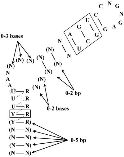
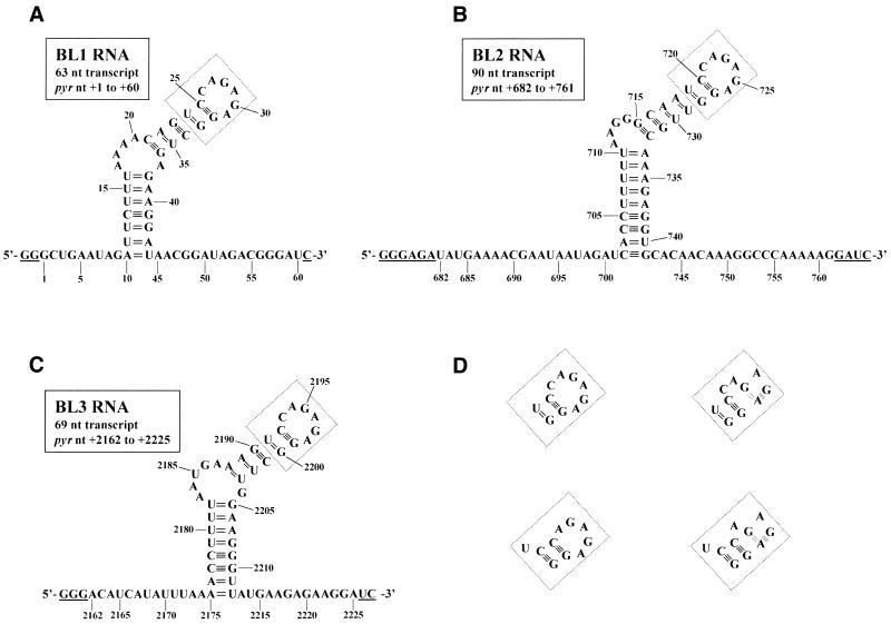
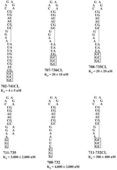
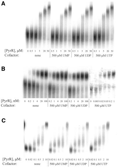
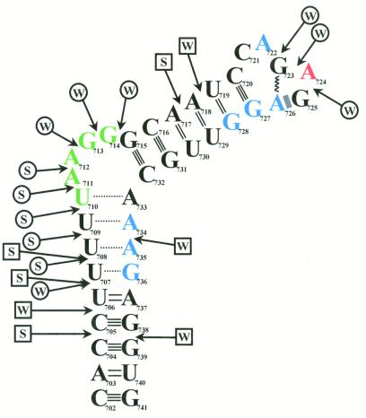
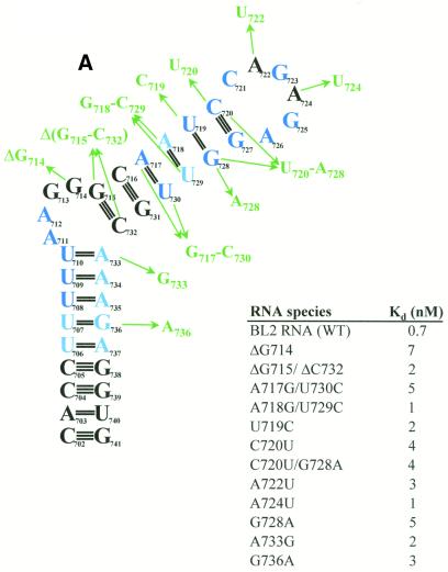
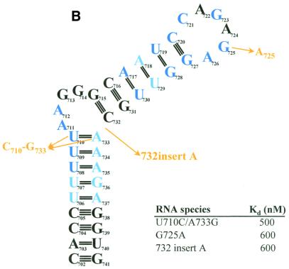
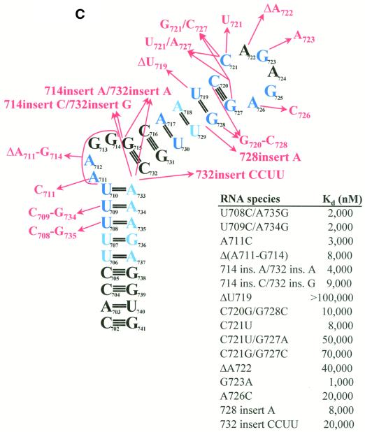
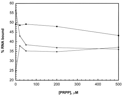
The pyrimidine nucleotide biosynthesis (pyr) operon in Bacillus subtilis is regulated by transcriptional attenuation. The PyrR protein binds in a uridine nucleotide-dependent manner to three attenuation sites at the 5'-end of pyr mRNA. PyrR binds an RNA-binding loop, allowing a terminator hairpin to form and repressing the downstream genes. The binding of PyrR to defined RNA molecules was characterized by a gel mobility shift assay. Titration indicated that PyrR binds RNA in an equimolar ratio. PyrR bound more tightly to the binding loops from the second (BL2 RNA) and third (BL3 RNA) attenuation sites than to the binding loop from the first (BL1 RNA) attenuation site. PyrR bound BL2 RNA 4-5-fold tighter in the presence of saturating UMP or UDP and 150- fold tighter with saturating UTP, suggesting that UTP is the more important co-regulator. The minimal RNA that bound tightly to PyrR was 28 nt long. Thirty-one structural variants of BL2 RNA were tested for PyrR binding affinity. Two highly conserved regions of the RNA, the terminal loop and top of the upper stem and a purine-rich internal bulge and the base pairs below it, were crucial for tight binding. Conserved elements of RNA secondary structure were also required for tight binding. PyrR protected conserved areas of the binding loop in hydroxyl radical footprinting experiments. PyrR likely recognizes conserved RNA sequences, but only if they are properly positioned in the correct secondary structure.


Figures

Figure 1
Figure 1
Consensus PyrR-binding loop. The consensus sequence and secondary structure for the 20 known PyrR-binding loops from B.caldolyticus, B.subtilis, E.faecalis, L.lactis, Lactobacillus leichmanii, L.plantarum, Streptococcus pneumoniae, Streptococcus pyogenes and Thermus ZO5 are shown. R = A or G; Y = C or U; N = any nucleotide. Parentheses indicate bases that are present in some sequences. The secondary structure pictured is formed in 12 of the 20 known PyrR-binding loops; in the other eight, the uridine nucleotide in the dashed circle initiates the purine-rich internal bulge, with the resulting base pairs indicated by dashed lines. The base pairs shown in solid boxes are conserved in 18 of the 20 examples, except for the A-U base pair in the upper stem, which is replaced by other base pairs in seven of the 20 examples.
Figure 2
Figure 2
Nucleotide sequences and predicted secondary structure of pyr mRNA BL1, BL2 and BL3. The nucleotide sequences and MFOLD v.3.1 predicted secondary structures for the transcripts from pBSBL1/BamHI (A), pBSBL2/BamHI (B) and pBSBL3/BamHI (C) are shown. Numbers denote the pyr nucleotide number, where +1 is the transcriptional start site for the operon (2). The underlined nucleotides are not pyr sequences but are found in the transcript as a result of the method used to obtain T7 RNA polymerase transcripts of defined sequence. The boxed region in each panel could fold in several plausible ways, which are shown in (D). When the hexaloop/pentaloop is folded into the putative GNRA tetraloop structure, the double dashed line represents a reverse Hoogsteen G-A base pair and the shaded rectangle represents purine-adenosine stacking.
Figure 3
Figure 3
RNA sequences used to determine the minimal BL2 RNA that binds to PyrR. RNA sequences used to determine the minimal RNA necessary for PyrR binding are shown. Boxed regions denote nucleotides not found in the native pyr transcript. BL2 RNA from pBSBL2/BamHI has already been described (Fig. 2B). Shorter RNAs are 702–741CL, 707–736CL, 708–735CL, 711–735, 708–732 and 711–732CL, where CL indicates that three C nucleotides were added to the 3′-end of the transcript to base pair to the three G nucleotides at the 5′-end of the transcript.
Figure 4
Figure 4
Apparent dissociation constants for the interaction of PyrR with BL1, BL2 and BL3 RNA were determined in the presence of no cofactor, 500 µM UMP, 500 µM UDP or 500 µM UTP; these constants are listed in Table 1. Representative gel mobility shift experiments for BL1 (A), BL2 (B) and BL3 (C) are shown here. All reactions contained 50 pM of the indicated RNA, except for the PyrR–BL2 binding reactions containing 500 µM UTP; these reactions used 1 pM BL2 RNA due to the extremely low PyrR concentrations needed to determine the apparent dissociation constant. As these reactions contained much less radioactivity than all other BL2 reactions, they were scanned separately to improve visibility. ‘Super-shifted’ bands were observed with all RNA species when the PyrR concentration was ≥5 µM (evident here in the BL1 and BL3 reactions); this phenomenon is discussed in the text. ‘Smeared’ RNA between the free RNA and PyrR–RNA species was counted as bound RNA, as was ‘super-shifted’ RNA, for calculation of apparent dissociation constants.
Figure 5
Figure 5
Titration of BL2 mRNA with PyrR. Gel mobility shift assays were performed with BL2 RNA held at 100 nM, which was over 100 times greater than the observed apparent dissociation constant for the BL2–PyrR interaction. Increasing amounts of PyrR were added. The binding data were fitted to two straight lines using linear least squares methods. The lines intersect at [PyrR]/[RNA] = 1.0 and RNA bound = 92%, indicating that one PyrR subunit binds to one molecule of pyr mRNA. Data averaged from three independent experiments are shown, two with 500 µM UTP and one with 500 µM UMP.
Figure 6
Figure 6
Autoradiogram of hydroxyl radical footprinting and nuclease digestion experiments. End-labeled BL2 RNA was checked for degradation or radiolysis both in the absence of any other factors (lane 1) and also in the presence of PyrR and UMP (lane 2). BL2 RNA was reacted with RNase T1 (lane 3) to produce a ladder of fragments that terminate at each G in the transcript and subjected to partial alkaline hydrolysis (lanes 4, 8 and 13) to produce a ladder of fragments that terminate at every position in the transcript. BL2 RNA was reacted with hydroxyl radicals in the absence of any other factors (lane 5), in the presence of PyrR alone (lane 6) or in the presence of PyrR and UMP (lane 7). BL2 RNA was reacted with RNase I in the absence of any other factors (lane 9) or in the presence of PyrR and UMP (lane 10). BL2 RNA was reacted with RNase V1 in the absence of any other factors (lane 11) or in the presence of PyrR and UMP (lane 12). The sequence diagram on the right has every fifth nucleotide numbered and predicted secondary structure features of the RNA noted. The predicted single-stranded regions of RNA that flank the BL2 stem–loop are denoted by 5′-SS and 3′-SS.
Figure 7
Figure 7
Map of hydroxyl radical footprinting and nuclease digestion results. End-labeled BL2 RNA was exposed to hydroxyl radicals either in the presence or absence of PyrR. In the presence of PyrR, nucleotides in blue were strongly protected, nucleotides in red were moderately protected and nucleotides in green were weakly protected from hydroxyl radical cleavage (see Fig. 6, lanes 6 and 7). The secondary structure of the RNA was mapped using end-labeled RNA, RNase I (specific for single-stranded RNA) and RNase V1 (specific for double-stranded RNA). A speculative model for the secondary structure of BL2 RNA is shown, based on the structure predicted by MFOLD v.3.1, as well as the nuclease digestion pattern that was observed. Locations of nuclease cuts are shown by arrows, with circle-headed arrows representing single-strand-specific cuts and square-headed arrows representing double-strand-specific cuts (see Fig. 6, lanes 9 and 11). S and W represent strong and weak cleavages, respectively. The dashed lines in the lower stem represent base pairs that are predicted to form transiently or not at all, due to strong cleavage by RNase I in this region. The hexaloop is folded into a GNRA tetraloop structure with the first two bases of the hexaloop extruded. The zigzag line represents a reverse Hoogsteen G-A base pair and the shaded rectangle represents purine-adenosine stacking. No direct evidence exists for a GNRA tetraloop structure actually forming in vivo or in vitro.
Figure 8
Figure 8
Effects of structural variants of BL2 on binding to PyrR. The effects of various changes in BL2 RNA sequence on the BL2–PyrR interaction were measured using the gel mobility shift assay (500 µM UMP was used) and are shown as apparent dissociation constants for each RNA. (A) Sequence changes in green had little effect on binding (apparent Kd < 10 nM; apparent Kd for BL2 = 0.7 nM). (B) Sequence changes in yellow had a moderate effect on binding (10 nM < apparent Kd < 1000 nM). (C) Sequence changes in red had a severe effect on binding (apparent Kd > 1000 nM). Nucleotides in dark blue are highly conserved among pyr anti-antiterminator sequences from various bacteria, while nucleotides in light blue denote positions of pyrimidine or purine conservation.
Figure 8
Figure 8
Effects of structural variants of BL2 on binding to PyrR. The effects of various changes in BL2 RNA sequence on the BL2–PyrR interaction were measured using the gel mobility shift assay (500 µM UMP was used) and are shown as apparent dissociation constants for each RNA. (A) Sequence changes in green had little effect on binding (apparent Kd < 10 nM; apparent Kd for BL2 = 0.7 nM). (B) Sequence changes in yellow had a moderate effect on binding (10 nM < apparent Kd < 1000 nM). (C) Sequence changes in red had a severe effect on binding (apparent Kd > 1000 nM). Nucleotides in dark blue are highly conserved among pyr anti-antiterminator sequences from various bacteria, while nucleotides in light blue denote positions of pyrimidine or purine conservation.
Figure 9
Figure 9
Effect of PRPP on PyrR–BL2 RNA interaction. PyrR (1 nM) and BL2 RNA (50 pM) concentrations were held constant. The concentration of PRPP was increased from 0 to 500 µM in the presence of no cofactor (filled circles), 200 µM UMP (filled squares) or 200 µM UTP (filled triangles).

References

    1. Turner R.J., Lu,Y. and Switzer,R.L. (1994) Regulation of the Bacillus subtilis pyrimidine biosynthetic (pyr) gene cluster by an autogenous transcriptional attenuation mechanism. J. Bacteriol., 176, 3708–3722. - PMC - PubMed
    1. Quinn C.L., Stephenson,B.T. and Switzer,R.L. (1991) Functional organization and nucleotide sequence of the Bacillus subtilis pyrimidine biosynthetic operon. J. Biol. Chem., 266, 9113–9127. - PubMed
    1. Kahler A.E. and Switzer,R.L. (1996) Identification of a novel gene of pyrimidine nucleotide biosynthesis, pyrDII, that is required for dihydroorotate dehydrogenase activity in Bacillus subtilis. J. Bacteriol., 178, 5013–5016. - PMC - PubMed
    1. Lu Y. and Switzer,R.L. (1996) Evidence that the Bacillus subtilis pyrimidine regulatory protein PyrR acts by binding to pyr mRNA at three sites in vivo. J. Bacteriol., 178, 5806–5809. - PMC - PubMed
    1. Lu Y., Turner,R.J. and Switzer,R.L. (1996) Function of RNA secondary structures in transcriptional attenuation of the Bacillus subtilis pyr operon. Proc. Natl Acad. Sci. USA, 93, 14462–14467. - PMC - PubMed

Publication types

MeSH terms