Review
. 2001 Dec;65(4):523-69, table of contents.
doi: 10.1128/MMBR.65.4.523-569.2001.

Biodegradation of aromatic compounds by Escherichia coli

Affiliations
Review

Biodegradation of aromatic compounds by Escherichia coli

E Díaz et al. Microbiol Mol Biol Rev. 2001 Dec.

Abstract

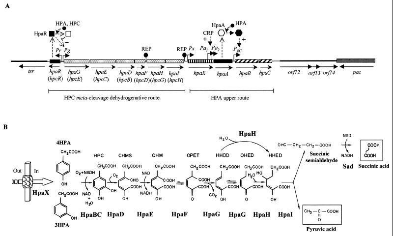
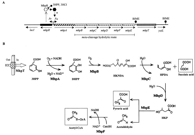
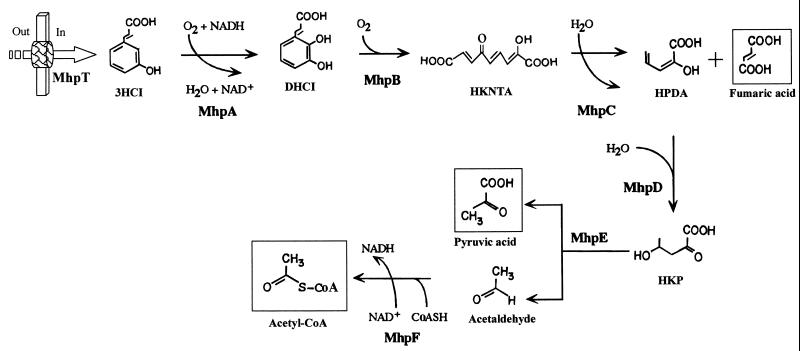
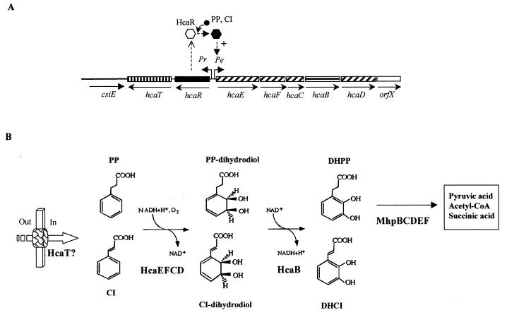
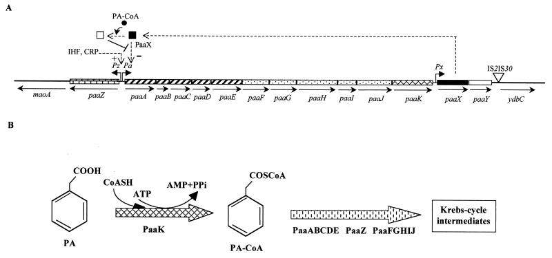
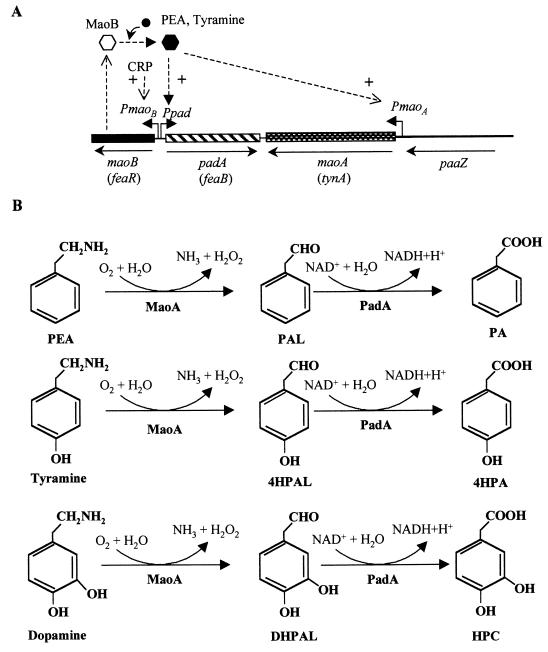
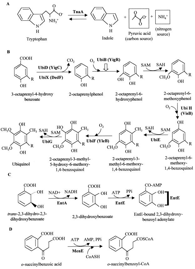
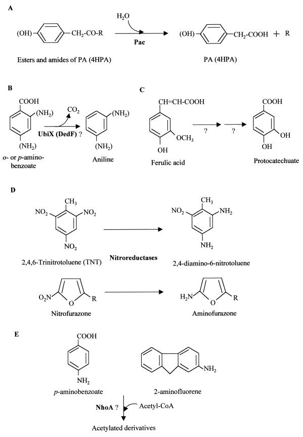
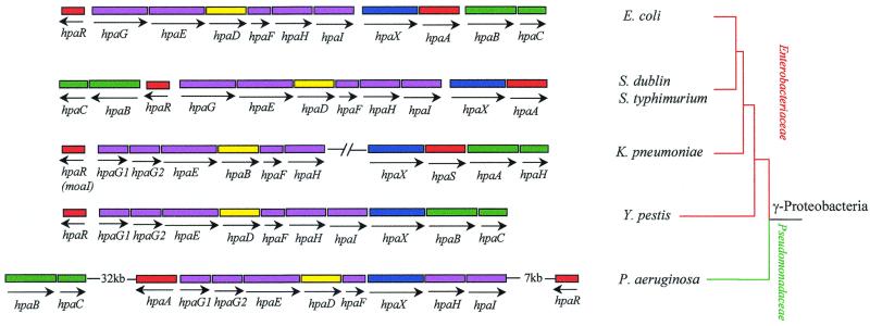
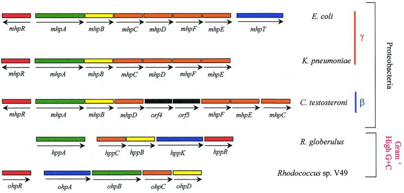
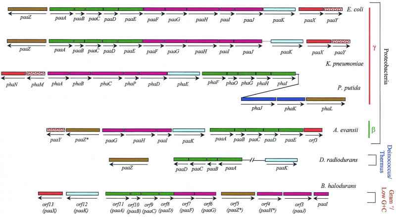
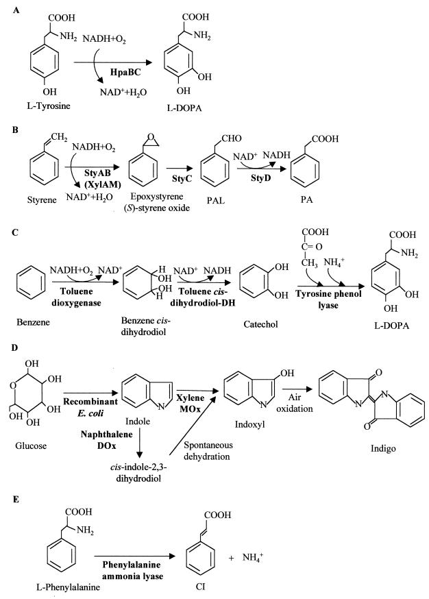
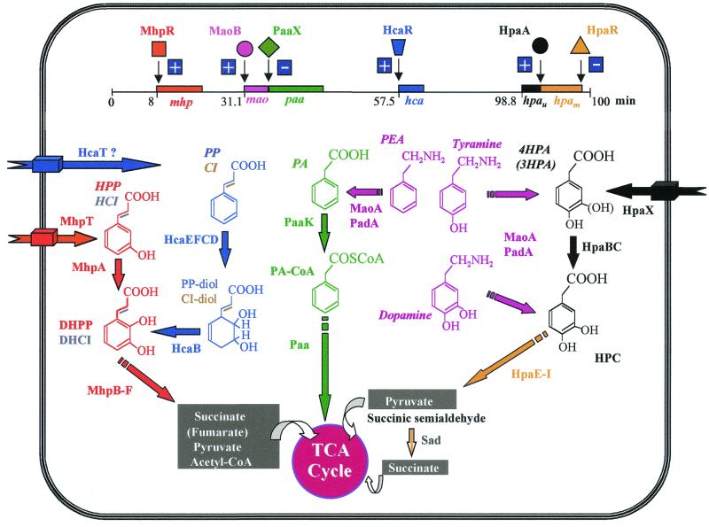
Although Escherichia coli has long been recognized as the best-understood living organism, little was known about its abilities to use aromatic compounds as sole carbon and energy sources. This review gives an extensive overview of the current knowledge of the catabolism of aromatic compounds by E. coli. After giving a general overview of the aromatic compounds that E. coli strains encounter and mineralize in the different habitats that they colonize, we provide an up-to-date status report on the genes and proteins involved in the catabolism of such compounds, namely, several aromatic acids (phenylacetic acid, 3- and 4-hydroxyphenylacetic acid, phenylpropionic acid, 3-hydroxyphenylpropionic acid, and 3-hydroxycinnamic acid) and amines (phenylethylamine, tyramine, and dopamine). Other enzymatic activities acting on aromatic compounds in E. coli are also reviewed and evaluated. The review also reflects the present impact of genomic research and how the analysis of the whole E. coli genome reveals novel aromatic catabolic functions. Moreover, evolutionary considerations derived from sequence comparisons between the aromatic catabolic clusters of E. coli and homologous clusters from an increasing number of bacteria are also discussed. The recent progress in the understanding of the fundamentals that govern the degradation of aromatic compounds in E. coli makes this bacterium a very useful model system to decipher biochemical, genetic, evolutionary, and ecological aspects of the catabolism of such compounds. In the last part of the review, we discuss strategies and concepts to metabolically engineer E. coli to suit specific needs for biodegradation and biotransformation of aromatics and we provide several examples based on selected studies. Finally, conclusions derived from this review may serve as a lead for future research and applications.


Figures

FIG. 1
FIG. 1
Pathway for the catabolism of HPA (4HPA and 3HPA) in E. coli. (A) Genetic map of the chromosomal hpa (in E. coli W) and hpc (in E. coli C) regions. Relevant genes are indicated by blocks: genes with similar shading participate in the same enzymatic step or in the same functional unit (route) of the pathway. The hpc genes are indicated in brackets. Regulatory and transport genes are shown by solid and vertically striped blocks, respectively. The genes flanking the hpa cluster (tsr, orf12, orf13, and orf14) are contiguous in the genome of E. coli K-12 and are represented by thick lines. orf12 and orf13 correspond to the yjiY gene from E. coli K-12. orf14 corresponds to the yjiA gene from E. coli K-12. The arrows show the directions of gene transcription. Bent arrows represent the Pr, Pg, Px, Pa1, Pa2 and PBC promoters. REP sequences are shown. The HpaR repressor and HpaA activator are represented by a square and hexagon, respectively; empty and solid symbols indicate inactive and active regulators, respectively; − and + indicate transcriptional repression and activation, respectively. The inducer molecule (HPA and HPC) is represented by a solid circle. (B) Biochemistry of the HPA catabolic pathway. The metabolites are 4HPA and 3HPA, HPC (homoprotocatechuate), CHMS (5-carboxymethyl-2-hydroxy-muconic semialdehyde), CHM (5-carboxymethyl-2-hydroxy-muconic acid), OPET (5-oxo-pent-3-ene-1,2,5-tricarboxylic acid), HHDD (2-hydroxy-hept-2,4-diene-1,7-dioic acid), OHED (2-oxo-hept-3-ene-1,7-dioic acid), and HHED (2,4-dihydroxy-hept-2-ene-1,7-dioic acid). The enzymes are HpaBC (HPA monooxygenase), HpaD (HPC 2,3-dioxygenase), HpaE (CHMS dehydrogenase), HpaF (CHM isomerase), HpaG (OPET decarboxylase), HpaH (hydratase), HpaI (HHED aldolase), and Sad (succinic semialdehyde dehydrogenase). The HPA transport protein (HpaX) is represented by a thick arrow. Out and In indicate outside and inside the cell, respectively.
FIG. 2
FIG. 2
Pathway for the catabolism of 3HPP in E. coli. (A) Genetic map of the chromosomal mhp cluster. Relevant genes are indicated by blocks: genes with similar shading participate in the same enzymatic step or in the same functional unit (route) of the pathway. Regulatory and transport genes are shown by solid and vertically striped blocks, respectively. Genes flanking the mhp cluster (lacI and yaiL) are represented by thick lines. The arrows show the directions of gene transcription. Bent arrows represent the Pr and Pa promoters. The location of the BIME is shown. The inactive and active forms of the MhpR activator are represented by empty and solid hexagons, respectively. + indicates transcriptional activation. The inducer molecule (3HPP and 3HCI) is represented by a solid circle. (B) Biochemistry of the 3HPP catabolic pathway. The metabolites are 3HPP, DHPP (2,3-dihydroxyphenlypropionic acid), HKNDA (2-hydroxy-6-keto-nona-2,4-diene 1,9-dioic acid), HPDA (2-hydroxy-penta-2,4-dienoic acid), and HKP (4-hydroxy-2-ketopentanoic acid). The enzymes are MhpA (3HPP monooxygenase), MhpB, (DHPP 1,2 dioxygenase), MhpC (HKNDA hydrolase), MhpD (HPDA hydratase), MhpE (HKP aldolase), and MhpF (acetaldehyde dehydrogenase [acylating]). The 3HPP transport protein (MhpT) is represented by a thick arrow. Out and In indicate outside and inside the cell, respectively.
FIG. 3
FIG. 3
Biochemistry of the 3HCI catabolic pathway. The metabolites are 3HCI, DHCI (2,3-dihydroxycinnamic acid), HKNTA (2-hydroxy-6-keto-nona-2,4,7-triene 1,9-dioic acid), HPDA (2-hydroxy-penta-2,4-dienoic acid) and HKP (4-hydroxy-2-ketopentanoic acid). The enzymes are MhpA (3HCI monooxygenase), MhpB, (DHCI 1,2-dioxygenase), MhpC (HKNTA hydrolase), MhpD (HPDA hydratase), MhpE (HKP aldolase), and MhpF (acetaldehyde dehydrogenase [acylating]). The 3HCI transport protein (MhpT) is represented by a thick arrow. Out and In indicate outside and inside the cell, respectively.
FIG. 4
FIG. 4
Pathway for the catabolism of PP in E. coli. (A) Genetic map of the chromosomal hca cluster. Relevant genes are indicated by blocks: genes with similar shading encode the subunits of the PP dioxygenase. Regulatory (solid block) and putative transport (vertically striped block) genes are also shown. The horizontally striped block indicates the gene encoding the PP dihydrodiol dehydrogenase. The empty block represents a gene of unknown function. The csiE gene flanking the hca cluster is represented by a thick line. The arrows show the directions of gene transcription. Bent arrows represent the Pr and Pe promoters. The inactive and active forms of the HcaR activator are represented by empty and solid hexagons, respectively. + indicates transcriptional activation. The inducer molecule (PP and CI) is represented by a solid circle. (B) Biochemistry of the PP catabolic pathway. The metabolites are PP, CI, PP dihydrodiol, CI-dihydrodiol, DHPP, and DHCI (see the legends to Fig. 2 and 3). The enzymes are HcaEFCD (PP dioxygenase), HcaB (PP-dihydrodiol dehydrogenase), and MhpBCDEF (see the legend to Fig. 2). The putative PP/CI transport protein (HcaT) is represented by a thick arrow. Out and In indicate outside and inside the cell, respectively.
FIG. 5
FIG. 5
Pathway for the catabolism of PA in E. coli. (A) Genetic map of the chromosomal paa cluster. Relevant genes are indicated by blocks: genes with similar shading participate in the same enzymatic step or in the same functional unit of the pathway. Genes flanking the paa cluster (maoA and ydbC) are represented by thick lines. The arrows show the directions of gene transcription. Bent arrows represent the Pz, Pa, and Px promoters. The locations of the IS2 and IS30 insertion sequences within ydbA in E. coli K-12 are shown. The regulatory gene (paaX) is represented by a black block. The inactive and active forms of the PaaX repressor are indicated by empty and solid squares, respectively. − and + indicate transcriptional repression and activation, respectively. The inducer molecule (PA-CoA) is represented by a solid circle. (B) Biochemistry of the PA catabolic pathway. The first intermediate of the pathway is PA-CoA (phenylacetyl CoA). The enzymes are PaaK (PA-CoA ligase), PaaABCDE (putative multicomponent oxygenase), PaaZ (putative ring-cleavage enzyme), and PaaFGHIJ (putative β-oxidation-like enzymatic system).
FIG. 6
FIG. 6
Upper pathway for the catabolism of aromatic amines (PEA, tyramine, and dopamine) in E. coli. (A) Genetic map of the chromosomal cluster for the initial catabolism of aromatic amines. Relevant genes are indicated by blocks. Alternative gene names are in parentheses. The paaZ gene from the paa cluster (see Fig. 5) is indicated by a thick line. The arrows show the directions of gene transcription. Bent arrows represent the PmaoB PmaoA and Ppad promoters. The regulatory gene maoB (feaR) is shown by a solid block. The inactive and active forms of the MaoB activator are represented by empty and solid hexagons, respectively. + indicates transcriptional activation. The inducer molecule (PEA, tyramine) is represented by a solid circle. (B) Biochemistry of the initial catabolism of aromatic amines. The metabolites are PAL (phenylacetaldehyde), 4HPAL (4-hydroxyphenylacetaldehyde), DHPAL (3,4-dihydroxyphenylacetaldehyde), PA, 4HPA, and HPC (homoprotocatechuate). The enzymes are MaoA (monoamine oxidase) and PadA (phenylacetaldehyde dehydrogenase).
FIG. 7
FIG. 7
Relevant enzymatic activities of E. coli for the metabolism of chorismate-derived compounds. The enzymes catalyzing the different reactions are those described in Table 4. SAM, S-adenosylmethionine; SAH, S-adenosylhomocysteine; R, octaprenyl side chain.
FIG. 8
FIG. 8
Biotransformation activities of E. coli on some aromatic compounds. The known or putative proteins catalyzing the different enzymatic reactions are indicated in boldface type and are described in Table 4.
FIG. 9
FIG. 9
Comparison of hpa clusters involved in HPA catabolism in different proteobacteria from the γ subgroup. Arrows show the directions of gene transcription. Genes are indicated by blocks: red (regulatory genes), blue (transport genes), green (genes encoding the initial HPA monooxygenase), yellow (ring cleavage dioxygenase gene), and purple (genes encoding the meta-cleavage dehydrogenative route). A broken line indicates an unknown distance. The hpaR gene from K. pneumoniae corresponds to the previously characterized moaI gene (in parentheses). References of the sequences are as follows: E. coli (strain W) (accession no. Z37980), S. enterica serovar Dublin (accession no. AF144422), S. enterica serovar Typhimurium and Y. pestis (obtained from the ERGO database website) K. pneumoniae (accession no. L41068 and AJ000054 and ERGO database), and P. aeruginosa PAO1 (Pseudomonas Genome Project, at http://www.pseudomonas.com/).
FIG. 10
FIG. 10
Comparison of gene clusters involved in HPP catabolism in different bacteria. Arrows show the directions of gene transcription. Genes are indicated by blocks: red (regulatory genes), blue (transport genes), green (genes encoding the HPP monooxygenase), yellow (ring cleavage dioxygenase gene), orange (genes encoding the meta-cleavage hydrolytic route), and black (genes of unknown function). The ohpR gene from Rhodococcus sp. strain V49 encodes a regulator that, in contrast to the regulators of the other four gene clusters, does not belong to the IclR protein family. β and γ indicate the β and γ subgroups of proteobacteria. The references of the sequences are as follows: E. coli strain K-12 (EcoGene database), K. pneumoniae (ERGO database), C. testosteroni TA441 (accession no. AB024335), R. globerulus PWD1 (accession no. U89712), and Rhodococcus sp. strain V49 (accession no. AF274045).
FIG. 11
FIG. 11
Comparison of gene clusters involved in PA catabolism in different bacteria. Arrows show the directions of gene transcription. Genes are indicated by blocks: red (regulatory genes), dark blue (transport genes), light blue (genes encoding the PA-CoA ligase), green (genes encoding a putative multicomponent oxygenase), brown (genes encoding a putative ring cleavage enzyme), purple (genes encoding a putative β-oxidation-like pathway), and dotted (genes of unknown function). The paaI ortholog in P. putida U has not been described previously (182, 217), and it was named phaP here. orf3 is an incomplete orf gene that encodes a putative transcriptional regulator of the TetR family. An asterisk indicates a truncated gene. A discontinuous line means that the sequence has not been yet reported. The broken line shows that the genes are not adjacent. γ and β represent the γ and β subgroups of proteobacteria. The references of the sequences are as follows: E. coli (strain W) (accession no. X97452), K. pneumoniae (ERGO database), P. putida U and KT2440 (accession no. AF029714 and database referenced at website http: //www.ncbi.nlm.nih.gov/Microb_blast/unfinishedgenome.html, respectively), A. evansii KB740 (paa or pac genes under accession no. AF176259 or AJ278756, respectively), D. radiodurans R1 (accession no. AE002069), and B. halodurans C-125 (accession no. AB011837).
FIG. 12
FIG. 12
Selected biotransformations of aromatic compounds in recombinant E. coli strains. The enzymes catalyzing the different reactions are indicated in boldface type. (A) Production of 3,4-dihydroxyphenylalanine (l-Dopa) from tyrosine through the 4HPA monooxygenase (HpaBC) from E. coli W. (B) Biotransformation of styrene into (S)-styrene oxide (epoxystyrene), phenylacetaldehyde (PAL), and PA by the StyAB, StyC, and StyD enzymes from different Pseudomonas strains. Formation of (S)-styrene oxide from styrene was also reported using the xylene monooxygenase (XylAM) from the TOL plasmid of P. putida. (C) Conversion of benzene into l-Dopa using the toluene dioxygenase and toluene cis-dihydrodiol dehydrogenase (DH) from P. putida F1 and the tyrosine phenol-lyase from C. freundii. (D) Biotransformation of glucose into the dye indigo in a recombinant E. coli strain that converts glucose into indole and then oxidizes the latter through naphthalene dioxygenase (DOx) or xylene monooxygenase (MOx) from P. putida. (E) Conversion of l-phenylalanine to cinnamic acid (CI) and ammonia through the phenylalanine ammonia-lyase of R. toruloides.
FIG. 13
FIG. 13
Schematic representation of the gene clusters and the encoded catabolic pathways for the aerobic degradation of aromatic compounds in E. coli. The gene clusters mhp, mao, paa, hca, hpau (upper route), and hpam (meta-cleavage route) are indicated by blocks and correspond to those described in Fig. 1 to 6. The locations of the clusters refer to the E. coli K-12 linkage map (the hpa cluster is absent in E. coli K-12). The regulatory proteins are indicated by different symbols that reflect the different regulatory protein families. + and − indicate transcriptional activation and repression, respectively. The transporters are represented by thick arrows spanning the cellular envelope. Abbreviations of the metabolites and enzymes are the same than those used in Fig. 1 to 6. The different colors correspond to the different catabolic pathways (or to different routes within the same pathway). A discontinuous arrow indicates more than one enzymatic step. TCA, tricarboxylic acids.

References

    1. Abdulrashid N, Clark D P. Isolation and genetic analysis of mutations allowing the degradation of furans and thiophenes by Escherichia coli. J Bacteriol. 1987;169:1267–1271. - PMC - PubMed
    1. Alam K Y, Clark D P. Molecular cloning and sequence of the thdF gene, which is involved in thiophene and furan oxidation by Escherichia coli. J Bacteriol. 1991;173:6018–6024. - PMC - PubMed
    1. Alekshun M N, Kim Y S, Levy S B. Mutational analysis of MarR, the negative regulator of marRAB expression in Escherichia coli, suggests the presence of two regions required for DNA binding. Mol Microbiol. 2000;35:1394–1404. - PubMed
    1. Allende J L, Gibello A, Martin M, Garrido-Pertierra A. Transport of 4-hydroxyphenylacetic acid in Klebsiella pneumoniae. Arch Biochem Biophys. 1992;292:583–588. - PubMed
    1. Altenschmidt U, Oswald B, Steiner E, Herrmann H, Fuchs G. New aerobic benzoate oxidation pathway via benzoyl-coenzyme A and 3-hydroxybenzoyl-coenzyme A in a denitrifying Pseudomonas sp. J Bacteriol. 1993;175:4851–4858. - PMC - PubMed

Publication types

MeSH terms

LinkOut - more resources