. 2001 Dec 1;15(23):3104-17.
doi: 10.1101/gad.926401.

Degradation of p53 by adenovirus E4orf6 and E1B55K proteins occurs via a novel mechanism involving a Cullin-containing complex

Affiliations

Degradation of p53 by adenovirus E4orf6 and E1B55K proteins occurs via a novel mechanism involving a Cullin-containing complex

E Querido et al. Genes Dev. .

Abstract

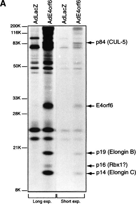
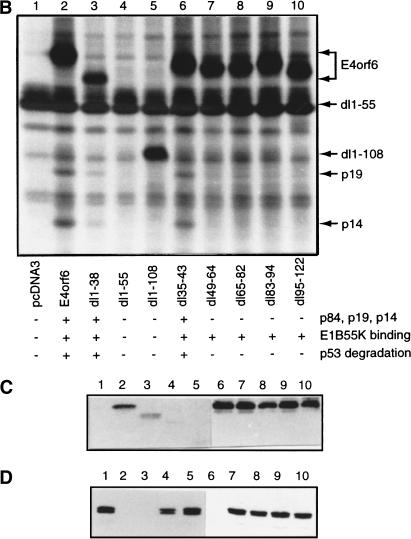
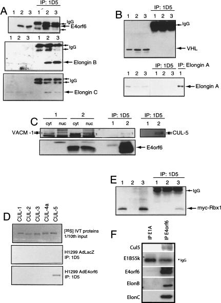
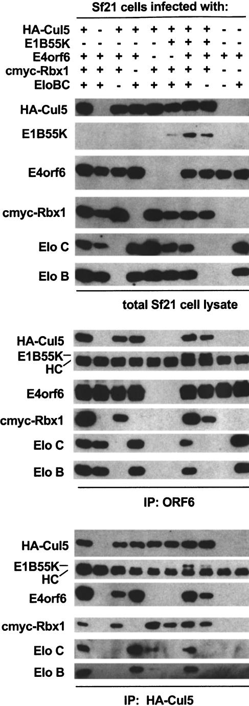
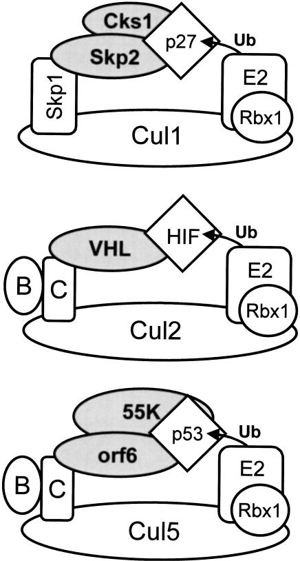
Although MDM2 plays a major role in regulating the stability of the p53 tumor suppressor protein, other poorly understood MDM2-independent pathways also exist. Human adenoviruses have evolved strategies to regulate p53 function and stability to permit efficient viral replication. One mechanism involves adenovirus E1B55K and E4orf6 proteins, which collaborate to target p53 for degradation. To determine the mechanism of this process, a multiprotein E4orf6-associated complex was purified and shown to contain a novel Cullin-containing E3 ubiquitin ligase that is (1) composed of Cullin family member Cul5, Elongins B and C, and the RING-H2 finger protein Rbx1(ROC1); (2) remarkably similar to the von Hippel-Lindau tumor suppressor and SCF (Skp1-Cul1/Cdc53-F-box) E3 ubiquitin ligase complexes; and (3) capable of stimulating ubiquitination of p53 in vitro in the presence of E1/E2 ubiquitin-activating and -conjugating enzymes. Cullins are activated by NEDD8 modification; therefore, to determine whether Cullin complexes are required for adenovirus-induced p53 degradation, studies were conducted in ts41 Chinese hamster ovary cells that are temperature sensitive for the NEDD8 pathway. E4orf6/E1B55K failed to induce the degradation of p53 at the nonpermissive temperature. Thus, our results identify a novel role for the Cullin-based machinery in regulation of p53.


Figures

Figure 1
Figure 1
Analysis of E4orf6 complex formation. (A) Detection of E4orf6 complexes. Extracts from H1299 cells infected with AdLacZ or AdE4orf6 and labeled with 35S were immunoprecipitated with 1D5 anti-E4orf6 antibody followed by SDS-PAGE and autoradiography. Two exposures have been included: positions of migration of prestained standard size markers (left) and those of E4orf6 and coimmunoprecipitating proteins (right). Identities for these species were derived from mass spectroscopy analysis of tryptic peptides. (B) Analysis of complex formation using E4orf6 deletion mutants. H1299 cells were transfected with plasmid DNAs encoding wild-type or mutant E4orf6, and extracts were immunoprecipitated and analyzed as in A. Coimmunoprecipitation of p19 and p14 cellular proteins with E4orf6 has been summarized below as + and −. Binding of p84 showed a similar pattern, as determined from other exposures of the gel (data not shown). Binding of E4orf6 to E1B55K (shown in C) and p53 turnover experiments with E4orf6 and E1B55K (D) have also been indicated as + and −. (C) Complex formation between E4orf6 mutants and E1B55K. Extracts from H1299 cells transfected with plasmid DNAs expressing E1B55K (pCA14 HH55K) and wild-type or mutant E4orf6 proteins (pcDNA3E4orf6) were immunoprecipitated with anti-E1B55K antibody (2A6), and membranes were immunoblotted with anti-E4orf6 antibody (1807). (D) E4orf6/E1B55K-induced turnover of p53. H1299 cells were transfected with plasmid DNAs pcDNA3 p53wt (p53), pCA14 HH55K (E1B-55K), and pcDNA3E4orf6 (wild-type or mutant E4orf6), and extracts were analyzed by Western blotting using anti-p53 antibodies 1801 and 421.
Figure 1
Figure 1
Analysis of E4orf6 complex formation. (A) Detection of E4orf6 complexes. Extracts from H1299 cells infected with AdLacZ or AdE4orf6 and labeled with 35S were immunoprecipitated with 1D5 anti-E4orf6 antibody followed by SDS-PAGE and autoradiography. Two exposures have been included: positions of migration of prestained standard size markers (left) and those of E4orf6 and coimmunoprecipitating proteins (right). Identities for these species were derived from mass spectroscopy analysis of tryptic peptides. (B) Analysis of complex formation using E4orf6 deletion mutants. H1299 cells were transfected with plasmid DNAs encoding wild-type or mutant E4orf6, and extracts were immunoprecipitated and analyzed as in A. Coimmunoprecipitation of p19 and p14 cellular proteins with E4orf6 has been summarized below as + and −. Binding of p84 showed a similar pattern, as determined from other exposures of the gel (data not shown). Binding of E4orf6 to E1B55K (shown in C) and p53 turnover experiments with E4orf6 and E1B55K (D) have also been indicated as + and −. (C) Complex formation between E4orf6 mutants and E1B55K. Extracts from H1299 cells transfected with plasmid DNAs expressing E1B55K (pCA14 HH55K) and wild-type or mutant E4orf6 proteins (pcDNA3E4orf6) were immunoprecipitated with anti-E1B55K antibody (2A6), and membranes were immunoblotted with anti-E4orf6 antibody (1807). (D) E4orf6/E1B55K-induced turnover of p53. H1299 cells were transfected with plasmid DNAs pcDNA3 p53wt (p53), pCA14 HH55K (E1B-55K), and pcDNA3E4orf6 (wild-type or mutant E4orf6), and extracts were analyzed by Western blotting using anti-p53 antibodies 1801 and 421.
Figure 2
Figure 2
Identification of E4orf6 binding proteins. (A) H1299 cells were infected with AdLacZ (lane 1), AdE4orf6 (lane 2), or AdE4orf6 plus AdHH55K plus Adp53wt (lane 3). Whole cell extracts (left three lanes) and 1D5 immunoprecipitates (right three lanes) were analyzed by Western blotting using anti-E4orf6 antibody 1807 (top), anti-Elongin B antibody (middle), and anti-Elongin C antibody (bottom). (B) Same as Figure 1A except that immunoblotting was performed with anti-VHL Ig32 antibody (top) or anti-Elongin A antibody (bottom). At the right (lane 1) is shown an immunoprecipitate prepared with anti-Elongin A and blotted with the same antibody. (C) H1299 cells were infected with AdLacZ (lane 1) or AdE4orf6 (lane 2), and cytoplasmic and nuclear extracts as well as whole cell extracts were prepared. Whole cell extracts immunoprecipitated with 1D5 anti-E4orf6 antibody were analyzed by Western blotting using either a polyclonal antibody to rabbit VACM-1 (left six lanes) or a polyclonal antibody against human Cul5 (right two lanes). E4orf6 (bottom) was detected using 1807 antibody. (D) Human Cul1, Cul2, Cul3, and Cul5 and mouse Cul4A were synthesized (top) by in vitro transcription/translation, and aliquots of these 35S-labeled proteins were combined with 1D5 immunoprecipitates from AdLacZ-infected (middle) or AdE4orf6-infected (bottom) H1299 cells. (E) H1299 cells were transfected with plasmid DNAs expressing Myc–Rbx1 (lane 1), E4orf6 (lane 2), and Myc–Rbx1 plus E4orf6 (lane 3). Whole cell extracts (left lanes) or 1D5 immunoprecipitates (right lanes) were analyzed by Western blotting with anti-Myc 9E10 antibody. (F) Human 293 cells were infected with wild-type Ad5 at a MOI of 40, and at 15 h postinfection, cell extracts were prepared and immunoprecipitated with either anti-E4orf6 (1809) or anti-E1A (M73) antibodies. The pattern of associated proteins determined by Western blotting as above.
Figure 3
Figure 3
Reconstitution of E4orf6/E1B55K/Cul5/Rbx1/Elongin B and C complexes in Sf21 cells. Sf21 cells were infected with baculoviruses encoding the proteins indicated. The lysates were immunoprecipitated using either anti-HA (12CA5) or anti-E4orf6 (1D5) antibody. Crude lysates (top) and washed immune complexes (middle, bottom) were separated by SDS-PAGE and immunoblotted with the indicated antibodies.
Figure 4
Figure 4
Intracellular colocalization of E4orf6 and Cul5. H1299 cells were transfected with plasmid DNAs expressing HA–Cul2, HA–Cul5, or E4orf6, and cells were fixed and examined by laser confocal microscopy. E4orf6 was detected using rabbit polyclonal antibody (1807) and Alexa 594 (red) anti-rabbit IgG antibody. HA-tagged human Cul2 and Cul5 were detected using mouse monoclonal anti-HA antibody (HA.11) and Alexa 488 (green) anti-mouse IgG antibody. (AC) HA–Cul5 and E4orf6. (DF) HA–Cul2 and E4orf6. C and F were generated by scanning for both the red and the green signal to detect any colocalization as bright yellow. (G) HA–Cul2 alone. Bar, 20 μm; cells shown are representative. (H) HA–Cul5 alone. (I) E4orf6 alone.
Figure 5
Figure 5
In vitro ubiquitination of p53 by E4orf6 complexes. (A) Lysates from Sf21 cells infected with baculoviruses encoding E1B55K, Elongin B, Elongin C, HA–Cul5, and Myc–Rbx1, with or without baculovirus encoding E4orf6, were immunoprecipitated with anti-E4orf6 antibody (1D5). Increasing amounts of purified p53 protein were incubated with the immunoprecipitates and with yUba1, hUbc5a, and GST–UbK48R in ubiquitination reaction buffer. Reaction mixtures were incubated for 30 min at 25°C and analyzed by Western blotting using anti-p53 antibody (421). (B) p53 was incubated with immunoprecipitates and with various combinations of yUba1, hUbc5a, and GST–UbK48R. The indicated components were omitted from reactions: E1, yUba1; E2, hUBC5a; GST-Ub, GST-UbK48R. HC indicates heavy chain.
Figure 5
Figure 5
In vitro ubiquitination of p53 by E4orf6 complexes. (A) Lysates from Sf21 cells infected with baculoviruses encoding E1B55K, Elongin B, Elongin C, HA–Cul5, and Myc–Rbx1, with or without baculovirus encoding E4orf6, were immunoprecipitated with anti-E4orf6 antibody (1D5). Increasing amounts of purified p53 protein were incubated with the immunoprecipitates and with yUba1, hUbc5a, and GST–UbK48R in ubiquitination reaction buffer. Reaction mixtures were incubated for 30 min at 25°C and analyzed by Western blotting using anti-p53 antibody (421). (B) p53 was incubated with immunoprecipitates and with various combinations of yUba1, hUbc5a, and GST–UbK48R. The indicated components were omitted from reactions: E1, yUba1; E2, hUBC5a; GST-Ub, GST-UbK48R. HC indicates heavy chain.
Figure 6
Figure 6
E4orf6/E1B55K require NEDDylated Cullins to degrade p53. (A) Wild-type and ts41 Chinese hamster ovary (CHO) cells maintained at 33°C were infected with various combinations of adenovirus vectors for 48 h (see + at top). Vectors included AdrtTA (vector control), Adp53wt, AdE4orf6, and AdHH55K. Some cultures were shifted to the 39°C for the last 15 h of incubation. Whole cell extracts analyzed by Western blotting using to detect p53 (1801), E4orf6 (1807), E1B55K (2A6), and actin. (B) CHO cells infected with various combinations of adenovirus vectors (as in A). Proteasome inhibitors MG132, lactacystin, or their solvents were added for the last 6 h of infection. Cell extracts were analyzed as in A.
Figure 7
Figure 7
Parallels between the SCF, VHL, and E4orf6 ubiquitin ligase complexes. The SCF (top), VHL (middle), and E4orf6 (bottom) ubiquitin ligase complexes have been illustrated. (B) Elongin B; (C) Elongin C; (p27) p27Kip1; (Ub) ubiquitin; (orf6) Ad5 E4orf6; (55K) Ad5 E1B55K; (Cul1) the mammalian homolog of Cdc53; and (Rbx1) Rbx1/ROC1. The architecture of the SCF complex shown is based on the X-ray structure of a Skp1/Skp2/Cul1/Rbx1 crystal determined recently (N. Zheng and N.P. Pavletich, pers. comm.). Models of the VHL and E4orf6 complexes are theoretical only, and the interaction interface between E4orf6 and the Cul5/Elongin BC/Rbx1 complex has not yet been determined.

Similar articles

Cited by

References

    1. Aso T, Haque D, Barstead RJ, Conaway RC, Conaway JW. The inducible elongin A elongation activation domain: Structure, function, and interaction with the elongin BC complex. EMBO J. 1996;15:5557–5566. - PMC - PubMed
    1. Bacchetti S, Graham FL. Inhibition of cell proliferation by an adenovirus vector expressing the human wild type p53 protein. Int J Oncol. 1993;3:781–788. - PubMed
    1. Bargonetti J, Reynisdottir I, Friedman PN, Prives C. Site-specific binding of wild-type p53 to cellular DNA is inhibited by SV40 T antigen and mutant p53. Genes & Dev. 1992;6:1886–1898. - PubMed
    1. Bartek J, Lukas J. p27 destruction: Cks1 pulls the trigger. Nat Cell Biol. 2001;3:E95–E98. - PubMed
    1. Bech-Otschir D, Kraft R, Huang X, Henklein P, Kapelari B, Pollmann C, Dubiel W. COP9 signalosome-specific phosphorylation targets p53 to degradation by the ubiquitin system. EMBO J. 2001;20:1630–1639. - PMC - PubMed

Publication types

MeSH terms