. 2001 Dec;11(12):2059-65.
doi: 10.1101/gr.205701.

Twin priming: a proposed mechanism for the creation of inversions in L1 retrotransposition

Affiliations

Twin priming: a proposed mechanism for the creation of inversions in L1 retrotransposition

E M Ostertag et al. Genome Res. 2001 Dec.

Abstract

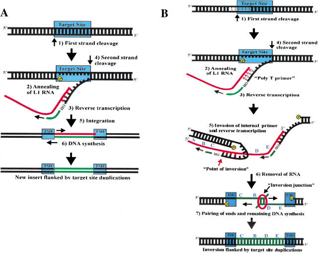
L1 retrotransposons are pervasive in the human genome. Approximately 25% of recent L1 insertions in the genome are inverted and truncated at the 5' end of the element, but the mechanism of L1 inversion has been a complete mystery. We analyzed recent L1 inversions from the genomic database and discovered several findings that suggested a mechanism for the creation of L1 inversions, which we call twin priming. Twin priming is a consequence of target primed reverse transcription (TPRT), a coupled reverse transcription/integration reaction that L1 elements are thought to use during their retrotransposition. In TPRT, the L1 endonuclease cleaves DNA at its target site to produce a double-strand break with two single-strand overhangs. During twin priming, one of the overhangs anneals to the poly(A) tail of the L1 RNA, and the other overhang anneals internally on the RNA. The overhangs then serve as primers for reverse transcription. The data further indicate that a process identical to microhomology-driven single-strand annealing resolves L1 inversion intermediates.


Figures

Figure 1
Figure 1
Target primed reverse transcription and twin priming. (A) This is a schematic of target primed reverse transcription (TPRT), based on in vitro studies of the R2 element from Bombyx mori (Luan et al. 1993). TPRT involves the following steps: (1) Cleavage of first DNA strand at the target site by the retrotransposon endonuclease (EN). (2) Annealing of retrotransposon RNA at the nick. (3) Reverse transcription from the free 3′-hydroxyl by the retrotransposon reverse transcriptase (RT). (4) Cleavage of second DNA strand. (5) Integration at the double-strand break. (6) Removal of RNA and completion of DNA synthesis. The TPRT process produces target site duplications (TSDs) at the flanks of the newly integrated retrotransposon. (B) Twin priming is a modification of the TPRT reaction with the following steps: (1) The L1 EN cleaves one strand of its DNA target site, producing the poly T primer. (2) The poly(A) tail of the L1 RNA anneals on the poly T primer. (3) L1 RT uses the L1 RNA as a template and the poly T primer to initiate reverse transcription. (4) The L1 EN cleaves the second DNA strand before reverse transcription has been completed, producing the internal primer. (5) The internal primer invades the L1 RNA and primes reverse transcription. (6) The RNA is removed from the RNA/cDNA structure. (7) The single-stranded cDNAs pair at a region of limited complementarity, and the remaining DNA synthesis is completed. The entire process results in an L1 inversion flanked by perfect target site duplications. The L1 RNA sequence is represented by 5′-A-B-C-D-E-3′. After the inversion, the insertion sequence is 5′-C-B-D-E-3′.
Figure 2
Figure 2
Complementarity of the primers. (A) The internal primer and the poly T primer were analyzed for complementarity to their predicted binding sites on the L1 RNA. The first six nucleotides, numbered from the 3′-hydroxyl, are listed. Nucleotides are highlighted in yellow if they are complementary to the corresponding nucleotide on the L1 RNA. The last row lists the number of complementary nucleotides at each position, out of a possible total of seventeen. (B) The number of matches (r) at each position and the corresponding P-values, representing the likelihood of obtaining r matches or greater by chance alone.
Figure 3
Figure 3
Clustering of the inversion points. The L1 RNA is represented by a black line ending in a poly(A) tail (A(n)). The position on the RNA is numbered from the first nucleotide (1) to the end of the AATAAA poly(A) signal (6022). The region of the L1 RNA from nucleotides 4000–6000 is expanded on the line above. A black arrow represents the inversion point of each element. Each inversion is labeled with its accession number and the position of its inversion point.
Figure 4
Figure 4
The structure of each inversion. A full-length L1 element is represented by the hashed line, numbered from the first nucleotide (1) to nucleotide 6000 near the end of the sequence. The structure of each inversion is represented below the schematic L1. A left-facing arrow represents noninverted sequence with the length denoted above (distance from the AATAAA poly(A) signal to the end of the noninverted sequence). A right-facing arrow represents inverted sequence with the length denoted above (distance from the inversion point to the target site duplication). For example, the sequence at the tail of the inverted sequence from Z95325 is represented on the L1 consensus sequence near nucleotide 5450, and the sequence at the head of the arrow is represented near nucleotide 1380. Each inversion has nucleotides from the consensus sequence either deleted (del) or duplicated (dup) at the end of the noninverted sequence (head of left-facing arrow). The size of the deletion or duplication is indicated above its corresponding location relative to the L1 consensus sequence. Note that in the case of a duplication, the duplicated sequence is located both in its usual univerted position at the end of the noninverted sequence (head of the left-facing arrow) and also is inverted at the beginning of the inverted sequence (tail of right-facing arrow). Most inversions have evidence of complementary nucleotides at the inversion junction that could be associated with either the end of the noninverted sequence (head of left-facing arrow) or the end of the inverted sequence (head of right-facing arrow). The number of complementary nucleotides is indicated in brackets under the column labeled “Overlap.” In the case of complementary nucleotides, the sizes of the inverted sequence, the noninverted sequence, and the deletion or duplication become variable, as indicated by the numbering. In one case there are no complementary nucleotides [0], and in two cases there are a number of nontemplated nucleotides. A negative number in brackets indicates the number of nontemplated nucleotides, and the nontemplated nucleotides are listed in parentheses.

References

    1. Altschul SF, Gish W, Miller W, Myers EW, Lipman DJ. Basic local alignment search tool. J Mol Biol. 1990;215:403–410. - PubMed
    1. Benson DA, Karsch-Mizrachi I, Lipman DJ, Ostell J, Rapp BA, Wheeler DL. GenBank. Nucleic Acids Res. 2000;28:15–18. - PMC - PubMed
    1. Boissinot S, Entezam A, Furano AV. Selection against deleterious LINE-1-containing loci in the human lineage. Mol Biol Evol. 2001;18:926–935. - PubMed
    1. Choi Y, Ishiguro N, Shinagawa M, Kim CJ, Okamoto Y, Minami S, Ogihara K. Molecular structure of canine LINE-1 elements in canine transmissible venereal tumor. Anim Genet. 1999;30:51–53. - PubMed
    1. Cost GJ, Boeke JD. Targeting of human retrotransposon integration is directed by the specificity of the L1 endonuclease for regions of unusual DNA structure. Biochemistry. 1998;37:18081–18093. - PubMed

Publication types

LinkOut - more resources