Review
. 2001 Dec 1;537(Pt 2):333-45.
doi: 10.1111/j.1469-7793.2001.00333.x.

Muscle damage from eccentric exercise: mechanism, mechanical signs, adaptation and clinical applications

Affiliations
Review

Muscle damage from eccentric exercise: mechanism, mechanical signs, adaptation and clinical applications

U Proske et al. J Physiol. .

Abstract

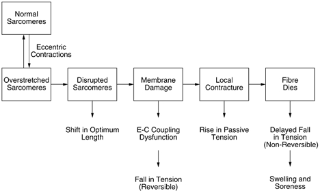
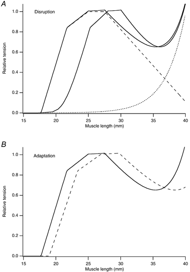
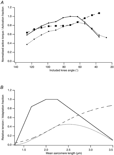
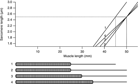
In eccentric exercise the contracting muscle is forcibly lengthened; in concentric exercise it shortens. While concentric contractions initiate movements, eccentric contractions slow or stop them. A unique feature of eccentric exercise is that untrained subjects become stiff and sore the day afterwards because of damage to muscle fibres. This review considers two possible initial events as responsible for the subsequent damage, damage to the excitation-contraction coupling system and disruption at the level of the sarcomeres. Other changes seen after eccentric exercise, a fall in active tension, shift in optimum length for active tension, and rise in passive tension, are seen, on balance, to favour sarcomere disruption as the starting point for the damage. As well as damage to muscle fibres there is evidence of disturbance of muscle sense organs and of proprioception. A second period of exercise, a week after the first, produces much less damage. This is the result of an adaptation process. One proposed mechanism for the adaptation is an increase in sarcomere number in muscle fibres. This leads to a secondary shift in the muscle's optimum length for active tension. The ability of muscle to rapidly adapt following the damage from eccentric exercise raises the possibility of clinical applications of mild eccentric exercise, such as for protecting a muscle against more major injuries.


Figures

Figure 1
Figure 1. Postulated series of events leading to muscle damage from eccentric exercise
During an active lengthening, longer, weaker sarcomeres are stretched onto the descending limb of their length-tension relation where they lengthen rapidly, uncontrollably, until they are beyond myofilament overlap and tension in passive structures has halted further lengthening. Repeated overextension of sarcomeres leads to their disruption. Muscle fibres with disrupted sarcomeres in series with still-functioning sarcomeres show a shift in optimum length for tension in the direction of longer muscle lengths. When the region of disruption is large enough it leads to membrane damage. This could be envisaged as a two-stage process, beginning with tearing of t-tubules. Any fall in tension at this point would be reversible with caffeine (see text). It would be followed by damage to the sarcoplasmic reticulum, uncontrolled Ca2+ release from its stores and triggering of a local injury contracture. That, in turn, would raise muscle passive tension. If the damage was extensive enough, parts of the fibre, or the whole fibre, would die. This fall in tension would not be recoverable with caffeine. Breakdown products of dead and dying cells would lead to a local inflammatory response associated with tissue oedema and soreness.
Figure 2
Figure 2. Postulated distribution of elastic filaments in sarcomeres
The top two diagrams consider a sarcomere with elastic filaments only linking the ends of the thick filaments to the Z-lines. The numbers indicate the distribution of tension. Total tension is set at 80 % of maximum to indicate that the sarcomere is on the descending limb of its length-tension relation. When one half-sarcomere becomes over-stretched, as a result of an eccentric contraction, tension borne by its myofilaments drops to zero and the full tension is borne by the elastic filament. The other half-sarcomere is unaffected since its isometric tension capability remains the same. When a second elastic element is included in the model, one which spans the full length of the sarcomere (lower 3 diagrams), overstretch of one half-sarcomere leads 50 % of the tension to be distributed to the elastic element in series with the thick filaments while, because of its proportionately smaller extension, that spanning the sarcomere bears less (30 %). It will also contribute 30 % of the tension to the other half-sarcomere, the remaining 50 % being distributed between the series elastic element (5%) and the cross-bridges (45 %). Since this half-sarcomere's isometric tension capability remains at 70 %, it shortens until tension in the series element has fallen to zero and myofilament overlap is somewhere on the ascending limb, generating 60 % of the tension, leaving 20 % in the elastic element spanning the sarcomere. This is what is observed under the electron microscope, one half-sarcomere over-stretched to beyond overlap, the other half very short. This kind of model indicates that passive tension in the whole sarcomere becomes significant when one half becomes over-stretched.
Figure 3
Figure 3. Changes in mechanical properties of muscle following a series of eccentric contractions
A, disruption of sarcomeres. Computer simulated sarcomere length-tension relations. The dashed line is the active length-tension relation taken from Gordon et al. (1966). The dotted line is an exponential curve representing passive tension; the continuous line is the total tension. Tension is normalised relative to the maximum active tension. Length is given as that of a postulated muscle fibre comprising 10 000 sarcomeres with a sarcomere length of 2.5 μm at optimum length. The control curve is the continuous curve on the left. After a series of eccentric contractions 10 % of the sarcomeres have their active force set to zero to simulate becoming disrupted, leading to a shift in optimum length of the total tension curve by 3 mm (continuous curve to the right). B, adaptation of the muscle fibre following injury from eccentric exercise. The continuous curve is the control total tension curve as in the upper panel, the dashed curve, that after the number of sarcomeres in series has been increased by 10 %, without changing the length of the tendon. It has led to an increase in optimum length by 2 mm.
Figure 4
Figure 4. Activation and the length-tension relation
A, torque-angle relationship for the vastus intermedius muscle of the anaesthetised rat. Torque-angle curves were measured before (continuous line) and after (dotted line) a series of eccentric contractions of the muscle. Here the muscle was stretched through 27 deg in 33 ms while being stimulated at 90 pulses per second. Stretches were arranged to start 5 deg short of the optimum angle and finished at 22 deg beyond optimum. Included knee angle is the angle subtended between knee and thigh. At each length, the ratio of torque before and after the contractions has been calculated (dashed line) giving an estimate of the activation fraction (modified from Allen, 1999). B, computer simulation of partial activation. The continuous curve is the fully activated sarcomere length-tension curve (Gordon et al. 1966). The dashed curve is a Hill plot (Hill, 1913), as a reasonable estimate of length dependence of the activation fraction. The dotted curve is the resulting partial activation curve. The partial activation simulation gives a realistic shift in optimum length for tension but is unable to simulate active tension seen at long lengths after a series of eccentric contractions. That is, at long lengths tension lies well below the control curve. Note also the greater fall in tension at short lengths.
Figure 5
Figure 5. Sarcomere length-muscle length relation
Relation between sarcomere length and muscle length for theoretical muscle fibres comprising different numbers of sarcomeres and different lengths of tendon. It is assumed that optimum sarcomere length is 2.5 μm. For a muscle fibre with 10 000 sarcomeres and 20 mm of tendon (fibre no. 1) tension begins to rise at 35 mm and the optimum is reached at 45 mm (dashed line). A shift in optimum length for active tension by 5 mm in the direction of longer lengths (dashed line) can be achieved by increasing the length of tendon to 25 mm (fibre no. 2). The drawback is that active tension is not developed until the muscle is stretched to 40 mm, that is, the working range of muscle lengths has been reduced. Increasing the number of sarcomeres from 10 000 to 12 000, and leaving tendon length at 20 mm (fibre no. 3) produces the required increase in optimum length and leads to a smaller reduction in the muscle's working range, where tension begins to rise at 39 mm. Increasing sarcomere number further to 14 000, while at the same time reducing the length of tendon to 15 mm (fibre no. 4) produces the most satisfactory result, the required shift in optimum length by 5 mm and reduction of the working range by only 1 mm.

References

    1. Allen DG. Eccentric muscle damage: mechanisms of early reduction of force. Acta Physiologica Scandanavica. 2001;171:311–319. - PubMed
    1. Allen TA. PhD Thesis. Clayton, Victoria, Australia: Monash University; 1999. Effect of eccentric contractions on the mechanical properties of skeletal muscle.
    1. Appell HJ, Soares JM, Duarte JA. Exercise, muscle damage and fatigue. Journal of Sports Medicine. 1992;13:108–115. - PubMed
    1. Armstrong RB, Ogilvie RW, Schwane JA. Eccentric exercise-induced injury to rat skeletal muscle. Journal of Applied Physiology. 1983;54:80–93. - PubMed
    1. Armstrong RB, Warren GL, Warren JA. Mechanisms of exercise-induced muscle fibre injury. Journal of Sports Medicine. 1991;12:184–207. - PubMed