

Spinothalamic lamina I neurones selectively responsive to cutaneous warming in cats
- PMID: 11731580
- PMCID: PMC2278968
- DOI: 10.1111/j.1469-7793.2001.00489.x
Spinothalamic lamina I neurones selectively responsive to cutaneous warming in cats
Abstract
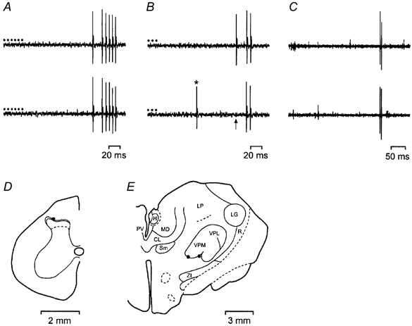
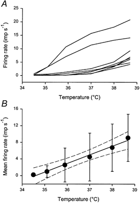
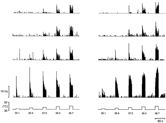
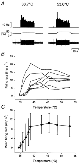
1. In order to further characterize the role of lamina I as the source of central ascending neural pathways for thermoreception and thermoregulation, experiments were performed on anaesthetized cats to determine the quantitative response characteristics of warming-specific lumbosacral spinothalamic lamina I neurones. 2. We identified 10 neurones out of 474 that were selectively excited by cutaneous warming (Warm cells). Their thresholds were all in the range 35-37 degrees C at a baseline of 34.5 degrees C, and their discharge linearly encoded the temperature of graded, innocuous warming stimuli with a sensitivity of 2.1 Hz x degrees C(-1). 3. The stimulus-response function of the Warm cells plateaued at temperatures that were in the noxious heat range. 4. The Warm cells were distinguished from other classes of spinothalamic lamina I neurones by their peripheral inputs, central conduction velocities and level of ongoing activity. 5. The discharge of Warm cells compares well with the known human psychophysics of warm sensibility, and these neurones are likely to be crucial to discriminative thermoreception. Additionally, a role in thermoregulation, a defining feature of mammalian homeostasis, is suggested.
Figures
References
-
- Andrew D, Craig AD. Spinothalamic lamina I neurons selectively sensitive to histamine: a central neural pathway for itch. Nature Neuroscience. 2001a;4:72–77. - PubMed
-
- Andrew D, Craig AD. The encoding of noxious mechanical stimuli by spinothalamic lamina I neurons. Society for Neuroscience Abstracts. 2001b;31 280.7.
-
- Christensen BN, Perl ER. Spinal neurons specifically excited by noxious or thermal stimuli: Marginal zone of the dorsal horn. Journal of Neurophysiology. 1970;33:293–311. - PubMed
-
- Craig AD. Spinal location of ascending lamina I axons in the macaque monkey. Journal of Pain. 2000;1:33–45.
-
- Craig AD, Chen K, Bandy D, Reiman EM. Thermosensory activation of insular cortex. Nature Neuroscience. 2000;3:184–190. - PubMed
Publication types
MeSH terms
Grants and funding
LinkOut - more resources
Full Text Sources
Other Literature Sources
Miscellaneous