

Case Reports
Delayed stroke secondary to increasing mass effect after endovascular treatment of a giant aneurysm by parent vessel occlusion
Affiliations
- PMID: 11733312
- PMCID: PMC7973838
Item in Clipboard
Case Reports
Delayed stroke secondary to increasing mass effect after endovascular treatment of a giant aneurysm by parent vessel occlusion
AJNR Am J Neuroradiol.
2001 Nov-Dec.
Abstract
A 47-year-old woman, who had lost vision in her left eye because of a giant left supraclinoid internal carotid artery aneurysm, was referred for endovascular treatment. Parent-vessel occlusion was performed to obtain circulatory exclusion of the aneurysm. Eight days after treatment, she became hemiparetic and dysphasic. Repeat angiography showed compression of the left middle cerebral artery by the swelling giant aneurysm. Preventive measures should be taken to avert worsening of mass effect when giant aneurysms become thrombotic.
Figures
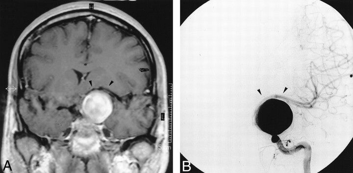
MR image and angiogram obtained prior to treatment. A, Coronal T1-weighted MR image after gadolinium chelate infusion shows the giant left supraclinoid aneurysm shifting the left MCA (arrowheads). B, Left ICA angiogram (frontal projection) depicts the left giant nonthrombosed supraclinoid aneurysm stretching the left MCA (arrowheads).
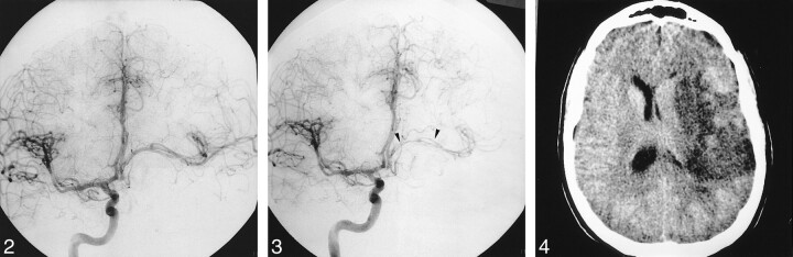
Right carotid angiogram (frontal projection), obtained immediately after left ICA occlusion, shows the absence of retrograde filling of the aneurysm and good cross flow via the ACoA. fig 3. Right carotid angiogram (frontal projection, late arterial phase), obtained 8 d after left ICA occlusion, shows that the stretching and shifting of the left MCA had increased (arrowheads). fig 4. Plain CT scan obtained 20 d after endovascular treatment shows a left MCA territory infarct.
Comment in
-
Device-mediated effect versus the healing response in interventional neuroradiology.AJNR Am J Neuroradiol. 2001 Nov-Dec;22(10):1810-1. AJNR Am J Neuroradiol. 2001. PMID: 11733306 Free PMC article. No abstract available.
-
Delayed stroke secondary to increasing mass effect after endovascular treatment of a giant aneurysm by parent vessel occlusion.AJNR Am J Neuroradiol. 2002 Sep;23(8):1438. AJNR Am J Neuroradiol. 2002. PMID: 12223395 Free PMC article. No abstract available.
Similar articles
-
Intramural thrombosis of giant aneurysms of the supraclinoid and subclinoid internal carotid artery. Report of 4 cases.Ital J Neurol Sci. 1987 Feb;8(1):35-41. doi: 10.1007/BF02361433. Ital J Neurol Sci. 1987. PMID: 3570721
-
Aneurysm formation after carotid occlusion.AJNR Am J Neuroradiol. 1995 Feb;16(2):329-31. AJNR Am J Neuroradiol. 1995. PMID: 7726081 Free PMC article.
-
Delayed intra-aneurysmal migration of a flow diverter construct after treatment of a giant aneurysm of the cavernous internal carotid artery.J Neuroradiol. 2020 May;47(3):233-236. doi: 10.1016/j.neurad.2019.01.092. Epub 2019 Jan 16. J Neuroradiol. 2020. PMID: 30659891 No abstract available.
-
Complex intracranial aneurysms: combined operative and endovascular approaches.Neurosurgery. 1998 Dec;43(6):1304-12; discussion 1312-3. doi: 10.1097/00006123-199812000-00020. Neurosurgery. 1998. PMID: 9848843 Review.
-
Treatment of Large or Giant Cavernous Aneurysm Associated with Persistent Trigeminal Artery: Case Report and Review of Literature.World Neurosurg. 2017 Dec;108:996.e11-996.e15. doi: 10.1016/j.wneu.2017.09.033. Epub 2017 Sep 14. World Neurosurg. 2017. PMID: 28919565 Review.
Cited by
-
Delayed stroke secondary to increasing mass effect after endovascular treatment of a giant aneurysm by parent vessel occlusion.AJNR Am J Neuroradiol. 2002 Sep;23(8):1438. AJNR Am J Neuroradiol. 2002. PMID: 12223395 Free PMC article. No abstract available.
-
Acute vasogenic edema induced by thrombosis of a giant intracranial aneurysm: a cause of pseudostroke after therapeutic occlusion of the parent vessel.AJNR Am J Neuroradiol. 2003 Jun-Jul;24(6):1237-9. AJNR Am J Neuroradiol. 2003. PMID: 12812962 Free PMC article.
-
Flow Diverter Performance Comparison of Different Wire Materials for Effective Intracranial Aneurysm Treatment.Bioengineering (Basel). 2024 Jan 12;11(1):76. doi: 10.3390/bioengineering11010076. Bioengineering (Basel). 2024. PMID: 38247953 Free PMC article.
-
Treatment of giant intracranial aneurysms: long-term outcomes in surgical versus endovascular management.Neurosurg Rev. 2022 Dec;45(6):3759-3770. doi: 10.1007/s10143-022-01884-3. Epub 2022 Oct 21. Neurosurg Rev. 2022. PMID: 36269463 Free PMC article.
-
Endovascular Treatment of Giant Intracranial Aneurysms.Cureus. 2020 May 26;12(5):e8290. doi: 10.7759/cureus.8290. Cureus. 2020. PMID: 32601564 Free PMC article.
References
-
- Gobin YP, Vinuela F, Gurian JH, et al. Treatment of large and giant fusiform intracranial aneurysms with Guglielmi detachable coils. J Neurosurg 1996;84:55-62 - PubMed
-
- Drake CG. Giant intracranial aneurysms: experience with surgical treatment in 174 patients. Clin Neurosurg 1979;26:12-95 - PubMed
-
- Halbach VV, Higashida RT, Dowd CF, et al. The efficacy of endosaccular aneurysm occlusion in alleviating neurological deficits produced by mass effect [see comments]. J Neurosurg 1994;80:659-666 - PubMed
-
- Hirasawa T, Tsubokawa T, Katayama Y, et al. Growth of a giant aneurysm following complete thrombosis by detachable balloon occlusion. Surg Neurol 1992;38:283-286 - PubMed
-
- Larson JJ, Tew JM Jr, Tomsick TA, van Loveren HR. Treatment of aneurysms of the internal carotid artery by intravascular balloon occlusion: long-term follow-up of 58 patients. Neurosurgery 1995;36:26-30 - PubMed
Publication types
MeSH terms
LinkOut - more resources
Full Text Sources
Medical