Case Reports
. 2001 Nov-Dec;22(10):1867-71.

Petrous apex cephaloceles

Affiliations
Case Reports

Petrous apex cephaloceles

K R Moore et al. AJNR Am J Neuroradiol. 2001 Nov-Dec.

Abstract

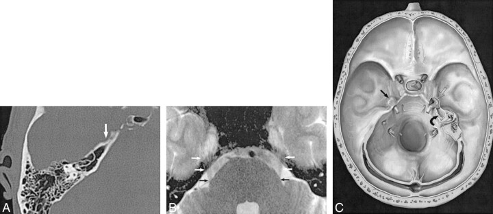
Background and purpose: Petrous apex cephaloceles (PACs) are uncommon lesions that are usually incidental but may be symptomatic. We reviewed MR and CT studies in 10 patients with PACs to identify characteristic imaging features that facilitate their diagnosis.

Methods: MR and CT studies from 10 patients with PACs were reviewed retrospectively. In each case the PAC was characterized by lesion center, signal intensity or attenuation, adjacent petrous apex pneumatization, and its relationship to Meckel's cave. Intraoperative findings were reviewed in the three cases in which surgery was performed.

Results: All 10 patients had lobulated expansile cystic petrous apex lesions centered along the posterolateral margin of Meckel's cave. All cysts were contiguous with Meckel's cave. Three patients had bilateral PACs. Four patients had symptoms that could potentially be explained by the PAC, while findings in the other six were incidental observations. Three patients underwent surgery, during which two lesions were diagnosed as meningoceles while the third was diagnosed as an arachnoid cyst protruding through a dural defect.

Conclusion: PACs represent a protrusion of meninges and CSF from the posterolateral portion of Meckel's cave into the petrous apex, which is their characteristic imaging appearance. PACs are usually incidental but may be symptomatic. Surgical intervention should be approached cautiously and undertaken only when symptoms are clearly linked to the presence of this lesion.


Figures

<sc>fig</sc> 1.
fig 1.
Normal appearance of Meckel's cave. A, CT scan of temporal bone shows smooth anterior petrous apex scalloping at the trigeminal notch and inferior border of the porus trigeminus that delimits the posterior margin of Meckel's cave. The trigeminal notch is a smoothly scalloped area of the petrous apex on bone windows (arrow) that encloses the posterior portion of Meckel's cave. B, High-resolution MR image shows CSF signal intensity in Meckel's cave and the contiguous porus trigeminus, the CSF portal by which the trigeminal nerve courses from the prepontine cistern over the petrous apex into Meckel's cave. This axial T2-weighted FSE (4000/94/4) MR image shows the cisternal trigeminal nerves (arrows) as they cross over the petrous apex into Meckel's cave to form the gasserian ganglion. C, Artist's rendering of temporal bone portrays the normal appearance of the right Meckel's cave (straight black arrow) with dural covering in place. The left petrous apex depicts a PAC. The dura has been removed to demonstrate the gasserian ganglion within Meckel's cave (open arrow) in direct contiguity with the CSF-filled PAC extending posteriorly into the petrous apex (curved arrow). (Reproduced with permission from Electronic Medical Education Resource Group, 2001.)
<sc>fig</sc> 2.
fig 2.
Cholesterol granuloma. A, Axial CT scan shows a smoothly marginated expansile lesion (arrows) centered within the petrous apex, in contradistinction to a PAC, in which the mass is centered within the posterior portion of Meckel's cave and secondarily extends into the petrous apex. B, Axial T1-weighted MR image confirms marked T1 shortening within the lesion (arrows), characteristic of cholesterol granuloma. Diagnosis was proved at surgical exploration.
<sc>fig</sc> 3.
fig 3.
Case 1: 25-year-old woman with meningocele. A, Axial bone algorithm CT scan reveals scalloping of the trigeminal notch and a sharply marginated lesion extending into the left petrous apex (asterisk). B, Axial contrast-enhanced fat-saturated T1-weighted SE (650/16/2) MR image reveals a low-signal-intensity cyst with mild rim enhancement within the left petrous apex (arrow). C, Axial T2-weighted FSE (4350/90/2) MR image confirms hyperintense fluid signal within the apex lesion (arrow).
<sc>fig</sc> 4.
fig 4.
Case 2: 5-year-old boy with arachnoid cyst. A, Axial T2-weighted FSE (4000/92/2) MR image reveals a cystic-appearing right petrous apex lesion with CSF signal intensity contiguous with Meckel's cave (black arrows). Also noted is an ipsilateral mastoid effusion (open arrow). B, Enhanced axial T1-weighted SE (450/12/2) MR image shows a lesion in the apex with CSF signal intensity (white arrows). Open arrow denotes complex effusion.
<sc>fig</sc> 5.
fig 5.
Case 3: 48-year-old woman with meningocele. A, Axial CT scan shows a sharply marginated, expansile left petrous apex mass (asterisk). B, Coronal T1-weighted (350/10/2) MR image shows a cyst of CSF signal intensity extending inferiorly into the petrous apex (arrows). C, Axial T2-weighted FSE (2800/90/1) MR image confirms that the expansile mass revealed on CT studies is a cyst with CSF signal intensity (asterisk) extending from Meckel's cave into the petrous apex.

References

    1. Moore K, Davidson H, Harnsberger H, Shelton C. A practical imaging approach to petrous apex lesions. IJNR Int J Neuroradiol 1999;5:166-184
    1. Moore KR, Harnsberger HR, Shelton C, Davidson HC. “Leave me alone” lesions of the petrous apex. AJNR Am J Neuroradiol 1998;19:733-738 - PMC - PubMed
    1. Cheung SW, Broberg TG, Jackler RK. Petrous apex arachnoid cyst: radiographic confusion with primary cholesteatoma. Am J Otol 1995;16:690-694 - PubMed
    1. Mulcahy MM, McMenomey SO, Talbot JM, Delashaw JB Jr. Congenital encephalocele of the medial skull base. Laryngoscope 1997;107:910-914 - PubMed
    1. Schick B, Draf W, Kahle G, Weber R, Wallenfang T. Occult malformations of the skull base. Arch Otolaryngol Head Neck Surg 1997;123:77-80 - PubMed

Publication types

LinkOut - more resources