. 2001 Nov-Dec;22(10):1901-14.

Etiology of cortical and white matter lesions in cyclosporin-A and FK-506 neurotoxicity

Affiliations

Etiology of cortical and white matter lesions in cyclosporin-A and FK-506 neurotoxicity

W S Bartynski et al. AJNR Am J Neuroradiol. 2001 Nov-Dec.

Abstract

Background and purpose: The etiology of the neurotoxicity associated with cyclosporin-A (CsA) and FK-506 treatment is not fully understood. At our institution, we noticed a distinct, abrupt change in the imaging characteristics of CsA and FK-506 neurotoxicity, which consisted of a shift in lesion morphology from a white matter abnormality to a mixed cortical and white matter pattern. The purpose of this study was to assess clinical parameters that might explain this change.

Methods: Twenty-two patients had a neurotoxic reaction and brain imaging changes while receiving CsA or FK-506. Nineteen patients received allogeneic bone marrow transplants, and three had aplastic marrow disorders. Fifty-one imaging studies (CT or MR imaging) were obtained, and lesion characteristics, locations, and time courses were evaluated along with relevant clinical data.

Results: Nine patients who had been conditioned for transplantation with cyclophosphamide and chemotherapy (busulfan or thiotepa) had a mixed pattern of cortical and white matter involvement (57 lesions). Isolated white matter involvement (62 lesions) developed in three nontransplant patients and 10 transplant patients conditioned with cyclophosphamide and total-body irradiation. All lesions occurred at typical brain watershed zones. Lesion enhancement was noted in two patients conditioned with chemotherapy. Initial images demonstrated characteristic lesions in 15 patients (68%). Initial images were normal in four patients (18%) and nonspecific in three patients (14%).

Conclusion: Lesion location in CsA and FK-506 neurotoxicity may depend on the presence or type of conditioning used before bone marrow transplantation. Nontransplant patients or those conditioned with total-body irradiation develop white matter lesions, whereas those conditioned with chemotherapy develop mixed cortical and white matter lesions.


Figures

None
fig. 1. Axial nonenhanced CT sections obtained 24 h after seizure in a patient (case 4) who was conditioned for transplantation with cyclophosphamide and TBI. She had a new-onset seizure 23 d after transplantation. At the onset of toxic reaction, she had grade 3 BMT-related TM, and her CsA level was within the normal therapeutic range. The initial head CT study obtained on the day of the seizure was normal. Her baseline blood pressure was 92/52 mm Hg but increased to 230/130 mm Hg at the time of toxic reaction and initial CT. A, Image at the level of lateral ventricles shows edema in the corpus callosum (arrow). B, Image at the level of the sentrum semiovale shows extensive areas of white matter edema in the parietal region bilaterally (arrow).
None
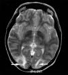
fig. 2. T2-weighted MR image (2500/84/2) obtained on the day of presentation shows typical areas of subcortical white matter abnormality without cortical involvement (arrows) in patient (case 2) who was conditioned for transplantation with cyclophosphamide and TBI and who presented with confusion and altered mental status 14 d after transplantation. She did not develop seizures. At the time of toxic reaction, she had grade 4 BMT-related TM and a CsA level within the normal therapeutic range. Her baseline blood pressure of 115/85 mm Hg increased to 167/104 mm Hg at the time of toxic reaction and MR imaging
None
fig. 3. MR images in a patient (case 14), conditioned for transplantation with cyclophosphamide and busulfan, who presented with new-onset seizure 176 d after transplantation. The patient had grade 3 BMT-related TM that rapidly progressed to grade 4. The CsA level was in the normal therapeutic range. His baseline blood pressure was 160/90 mm Hg, which increased to 170/110 mm Hg at the time of toxic reaction. The initial CT scan showed only nonspecific areas of low attenuation in the parietal region. MR images were obtained 6 h after CT. A–C, T2-weighted MR images from the midventricular level and high parietal region (2500/84/2) show extensive high-signal-intensity edema involving the cortex in the frontal and parietal regions bilaterally (arrowheads). D and E, Contrast-enhanced images corresponding to B and C show bilateral punctate enhancement in the cortex of the frontal region (arrows). These areas of enhancement correspond to the regions of cortical signal abnormality noted in B and C.
None
fig. 4. CT scans obtained at the time of toxic reaction shows areas of low attenuation extending centrally from the frontal lobe to the parietal region, involving the white matter and projecting to the cortical surface. Hemorrhages are noted in the right frontal lobe and right parietal area (arrows). The brain abnormalities are severe and appear to lie in a region that could easily coincide with the boundary between anterior cerebral and middle cerebral territories. Images were obtained in a patient (case 11), conditioned for transplantation with cyclophosphamide and busulfan, who presented with generalized tonic-clonic seizure activity 8 d after transplantation. At the onset of toxic reaction, the patient had grade 3 BMT-related TM that rapidly worsened to grade 4. The cyclosporin level at the time of toxic reaction was within the normal therapeutic range. Her baseline blood pressure was 110/70 mm Hg, which increased to 152/96 mm Hg at toxic reaction
None
fig. 5. Images in a patient (case 18), who was conditioned for transplantation with cyclophosphamide, thiotepa, and busulfan and who presented with binocular cortical blindness 15 d after transplantation. At the time of toxic reaction, the patient had grade 3 BMT-related TM, and the CsA level was within the normal therapeutic range. Her baseline blood pressure was 150/88 mm Hg, which increased to 180/100 mm Hg at toxic reaction. A, T2-weighted axial MR image (2500/84/2) shows areas of mixed cortical and white matter signal intensity abnormalities in the occipital pole region bilaterally. Abnormalities are greater on the left (arrow). At autopsy, vasogenic edema was present in the bilateral occipital lobes, with evidence of ischemic neuronal necrosis in the cortex at the occipital watershed region. B, T2-weighted sagittal MR image (2500/84/2) clearly shows signal intensity abnormality in the calcarine region (arrow). C, 3D TOF MR angiogram, collapsed view (46/6.9/1), shows vessel narrowing and irregularity in the left middle cerebral artery (curved arrow) and bilateral posterior cerebral arteries; these findings are more striking on the left (straight arrow). D, Postprocessed 3D TOF MR angiogram with background noise elimination shows a string-of-beads appearance in the posterior cerebral arteries; the finding is more severe on the left (arrow). At autopsy, these vessels appeared normal, without evidence of vasculitis.

References

    1. Joss DV, Barrett AJ, Kendra JR, Lucas CF, Desai S. Hypertension and convulsions in children receiving cyclosporin A. Lancet 1982;1:906 - PubMed
    1. Boogaerts MA, Zachee P, Verwilghen RL. Cyclosporin, methylprednisolone, and convulsions. Lancet 1982;2:1216-1217 - PubMed
    1. Dieperink H, Moller J. Ketoconazole and cyclosporin. Lancet 1982;2:1217 - PubMed
    1. Durrant S, Chipping PM, Palmer S, Gordon-Smith EC. Cyclosporin A, methylprednisolone, and convulsions. Lancet 1982;2:829-830 - PubMed
    1. Noll RB, Kulkarni R. Complex visual hallucinations and cyclosporine. Arch Neurol 1984;41:329-330 - PubMed

LinkOut - more resources