

Sequential MR imaging and proton MR spectroscopy in patients who underwent recent detoxification for chronic alcoholism: correlation with clinical and neuropsychological data
- PMID: 11733327
- PMCID: PMC7973857
Sequential MR imaging and proton MR spectroscopy in patients who underwent recent detoxification for chronic alcoholism: correlation with clinical and neuropsychological data
Abstract
Background and purpose: Chronic alcohol abuse may cause neuropsychological disorders and result in brain atrophy. The purpose of this study was to evaluate the metabolic, morphologic, and functional cerebral changes in the early stage of abstinence from chronic alcoholism.
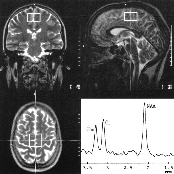
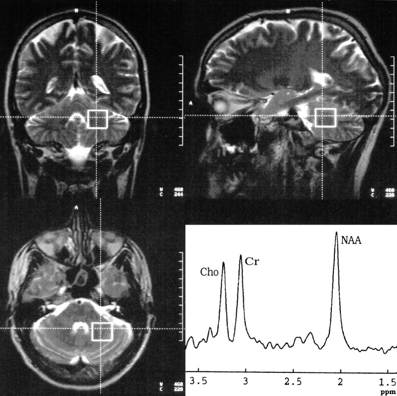
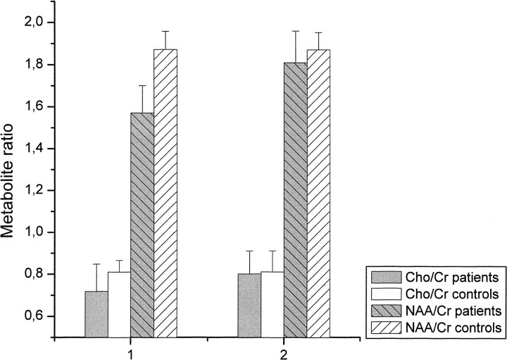
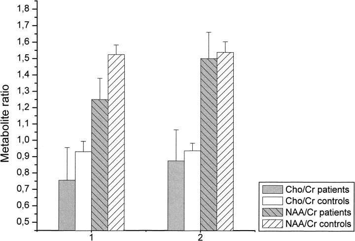
Methods: Seventeen alcohol-dependent patients underwent MR imaging and MR spectroscopy on days 1 through 3 and days 36 through 39 of abstinence. In addition, psychological performance measures testing intelligence, concentration, attention, and memory were applied. Neuropsychological data were correlated with spectroscopic and volumetric results by using a Pearson's product moment correlation. The same measurements were also performed in 12 healthy, age-matched control subjects. Peak integral values for N-acetylaspartate (NAA) and choline (Cho) were referred to the peak integral value of creatine (Cr) as the internal reference.
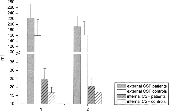
Results: NAA/Cr was decreased in the patients in both the frontal lobes and cerebellum immediately after cessation of drinking (days 1 through 3). After 36 to 39 days of abstinence, NAA/Cr had significantly increased in the patients and corresponded to performance on psychological tests. The Cho/Cr ratio was decreased in the cerebellum during early abstinence but was recovered on days 36 through 39. The patients had enlarged CSF spaces 1 to 3 days after detoxification, which decreased during sobriety. The extent of brain atrophy did not correspond to performance on psychological performance tests.
Conclusion: Regression of brain atrophy and metabolic recovery occurs at an early stage after abstinence from chronic alcohol abuse. MR spectroscopy findings return to normal metabolic levels within weeks after detoxification. The recovery of NAA/Cr is associated with improved performance on neuropsychological tests.
Figures
References
-
- Charness ME, Simon RP, Greenberg DA. Ethanol and the nervous system. N Engl J Med 1989;321:442-454 - PubMed
-
- Carlen PL, Wotzmann G, Holgate RC, Wilkinson DA, Rankin JG. Reversible brain atrophy in recently abstinent alcoholics measured by computed tomography scans. Science 1978;200:1076-1078 - PubMed
-
- Besson JAO, Glenn AIM, Foreman EL, et al. Nuclear magnetic resonance disorders in alcoholic disorder and the role of vasopressin. Lancet 1981;2:923-924 - PubMed
-
- Fein G, Meyerhoff DJ, Di Sclafani V, et al. 1H magnetic resonance spectroscopic imaging separates neuronal from glial changes in alcohol-related brain atrophy. In: Alcohol and Glial Cells, Research Monograph 27. Bethesda, MD: National Institutes of Health; 1994:227–241
-
- Jagannathan NR, Desai NG, Raghunathan P. Brain metabolite changes in alcoholism: an in vivo proton magnetic resonance spectroscopy (MRS) study. Magn Reson Imaging 1996;14:553-557 - PubMed
MeSH terms
Substances
LinkOut - more resources
Full Text Sources
Medical