

Neutrophil transmigration in inflammatory bowel disease is associated with differential expression of epithelial intercellular junction proteins
- PMID: 11733350
- PMCID: PMC1850599
- DOI: 10.1016/S0002-9440(10)63051-9
Neutrophil transmigration in inflammatory bowel disease is associated with differential expression of epithelial intercellular junction proteins
Abstract
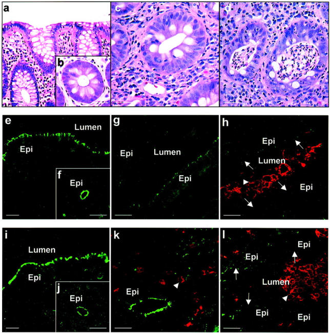
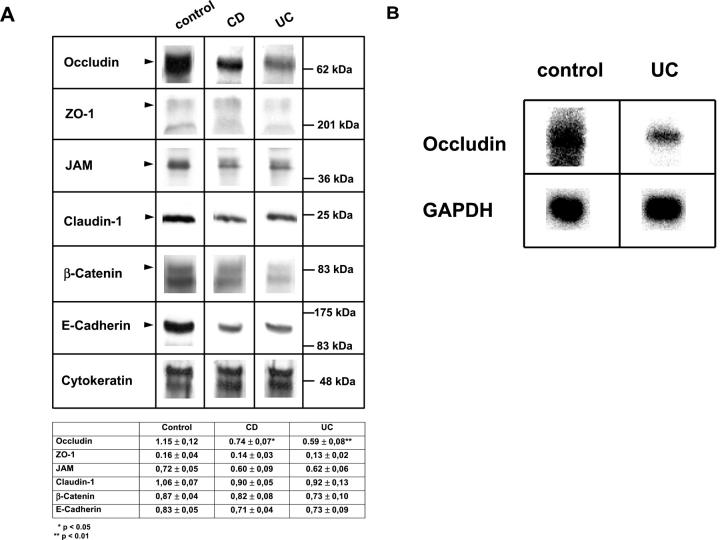
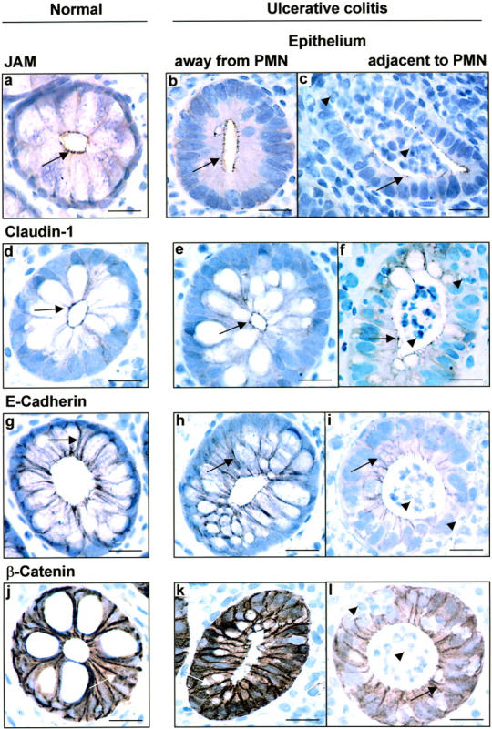
Inflammatory bowel disease (IBD) consisting of ulcerative colitis (UC) and Crohn's (CD) typically displays a waxing and waning course punctuated by disease flares that are characterized by transepithelial migration of neutrophils (PMN) and altered barrier function. Since epithelial barrier function is primarily regulated by the apical most intercellular junction referred to as the tight junction (TJ), our aim was to examine expression of TJ and adherens junction (AJ) proteins in relation to PMN infiltration in mucosal tissue samples from patients with active IBD. Expression of epithelial intercellular TJ proteins (occludin, ZO-1, claudin-1, and JAM) and subjacent AJ (beta-catenin and E-cadherin) proteins were examined by immunoflourescence/confocal microscopy, immunohistochemistry, and Western blotting. Colonic mucosa from patients with UC revealed dramatic, global down-regulation of the key TJ transmembrane protein occludin in regions of actively transmigrating PMN and in quiescent areas in the biopsy samples. Significant decreases in occludin expression were observed at the protein and mRNA levels by Western and Northern blotting. In contrast, expression of other TJ and AJ proteins such as ZO-1, claudin-1, JAM, beta-catenin, and E-cadherin were down-regulated only in epithelial cells immediately adjacent to transmigrating PMN. Analysis of inflamed mucosa from Crohn's disease patients mirrored the results obtained with UC patients. No change in TJ and AJ protein expression was observed in colonic epithelium from patients with collagenous colitis or lymphocytic colitis that are respectively characterized by a thickened subepithelial collagen plate and increased intraepithelial lymphocytes. These results suggest that occludin expression is diminished in IBD by mechanisms distinct from those regulating expression of other intercellular junction proteins. We speculate that down-regulation of epithelial occludin may play a role in enhanced paracellular permeability and PMN transmigration that is observed in active inflammatory bowel disease.
Figures
Similar articles
-
Constitutive activation of Rho proteins by CNF-1 influences tight junction structure and epithelial barrier function.J Cell Sci. 2003 Feb 15;116(Pt 4):725-42. doi: 10.1242/jcs.00300. J Cell Sci. 2003. PMID: 12538773
-
Interleukin-18 facilitates neutrophil transmigration via myosin light chain kinase-dependent disruption of occludin, without altering epithelial permeability.Am J Physiol Gastrointest Liver Physiol. 2012 Feb 1;302(3):G343-51. doi: 10.1152/ajpgi.00202.2011. Epub 2011 Dec 1. Am J Physiol Gastrointest Liver Physiol. 2012. PMID: 22135309
-
The epithelium in inflammatory bowel disease: potential role of endocytosis of junctional proteins in barrier disruption.Novartis Found Symp. 2004;263:115-24; discussion 124-32, 211-8. Novartis Found Symp. 2004. PMID: 15669638 Review.
-
Adherens junctions and tight junctions are regulated via different pathways by progastrin in epithelial cells.J Cell Sci. 2003 Apr 1;116(Pt 7):1187-97. doi: 10.1242/jcs.00321. J Cell Sci. 2003. PMID: 12615962
-
Tight junctions and cell-cell interactions.Methods Mol Biol. 2006;341:185-95. doi: 10.1385/1-59745-113-4:185. Methods Mol Biol. 2006. PMID: 16799199 Review.
Cited by
-
Relationship between pathogenic E.coli O78-induced intestinal epithelial barrier damage and Zonulin expression levels in yaks.Front Cell Infect Microbiol. 2024 Sep 23;14:1456356. doi: 10.3389/fcimb.2024.1456356. eCollection 2024. Front Cell Infect Microbiol. 2024. PMID: 39376662 Free PMC article.
-
Neutrophil-Epithelial Interactions: A Double-Edged Sword.Am J Pathol. 2016 Jun;186(6):1404-16. doi: 10.1016/j.ajpath.2016.02.001. Epub 2016 Apr 12. Am J Pathol. 2016. PMID: 27083514 Free PMC article. Review.
-
Cross-talk between intestinal epithelial cells and immune cells in inflammatory bowel disease.Sci Rep. 2016 Jul 15;6:29783. doi: 10.1038/srep29783. Sci Rep. 2016. PMID: 27417573 Free PMC article.
-
Mechanisms of intestinal dysbiosis: new insights into tuft cell functions.Gut Microbes. 2024 Jan-Dec;16(1):2379624. doi: 10.1080/19490976.2024.2379624. Epub 2024 Jul 23. Gut Microbes. 2024. PMID: 39042424 Free PMC article. Review.
-
Lipopolysaccharide induces CXCL2/macrophage inflammatory protein-2 gene expression in enterocytes via NF-kappaB activation: independence from endogenous TNF-alpha and platelet-activating factor.Immunology. 2006 Jun;118(2):153-63. doi: 10.1111/j.1365-2567.2006.02344.x. Immunology. 2006. PMID: 16771850 Free PMC article.
References
-
- Madara JL: Regulation of the movement of solutes across tight junctions. Annu Rev Physiol 1998, 60:143-159 - PubMed
-
- Katz KD, Hollander D, Vadheim CM, McElree C, Delahunty T, Dadufalza VD, Krugliak P, Rotter JI: Intestinal permeability in patients with Crohn’s disease and their healthy relatives. Gastroenterology 1989, 97:927-931 - PubMed
-
- Sandle GI, Higgs N, Crowe P, Marsh MN, Venkatesan S, Peters TJ: Cellular basis for defective electrolyte transport in inflamed human colon. Gastroenterology 1990, 99:97-105 - PubMed
-
- Hollander D: The intestinal permeability barrier: a hypothesis to its regulation and involvement in Crohn’s disease. Scand J Gastroenterol 1992, 27:721-726 - PubMed
-
- Söderholm JD, Peterson KH, Olaison G, Franzen LE, Weström B, Magnusson K-E, Sjödahl R: Epithelial permeability to proteins in the noninflamed ileum of Crohn’s disease? Gastroenterology 1999, 117:65-72 - PubMed
Publication types
MeSH terms
Substances
Grants and funding
LinkOut - more resources
Full Text Sources
Other Literature Sources
Miscellaneous
