. 2001 Dec;159(6):2045-54.
doi: 10.1016/s0002-9440(10)63056-8.

Cathepsin B knockout mice are resistant to tumor necrosis factor-alpha-mediated hepatocyte apoptosis and liver injury: implications for therapeutic applications

Affiliations

Cathepsin B knockout mice are resistant to tumor necrosis factor-alpha-mediated hepatocyte apoptosis and liver injury: implications for therapeutic applications

M E Guicciardi et al. Am J Pathol. 2001 Dec.

Abstract

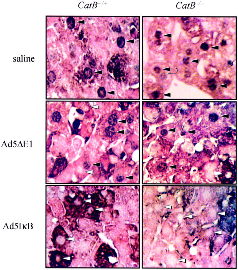
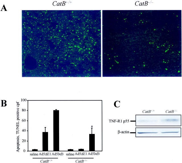
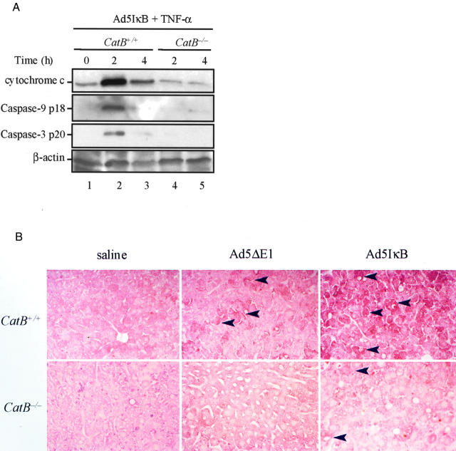
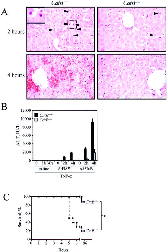
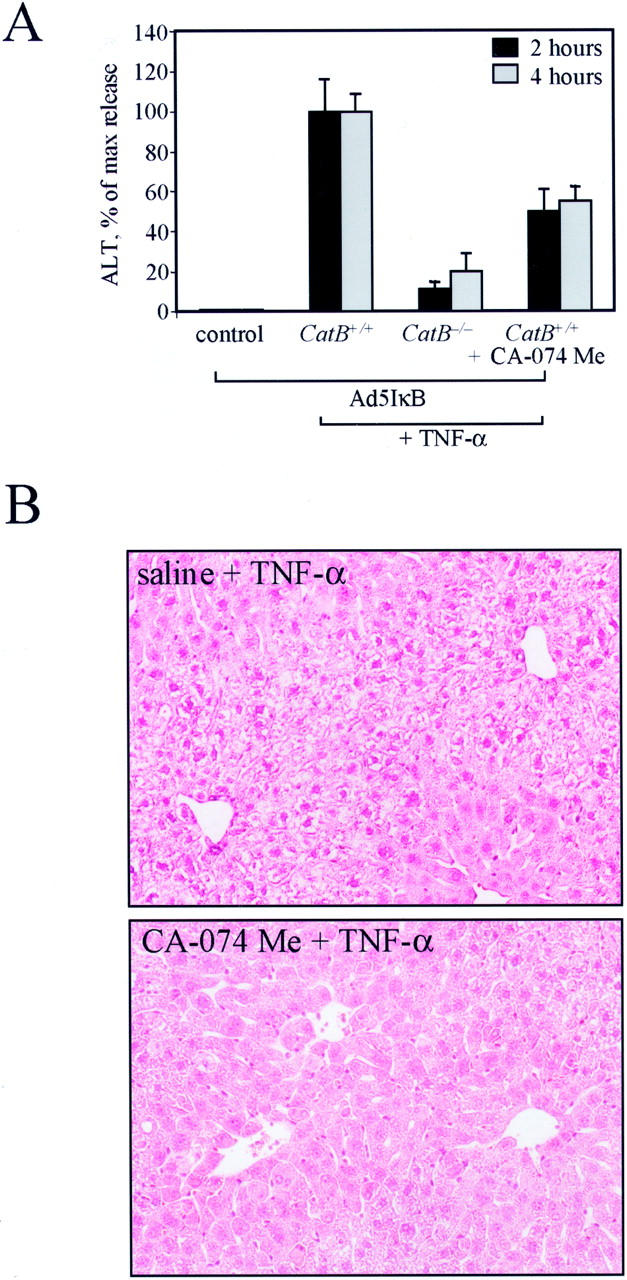
Tumor necrosis factor-alpha (TNF-alpha) contributes to liver injury by inducing hepatocyte apoptosis. Recent evidence suggests that cathepsin B (cat B) contributes to TNF-alpha-induced apoptosis in vitro. The aim of the present study was to determine whether cat B contributes to TNF-alpha-induced hepatocyte apoptosis and liver injury in vivo. Cat B knockout (catB(-/-)) and wild-type (catB(+/+)) mice were first infected with the adenovirus Ad5I kappa B expressing the I kappa B superrepressor to inhibit nuclear factor-kappa B-induced survival signals and then treated with murine recombinant TNF-alpha. Massive hepatocyte apoptosis with mitochondrial release of cytochrome c and activation of caspases 9 and 3 was detected in catB(+/+) mice 2 hours after the injection of TNF-alpha. In contrast, significantly less hepatocyte apoptosis and no detectable release of cytochrome c or caspase activation occurred in the livers of catB(-/-) mice. By 4 hours after TNF-alpha injection, only 20% of the catB(+/+) mice were alive as compared to 85% of catB(-/-) mice. Pharmacological inhibition of cat B in catB(+/+) mice with L-3-trans-(propylcarbamoyl)oxirane-2-carbonyl-L-isoleucyl-L-proline (CA-074 Me) also reduced TNF-alpha-induced liver damage. The present data demonstrate that a cat B-mitochondrial apoptotic pathway plays a pivotal role in TNF-alpha-induced hepatocyte apoptosis and liver injury.


Figures

Figure 1.
Figure 1.
Infection with the adenovirus Ad5IκB effectively prevents NF-κB translocation to the nucleus in mouse hepatocytes. Wild-type and catB−/− mice were injected via tail vein with the adenovirus Ad5IκB encoding for an IκB superrepressor. In control experiments, mice were injected with the empty adenovirus Ad5ΔE1 or with sterile saline. Twenty-four hours later, each mouse received a dose of 0.5 μg of murine recombinant TNF-α intravenously. Mice were killed after 2 hours of treatment with TNF-α, and immunohistochemistry for NF-κB was performed on paraffin-embedded tissue. Hepatocyte nuclei from control animals showed strongly positive staining for NF-κB (black arrowheads). Inhibition of NF-κB translocation from the cytosol to the nucleus was confirmed by the lack of immunohistochemical staining for NF-κB in the nuclei of both catB+/+ and catB−/− in mice infected with Ad5IκB (white arrowheads).
Figure 2.
Figure 2.
CatB−/− mice are more resistant to TNF-α-mediated hepatocyte apoptosis. Apoptosis was measured in situ by TUNEL assay in catB+/+and catB−/− mouse livers after a 2-hour treatment with TNF-α. TUNEL-positive cells were visualized by fluorescence microscopy using excitation and emission wavelengths of 490 and 520 nm, respectively. Representative fluorescence photomicrographs of low-power fields of liver of catB+/+ and catB−/− mice injected with Ad5IκB are shown (A) along with quantitation of apoptotic cells per high-power field in controls (saline, Ad5ΔE1) and Ad5IκB-injected animals (B). Original magnifications, ×40. C: Expression of TNF-R1 in catB+/+ and catB−/− mouse livers was evaluated by immunoblot. Aliquots of 60 μg of protein from whole-cell lysates were prepared as described in Materials and Methods and subjected to sodium dodecyl sulfate-polyacrylamide gel electrophoresis on a 10% acrylamide gel, transferred to nitrocellulose membrane, and blotted for TNF-R1. TNF-R1 was detected in both catB+/+ and catB−/− mouse livers after 2 to 4 hours of treatment with TNF-α. The blot for β-actin served as a control for protein loading.
Figure 3.
Figure 3.
The mitochondrial pathway of apoptosis is activated in catB+/+ but not in catB−/− mice. CatB+/+ and catB−/− mice were injected with the adenovirus Ad5IκB. Twenty-four hours later, mice were treated intravenously with TNF-α and sacrificed after 2 and 4 hours. A: At the indicated time points, cytosolic fractions were prepared as described in Materials and Methods and aliquots of 60 μg of protein were subjected to sodium dodecyl sulfate-polyacrylamide gel electrophoresis on a 15% acrylamide gel, transferred to nitrocellulose membrane, and sequentially blotted for cytochrome c, active caspase 9, and active caspase 3. Cytosolic cytochrome c and active caspase 9 and 3 were only detected in catB+/+-treated livers. Blot for β-actin served as a control for protein loading. B: Representative immunohistochemistry for active caspase 3 and 7 in catB+/+ and cat B−/− mice injected with saline, Ad5ΔE1 (controls), or Ad5IκB, after a 2-hour treatment with TNF-α. Strongly positive immunochemical staining reaction for active caspase 3 and 7 was detectable in the cytoplasm of hepatocytes from Ad5IκB-injected catB+/+ mice after treatment with TNF-α, but not from catB−/− mice or in controls. The tissue sections were counterstained with eosin. Original magnifications, ×25.
Figure 4.
Figure 4.
. TNF-α induces acute liver damage in catB+/+, but not in catB−/− mice. CatB+/+ and catB−/− mice were injected with the adenovirus Ad5IκB, treated intravenously 24 hours later with TNF-α and sacrificed after 2 and 4 hours. A: Representative microscopic photographs of the liver of catB+/+ and catB−/− mice after 2 and 4 hours of treatment with TNF-α are shown. CatB+/+ mice showed massive hepatocyte apoptosis at 2 hours and extensive hemorrhagic necrosis at 4 hours. Normal liver architecture and few apoptotic hepatocytes were observed in catB−/− mice both at 2 and 4 hours. Arrowheads point to apoptotic cells. Higher magnification of apoptotic cells is shown in the inset. Original magnification, ×40 (H&E). B: Measurement of serum ALT values in catB+/+ and catB−/− mice injected with saline, Ad5ΔE1 (controls), or Ad5IκB, and treated with TNF-α. Serum ALT values were significantly greater in catB+/+ than in catB−/− mice after 2 and 4 hours of treatment with TNF-α. Data are expressed as mean ± SEM, n = 6, *P < 0.01 catB+/+ versus catB−/− mice. C: Survival curves of catB+/+ (dotted line) and catB−/− (solid line) mice injected with Ad5IκB and treated with TNF-α (n = 10 catB+/+ and n = 8 catB−/−; *, P < 0.01).
Figure 5.
Figure 5.
Pharmacological inhibition of cat B reduces TNF-α-induced liver damage in catB+/+ mice. CatB+/+ mice were injected with the adenovirus Ad5IκB. Twenty-four hours later, mice were given an intraperitoneal injection of 0.9 ml of saline with or without CA-074 Me (4 mg/100 g) 30 minutes before the intravenous injection of TNF-α (0.5 μg), and sacrificed after 2 and 4 hours. A: Serum ALT values after 2 and 4 hours of treatment with TNF-α were significantly reduced in catB+/+ mice pretreated with CA-074 Me (CatB+/+ + CA-074 Me) as compared to catB+/+ mice pretreated with saline (CatB+/+). Measurement of serum ALT values in catB−/− mice (CatB−/−) and untreated catB +/+ mice (control) are also shown. Data are expressed as percentage of release (max release) obtained treating wild-type mice with TNF-α (mean ± SEM). B: Representative photomicrographs of livers of catB+/+ mice pretreated with saline (saline + TNF-α) or with CA-074 Me (CA-074 + TNF-α) and subsequently treated with TNF-α for 2 hours are shown. CatB+/+ mice pretreated with saline showed massive hygropic degeneration and architectural distortion. Liver architecture is preserved and minimal damage is observed in catB+/+ mice pretreated with CA-074 Me. Original magnification, ×40 (H&E).

References

    1. Wallach D, Varfolomeev EE, Malinin NL, Goltsev YV, Kovalenko AV, Boldin MP: Tumor necrosis factor receptor and Fas signaling mechanisms. Annu Rev Immunol 1999, 17:331-367 - PubMed
    1. Bradham CA, Plumpe J, Manns MP, Brenner DA, Trautwein C: Mechanisms of hepatic toxicity. I. TNF-induced liver injury. Am J Physiol 1998, 275:G387-G392 - PubMed
    1. McClain CJ, Cohen DA: Increased tumor necrosis factor production by monocytes in alcoholic hepatitis. Hepatology 1989, 9:349-351 - PubMed
    1. Colletti LM, Remick DG, Burtch GD, Kunkel SL, Strieter RM, Campbell DA, Jr: Role of tumor necrosis factor-alpha in the pathophysiologic alterations after hepatic ischemia/reperfusion injury in the rat. J Clin Invest 1990, 85:1936-1943 - PMC - PubMed
    1. Gonzalez-Amaro R, Garcia-Monzon C, Garcia-Buey L, Moreno-Otero R, Alonso JL, Yague E, Pivel JP, Lopez-Cabrera M, Fernandez-Ruiz E, Sanchez-Madrid F: Induction of tumor necrosis factor alpha production by human hepatocytes in chronic viral hepatitis. J Exp Med 1994, 179:841-848 - PMC - PubMed

Publication types

MeSH terms

Substances

LinkOut - more resources