. 2001 Dec;159(6):2055-69.
doi: 10.1016/S0002-9440(10)63057-X.

Effects of paramyxoviral infection on airway epithelial cell Foxj1 expression, ciliogenesis, and mucociliary function

Affiliations

Effects of paramyxoviral infection on airway epithelial cell Foxj1 expression, ciliogenesis, and mucociliary function

D C Look et al. Am J Pathol. 2001 Dec.

Abstract

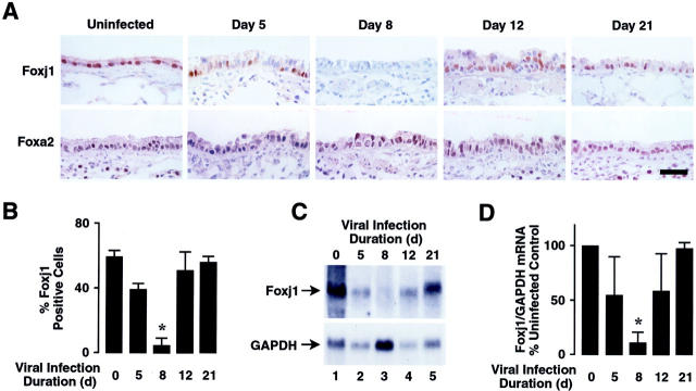
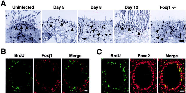
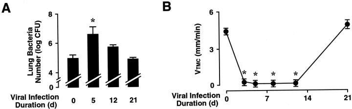
To elucidate molecular mechanisms underlying the association between respiratory viral infection and predisposition to subsequent bacterial infection, we used in vivo and in vitro models and human samples to characterize respiratory virus-induced changes in airway epithelial cell morphology, gene expression, and mucociliary function. Mouse paramyxoviral bronchitis resulted in airway epithelial cell infection and a distinct pattern of epithelial cell morphology changes and altered expression of the differentiation markers beta-tubulin-IV, Clara cell secretory protein, and Foxj1. Furthermore, changes in gene expression were recapitulated using an in vitro epithelial cell culture system and progressed independent of the host inflammatory response. Restoration of mature airway epithelium occurred in a pattern similar to epithelial cell differentiation and ciliogenesis in embryonic lung development characterized by sequential proliferation of undifferentiated cells, basal body production, Foxj1 expression, and beta-tubulin-IV expression. The effects of virus-induced alterations in morphology and gene expression on epithelial cell function were illustrated by decreased airway mucociliary velocity and impaired bacterial clearance. Similar changes in epithelial cell Foxj1 expression were also observed in human paramyxoviral respiratory infection. Taken together, these model systems of paramyxoviral respiratory infection mimic human pathology and identify epithelial cell Foxj1 expression as an early marker of epithelial cell differentiation, recovery, and function.


Figures

Figure 1.
Figure 1.
Respiratory paramyxoviral infection alters airway epithelial cell morphology and cilia. Wild-type C57BL/6J mice underwent intranasal inoculation with 5 × 10 EID50 of SdV. At the indicated times after inoculation, lungs were harvested for evaluation. A: Lungs were subjected to histopathological study in which 6-μm sections were immunostained for SdV protein and detected using alkaline phosphatase and a red chromagen. Representative sections are shown and no detectable staining was observed for nonimmune IgG. Scale bar, 20 μm. B: Tracheas were examined by scanning EM at low magnification (top row; scale bar, 12 μm), and high magnification (bottom row; scale bar, 4 μm).
Figure 2.
Figure 2.
Respiratory paramyxoviral infection alters expression of airway epithelial cell differentiation markers in vivo. A: Mice underwent intranasal inoculation with SdV as in Figure 1A ▶ . At the indicated times after inoculation, lungs were harvested for histopathological study in which 6-μm sections were immunostained for β-tubulin-IV using green FITC fluorescence or for CCSP or TTF-1 using horseradish peroxidase with DAB resulting in a brown product. Representative sections are shown and no detectable staining was observed for nonimmune IgG. Scale bar, 30 μm. The percentage of airway lumen cells that expressed β-tubulin-IV (B), CCSP (C), and TTF-1 (D) were calculated visually using samples from A. Values are expressed as mean ± SD (n = 3 to 10 regions from three to five different animals in each group), and a significant decrease compared to mice not infected with SdV is indicated by an asterisk.
Figure 3.
Figure 3.
Respiratory paramyxoviral infection causes loss of Foxj1 expression in vivo. A: Mice underwent intranasal inoculation with SdV as in Figure 1A ▶ . At the indicated times after inoculation, lungs were harvested for histopathological study in which 6-μm sections were immunostained for Foxj1 or Foxa2 using horseradish peroxidase and DAB resulting in a brown product. Representative sections are shown and no detectable staining was observed for nonimmune IgG. Scale bar, 30 μm. B: The percentage of airway lumen cells that expressed Foxj1 was calculated visually using samples from A. Values are expressed as mean ± SD (n = 8 regions from four different animals in each group), and a significant decrease compared to animals not infected with SdV is indicated by an asterisk. C: Mice were infected with SdV as in Figure 1A ▶ . After the indicated durations of infection, total cellular RNA was isolated from lungs. Levels of Foxj1 and GAPDH mRNA were determined by RNA blot analysis and the position of the 1.4-kb Foxj1 and 1.3-kb GAPDH mRNAs are indicated by arrows. D: Relative levels of Foxj1 mRNA were calculated by densitometry similar to C. Values are expressed as mean percent Foxj1/GAPDH RNA level compared to animals not infected with SdV ± SD (n = 3 different animals in each group), and a significant decrease compared to uninfected animals is indicated by an asterisk.
Figure 4.
Figure 4.
Respiratory paramyxoviral infection directly injures airway epithelial cells with loss of Foxj1 expression in vitro. Differentiated RTE cells grown on semipermeable supports under air liquid interface conditions were inoculated with 2 × 10 EID50 of SdV. At the indicated times after inoculation, RTE cell were evaluated for gene expressions and changes in morphology. A: RTE cells on membrane supports embedded in paraffin were subjected to histopathological study in which 6-μm sections were immunostained for SdV protein and detected using alkaline phosphatase and a red chromagen. Scale bar, 30 μm. RTE cells grown on semipermeable supports were fixed and examined en face by scanning EM. Scale bar, 4 μm. RTE cells in sections (as in A) were immunostained for β-tubulin-IV using green FITC fluorescence or Foxj1 and TTF-1 using a horseradish peroxidase-DAB resulting in a brown product. Representative sections are shown and no detectable staining was observed for nonimmune IgG. Scale bar, 30 μm. B: The percentage of airway cells expressing SdV proteins (open circles) calculated from RTE cell cytospin preparations using immunodetection of SdV as in A, the percentage of ciliated RTE cells (closed squares) calculated visually from low-power-scanning EM images as in A, and TTF-1 (shaded diamonds) calculated visually using samples from A. Values are expressed as mean ± SD (n = 3 to 6 regions from two to three different animals in each group), and a significant difference compared to animals not infected with SdV is indicated by an asterisk. C: Total cellular RNA was isolated from RTE cells, uninfected (Un) or inoculated with SdV, and levels of Foxj1 and GAPDH mRNA were determined by RNA blot analysis as in Figure 3C ▶ . Representative data of three independent experiments is shown.
Figure 5.
Figure 5.
Respiratory paramyxoviral infection induces airway epithelial cell proliferation. A: Mice underwent intranasal inoculation with SdV as in Figure 1A ▶ . At the indicated times after inoculation, lungs were harvested for examination by scanning EM. Luminal cell density was calculated visually in 104-μm areas. Values are expressed as mean ± SD (n = 5 regions from two to three different animals in each group), and a significant difference compared to animals not infected with SdV is indicated by an asterisk. B: Mice were infected with SdV as in Figure 1A ▶ , and 20 hours before evaluation animals were administered intraperitoneal BrdU. After the indicated duration of infection, lungs were harvested for histopathological study in which 6-μm sections were immunostained for BrdU using horseradish peroxidase and DAB to produce a brown product. Samples were counterstained with hematoxylin. Representative sections are shown and no detectable staining was observed for nonimmune IgG. Scale bar, 20 μm. C: The percentage of airway lumen cells that expressed BrdU was calculated visually using samples from B. Values are expressed as mean ± SD (n = 5 to 11 regions from two to three different animals in each group), and a significant increase compared to animals not infected with SdV is indicated by an asterisk.
Figure 6.
Figure 6.
Respiratory paramyxoviral infection leads to an ordered epithelial repair process. A: Wild-type C57BL/6J mice underwent intranasal inoculation with SdV as in Figure 1A ▶ . At the indicated times after inoculation, lungs were harvested for examination by transmission EM. Shown at the far right is a section from an uninfected Foxj1 null (−/−) mouse. Arrowheads indicate location of basal bodies. Scale bar, 1 μm. B: Mice were infected with SdV as in Figure 1A ▶ and administered BrdU as in Figure 5B ▶ . After 12 days, lungs were harvested for study in which 6-μm sections were immunostained for BrdU using green FITC fluorescence and Foxj1 using red Cy3 fluorescence. Scale bar, 30 μm. C: Samples obtained as described in B were immunostained for BrdU using green FITC fluorescence and Foxa2 using red Cy3 fluorescence. Scale bar, 30 μm.
Figure 7.
Figure 7.
Respiratory paramyxoviral infection impairs airway clearance. A: Mice were infected with SdV as in Figure 1A ▶ for the indicated durations, followed by tracheobronchial inoculation with 10 cfu of nontypable H. influenzae strain 12. After 16 hours of bacterial infection, lung homogenates were prepared for quantitation of H. influenzae. Values are expressed as mean ± SD (n = 3 to 6 in each group), and a significant increase compared to animals not infected with SdV is indicated by an asterisk. B: Mice were infected with SdV as in Figure 1A ▶ . At the indicated times after inoculation, mice were anesthetized, and the velocity of 0.2-μm polystyrene beads within the trachea was determined. Values are expressed as mean ± SD (n = 3 to 5 in each group), and a significant decrease compared to animals not infected with SdV is indicated by an asterisk.
Figure 8.
Figure 8.
Human respiratory paramyxoviral infection causes loss of Foxj1 expression. Samples from lungs obtained during diagnostic or postmortem examination of patients uninfected (control) or infected with RSV (patients 1 to 5) as demonstrated by immunostaining and or sample culture. Serial 6-μm sections were immunostained for Foxj1 and TTF-1 and detected using horseradish peroxidase and DAB to produce a brown product. Samples were counterstained with hematoxylin. Representative sections are shown and no detectable staining was observed for nonimmune IgG. Scale bar, 20 μm.

Similar articles

Cited by

References

    1. Zhang P, Summer WR, Bagby GJ, Nelson S: Innate immunity and pulmonary host defense. Immunol Rev 2000, 173:39-51 - PubMed
    1. Thomas LH, Wickremasinghe MI, Sharland M, Friedland JS: Synergistic upregulation of interleukin-8 secretion from pulmonary epithelial cells by direct and monocyte-dependent effects of respiratory syncytial virus infection. J Virol 2000, 74:8425-8433 - PMC - PubMed
    1. Frick AG, Joseph TD, Pang L, Rabe AM, St Geme JW, III, Look DC: Haemophilus influenzae stimulates ICAM-1 expression on respiratory epithelial cells. J Immunol 2000, 164:4185-4196 - PubMed
    1. Wanner A, Salathé M, O’Riordan TG: Mucociliary clearance in the airway. Am J Respir Crit Care Med 1996, 154:1868-1902 - PubMed
    1. Erjefalt JS, Erjefalt I, Sundler F, Persson CG: In vivo restitution of airway epithelium. Cell Tissue Res 1995, 281:305-316 - PubMed

Publication types

MeSH terms