. 2001 Dec;159(6):2215-25.
doi: 10.1016/s0002-9440(10)63072-6.

Selective insolubility of alpha-synuclein in human Lewy body diseases is recapitulated in a transgenic mouse model

Affiliations

Selective insolubility of alpha-synuclein in human Lewy body diseases is recapitulated in a transgenic mouse model

P J Kahle et al. Am J Pathol. 2001 Dec.

Abstract

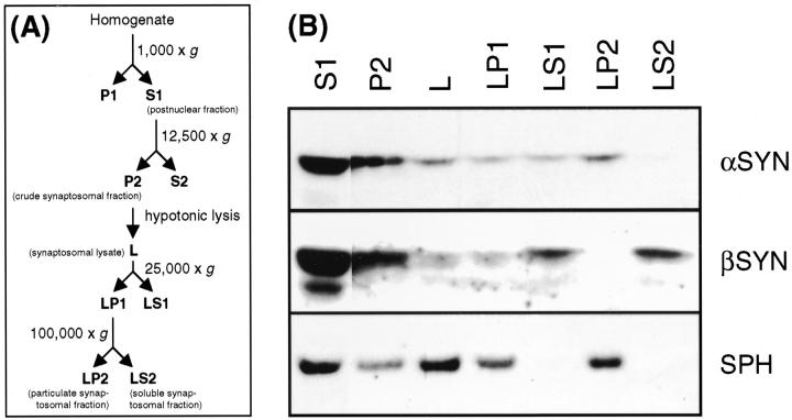
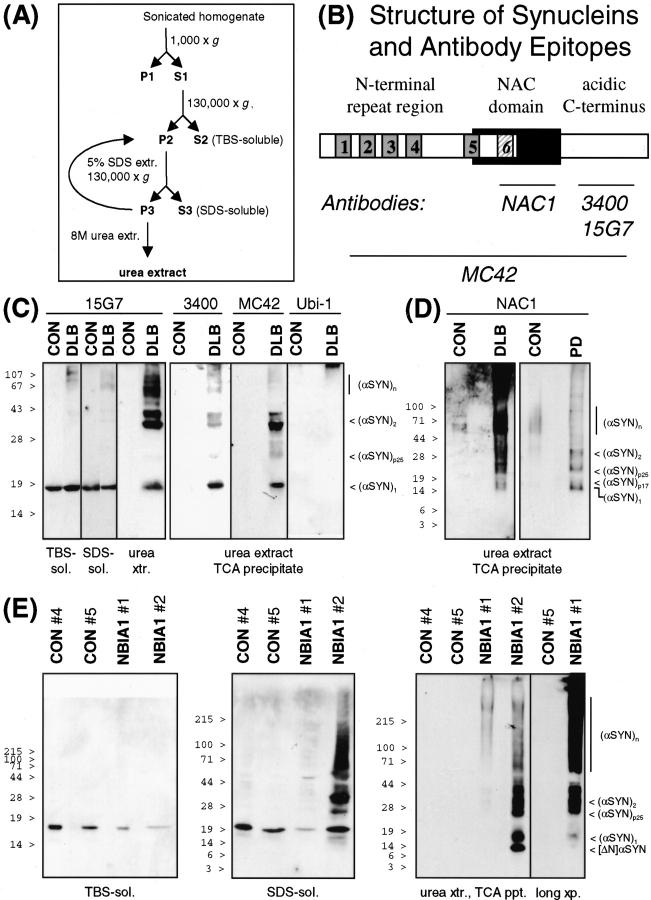
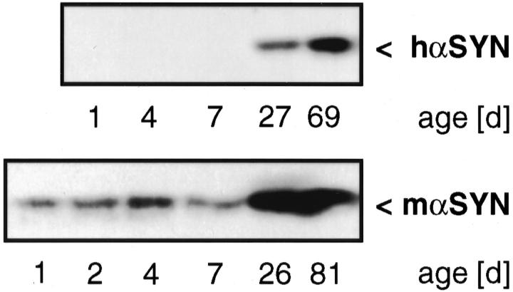
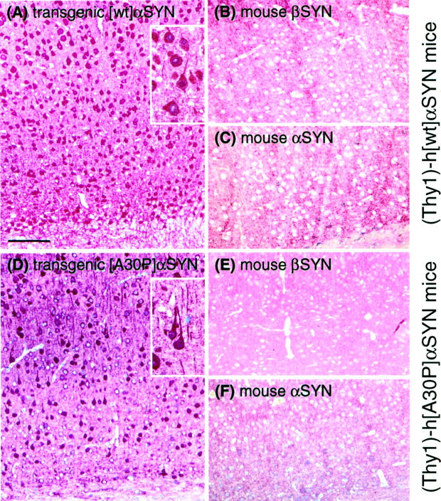
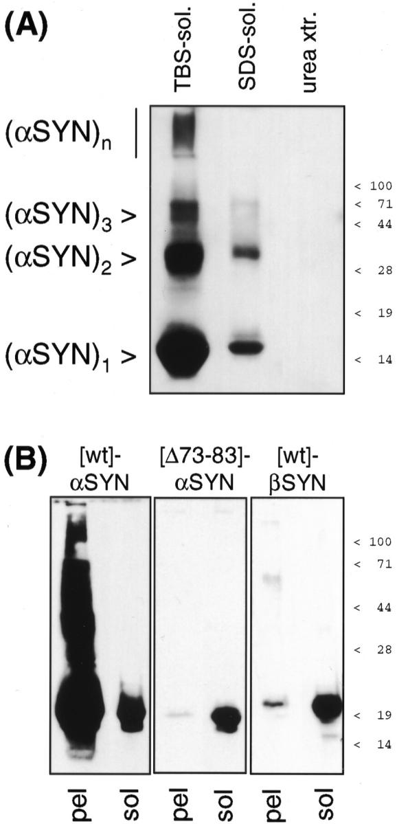
alpha-Synuclein (alpha-SYN) is deposited in intraneuronal cytoplasmic inclusions (Lewy bodies, LBs) characteristic for Parkinson's disease (PD) and LB dementias. alpha-SYN forms LB-like fibrils in vitro, in contrast to its homologue beta-SYN. Here we have investigated the solubility of SYNs in human LB diseases and in transgenic mice expressing human wild-type and PD-associated mutant [A30P]alpha-SYN driven by the brain neuron-specific promoter, Thy1. Distinct alpha-SYN species were detected in the detergent-insoluble fractions from brains of patients with PD, dementia with LBs, and neurodegeneration with brain iron accumulation type 1 (formerly known as Hallervorden-Spatz disease). Using the same extraction method, detergent-insolubility of human alpha-SYN was observed in brains of transgenic mice. In contrast, neither endogenous mouse alpha-SYN nor beta-SYN were detected in detergent-insoluble fractions from transgenic mouse brains. The nonamyloidogenic beta-SYN was incapable of forming insoluble fibrils because amino acids 73 to 83 in the central region of alpha-SYN are absent in beta-SYN. In conclusion, the specific accumulation of detergent-insoluble alpha-SYN in transgenic mice recapitulates a pivotal feature of human LB diseases.


Figures

Figure 1.
Figure 1.
Subcellular fractionation of frozen human brain tissue. A: Schematic representation of the subcellular fractionation steps, annotations in B correspond to the fractions outlined in A. B: Parietal cortex (0.4 g) from a human control individual was homogenized and subjected to subcellular fractionation. Twenty μg of each fraction was TCA precipitated (except postnuclear fraction S1 that was loaded directly) and subjected to denaturing 12.5% PAGE. Sequential Western probing was done with 15G7 anti-α-SYN, SY38 anti-synaptophysin (SPH), and 6584 anti-β-SYN, as indicated. Data are representative for three different control cortex fractionations.
Figure 2.
Figure 2.
SDS-insoluble α-SYN species in LB diseases. A: Schematic representation of the differential extraction steps. See Materials and Methods for details. B: Structure of α-SYN and epitopes recognized by anti-α-SYN antibodies. The imperfect KTKEGV repeats are numbered, the sixth repeat missing in β-SYN (see below) is stippled. C: Extracts from temporal cortex of control and DLB brain were prepared, and 10 μg of TBS-soluble material, 10 μg of SDS-soluble material, and 10 μl of urea extracts or TCA precipitates from 50-μl urea extracts (as indicated at the bottom) were subjected to denaturing 12.5% PAGE. Western blots were sequentially probed with three different antibodies against α-SYN (15G7, 3400, MC42) and anti-ubiquitin (Ubi-1), as indicated on the top. Immunoreactivity was visualized with SuperSignal (for 15G7) or ECLplus. D: TCA precipitates from 50-μl urea extracts from temporal cortex of control and DLB brain (left) and from parietal cortex of control and PD brain (right) were loaded on 10 to 20% Tris-tricine gels. The corresponding Western blots were probed with anti-NAC and developed with ECLplus. E: Parietal cortex samples from two controls and two NBIA1 patients were extracted in parallel. TBS-soluble material (10 μg, left), SDS-soluble material (25 μg, middle), and urea extracts (80 μl, right) were separated by SDS-PAGE (TBS-soluble, 15%; SDS and urea extracts, 4 to 20% gradient). MC42 and ECLplus were used for Western detection of α-SYN. Note that sample NBIA1 no. 2 with higher LB density than NBIA1 no. 1 had also much stronger α-SYN immunoreactivity in the urea extract. Nevertheless, the α-SYN immunoreactive band pattern of NBIA1 no. 1 was qualitatively the same as of NBIA1 no. 2, as evidenced by a longer exposure of the blot to the far right. Each experiment is representative for two to three independent extractions. Positions of prestained molecular weight standards are indicated to the left. See text for description of α-SYN species denoted to the right.
Figure 3.
Figure 3.
Developmental expression of α-SYN. Mouse brains were collected from mice at the indicated age, and Western blots prepared from 25-μg cytosolic extracts. Wild-type mouse blots were probed with MC42 (top), representative [(Thy1)-h[A30P]α-SYN line 18] transgenic mouse blots probed with 3400 (bottom), and developed with ECLplus.
Figure 4.
Figure 4.
Immunostainings of brain slices (motor cortex) show specific accumulation of α-SYN but not of β-SYN in transgenic mice. Animals expressing either [wt]α-SYN (A–C) or [A30P]α-SYN (D–F) showed a strong cytosolic labeling of neuronal cells with the human-specific α-SYN antibody 15G7 (A and D). A section from a 1-month-old [wt]α-SYN-expressing mouse is shown in A to demonstrate the early onset of accumulation of transgenic protein. In contrast, immunostainings with the β-SYN-specific antiserum (6485) and the murine-specific α-SYN antiserum (7544) only revealed a synaptic staining pattern. Neither accumulation of endogenous murine α-SYN (C and F) nor β-SYN (B and E) was detectable in neuronal cell bodies. Scale bar in A, 100 μm.
Figure 5.
Figure 5.
Detergent-insoluble α-SYN in transgenic mouse brains. Whole brains of transgenic mice (A: 3- to 4-month-old [wt]α-SYN lines 14 and 23, and [A30P]α-SYN lines 18 and 31; B: 1-month-old and 1-year-old [A30P]α-SYN lines 18 and 31; as indicated at the bottom) and age-matched nontransgenic littermates (lm) were differentially extracted. Buffer- and detergent-soluble proteins (10 μg for transgenic human α-SYN, 50 μg for endogenous mouse SYNs), and TCA precipitate of urea extracts were subjected to denaturing 15% PAGE. Western blots were probed with human (transgene)-specific anti-hα-SYN 15G7, endogenous mouse-specific anti-mα-SYN 7544, and anti-β-SYN 6485, as indicated to the left. 15G7-immunoreactive bands were developed with SuperSignal, polyclonal antibody immunoreactivity with ECLplus. A control lane on the urea extract blots contained 10 μg of cytosol from a transgenic [A30P]α-SYN mouse. The positions of prestained molecular weight markers are indicated to the right. Individual variance of transgenic α-SYN expression levels may account for the apparently higher amount of urea extractable mutant h[A30P]α-SYN compared to h[wt]α-SYN in one experiment (A, exp. 2), but not in two additional experiments (one of them shown as A, exp. 1), and for the apparent increase with age of SDS-soluble α-SYN (B) that was not seen in an additional experiment.
Figure 6.
Figure 6.
α-SYN, but not β-SYN aggregates in vitro because of a critical determinant in the NAC domain. A: Recombinant human α-SYN aggregates were collected by 100,000 × g centrifugation and redissolved in 15 volumes of TBS+. The buffer-insoluble material was extracted like the brain samples above. All fractions were TCA precipitated and separated by denaturing 15% PAGE. Western blots were probed with 3400 anti-α-SYN and developed with SuperSignal. This experiment was repeated three times with the same result. B: Solutions (2 mg/ml) of [wt]α-SYN (left), [Δ73–83]α-SYN (middle), and [wt]β-SYN (right) were aggregated for 7 days. After ultracentrifugation, the 100,000 × g pellets (pel) were subjected to denaturing 12.5% PAGE. α-SYN immunoblots were probed with MC42 and β-SYN immunoblots with 6485, and developed with SuperSignal until the band intensities of 1-μg freshly dissolved, nonaggregated protein (sol) were comparable. Note the typical retarded electrophoretic motility of recombinant β-SYN. Positions of prestained molecular weight markers are indicated to the right.

References

    1. Uéda K, Fukushima H, Masliah E, Xia Y, Iwai A, Yoshimoto M, Otero DA, Kondo J, Ihara Y, Saitoh T: Molecular cloning of cDNA encoding an unrecognized component of amyloid in Alzheimer disease. Proc Natl Acad Sci USA 1993, 90:11282-11286 - PMC - PubMed
    1. Spillantini MG, Schmidt ML, Lee VM-Y, Trojanowski JQ, Jakes R, Goedert M: α-Synuclein in Lewy bodies. Nature 1997, 388:839-840 - PubMed
    1. Wakabayashi K, Matsumoto K, Takayama K, Yoshimoto M, Takahashi H: NACP, a presynaptic protein, immunoreactivity in Lewy bodies in Parkinson’s disease. Neurosci Lett 1997, 239:45-48 - PubMed
    1. Takeda A, Mallory M, Sundsmo M, Honer W, Hansen L, Masliah E: Abnormal accumulation of NACP/α-synuclein in neurodegenerative disorders. Am J Pathol 1998, 152:367-372 - PMC - PubMed
    1. Irizarry MC, Growdon W, Gomez-Isla T, Newell K, George JM, Clayton DF, Hyman BT: Nigral and cortical Lewy bodies and dystrophic nigral neurites in Parkinson’s disease and cortical Lewy body disease contain α-synuclein immunoreactivity. J Neuropathol Exp Neurol 1998, 57:334-337 - PubMed

Publication types

MeSH terms